This protocol describes a survival procedure for safe and well-tolerated murine mesenteric lymphadenectomyat each of four stations along the mesenteric lymph node chain. This procedure is adaptable to allow for selective disruption of lymphatic communication with the distal colon, proximal colon, and/or small intestine.
A subscription to JoVE is required to view this content. Sign in or start your free trial.
Method Article
Murine Mesenteric Lymphadenectomy for Selective Disruption of Lymphatic Communication with Region-Specific Gut
In This Article
Summary
Abstract
Immunosurveillance versus antigen-tolerance is a fine balance within the gut microenvironment, during which the critical role of circulating T cell priming within local lymph nodes is well-known. Subsequent to T cell priming, recent literature has begun to demonstrate a unique, non-redundant role of non-circulating T cells residing in the draining lymph node of an organ. This may be particularly true in the gut, where lymph drainage is known to be highly specific. However, to truly understand the unique impact of these local cells versus systemic functional redundancy, the scientific community requires a well-described procedure to isolate and selectively perturb these nodes. Described here is a survival procedure for safe and well-tolerated murine mesenteric lymphadenectomy at each of four stations along the mesenteric lymph node chain. This procedure is adaptable to allow for selective disruption of lymphatic communication with the distal colon, proximal colon, and/or small intestine. Included are detailed instructions, visual aids, helpful tips for improved success, and troubleshooting for suboptimal survival. Resected lymph nodes are immediately ready for further downstream processing if desired. This technique is best suited for studies requiring selective disruption of lymph drainage from the distal colon, proximal colon, and/or small intestine.
Introduction
T cells are one of the major effector cell types in adaptive immunity, with described roles across diverse disease categories, including infections, autoimmunity, and malignancy1,2,3. A critical step for T cell functionality is the three-phase priming by antigen-presenting cells within secondary lymph organs, such as local lymph nodes4. In modern medicine, a mainstay of proper oncologic resection across multiple organ systems includes some degree of local sampling or en bloc lymph node resection5,6,7,8. For example, in colon cancer, consensus guidelines recommend that 12 lymph nodes be removed and examined in curative-intent resection due to a clear correlation with improved long-term survival6,7. However, can local lymph nodes truly be resected with impunity? Recent studies in mice have demonstrated that the tumor-draining lymph node contains a reservoir of antitumor T cell precursors, which may be protected from the terminal differentiation and T cell exhaustion that occurs within the tumor microenvironment9,10,11. This would suggest that removing such a critical population could dampen ongoing immunosurveillance capabilities.
The gut lymph drainage pattern in mice is known to be quite specialized along the mesenteric lymph node chain; for example, the proximal colon is almost exclusively drained by the first lymph node station, the small intestines by the second/third station, and the distal colon predominantly by the fourth lymph node station12 (Figure 1, Supplementary Figure 1). Thus, direct communication between segments of the gut and lymphatics can be disrupted with targeted ablation of these nodes. To block trafficking between lymph nodes and tissues, S1P receptor agonists, such as FTY720, and integrin blockade are commonly used; however, these drugs broadly inhibit trafficking and thus cannot be used to investigate the contribution of specific lymph nodes13,14. However, the ability of the wider scientific community to study the local effect of immune interactions within gut lymph nodes is limited by the absence of a well-described surgical resection for each station. With this gap in mind, a survival procedure for murine mesenteric lymphadenectomy at each node station was developed and is described here. The overall goal of this procedure is to achieve safe and reliable excision of the mesenteric lymph node station(s) in mice with high postoperative survival and tolerance.
This procedure has been tested and is easily performed in juvenile and adult age mice across multiple strains (i.e.,Balb/c, C57BL/6J, 129S4), but it is limited by the need for accessible and easily identifiable lymph nodes. Therefore, it may not be well-suited to combine with mouse models or genotypes with smaller than usual or imperceptible lymph nodes (e.g., lymphotoxin-alpha deficient mice)15. In addition, older mice (>15 weeks) tend to have more visceral adiposity, making lymph node identification and isolationmore difficult, and while this is not a contraindication, it can be success/procedure-limiting. Aside from these limitations, the mesenteric lymphadenectomy offers a highly controlled and highly selective perturbation of lymph nodes. This procedure would be best suited to unravel questions about the local effect of lymph nodes and their associated resident immune cells and interactions on segments of the gut (e.g., proximal colon, distal colon, small intestine).

Figure 1: Anatomical landmarks of stations in the mesenteric lymph node chain. (A) Unlabeled and (B) labeled depiction of four major mesenteric lymph node stations in situ. Proximal colon drainage is predominantly via station 1, small intestine is predominantly station 2/3, and distal colon is predominantly station 4. (DC= distal colon, ICV= ileocolic vessels, PC= proximal colon, CEC= cecum, IV/JV= ileal vessels/jejunal vessels, SI= small intestine). Scale bars: 1.5 cm. Please click here to view a larger version of this figure.
Access restricted. Please log in or start a trial to view this content.
Protocol
All procedures are performed in compliance with Yale Institutional Animal Care and Use Committee protocols. To develop this procedure, 8-12 week-old C57BL/6 strain, wild-type mice, and age/sex/genotype-matched sham controls were utilized. Both male and female mice were included. Body weight ranged from 18-25 g. The reagents and the equipment used are listed in the Table of Materials.
1. Preparation of instruments, animals, and the operating room
- Sterilize all surgical instruments. Ensure the operating surface and anesthetic nose cone are cleansed with 70% ethanol.
- Set up the inhaled anesthetic vaporizer machine with a nose cone and suction for excess anesthetic scavenging. Set up an operating surface with a warm-water circulating pad to maintain animal body temperature and supplies in a convenient, sterile location.
NOTE: This procedure does not require an operating microscope; however, consider including this instrument to improve visualization. - Induce anesthesia by administering 2%-3% inhaled isoflurane with an oxygen flow rate of 2 L/min via nose cone from an inhaled anesthetic vaporizer machine (following institutionally approved protocols).
- Ensure appropriate plane of anesthesia by confirming a respiratory rate of approximately 30-40 breaths/min and lack of response to hindfoot pinch. Periodically recheck for the appropriate plane of anesthesia and adjust the isoflurane flow rate accordingly.
NOTE: Alternatively, an injectable anesthetic is also appropriate.
- Ensure appropriate plane of anesthesia by confirming a respiratory rate of approximately 30-40 breaths/min and lack of response to hindfoot pinch. Periodically recheck for the appropriate plane of anesthesia and adjust the isoflurane flow rate accordingly.
- Follow one's institute guidelines for appropriate analgesia and local anesthesia. For example, administer a long-acting analgesic such as 3.25 mg/kg buprenorphine XR subcutaneously and administer up to 0.04 mL/5g body weight of 1mg/mL lidocaine solution subcutaneously at the planned incision.
- Prepare the animal for procedure by positioning supine, applying eye lubricant, plucking or clipping abdominal fur, and sterilizing the abdomen with povidone/iodine solution, followed by 70% ethanol (Figure 2A).
- Use sterile gloves and apply sterile drapes over the animal with a window to the operative field. Maintain sterility throughout the procedure.
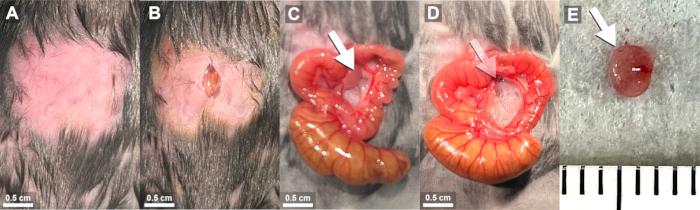
Figure 2: Representative images of major procedural steps in mesenteric lymphadenectomy. (A) Plucked and sterilized abdominal window. (B) 0.5 cm incision through the skin and linea alba. (C) Exposure of the mesenteric lymph node chain by orienting the ileum right, proximal colon left, and cecum inferior. The first mesenteric lymph node lies along the ileocolic artery (white arrow). (D) Excised mesenteric lymph node (faded arrow). Scale bars: 0.5 cm. (E) Ex vivo mesenteric lymph node (white arrow; 2.5 mm x 3 mm). Ticks = 1 mm. Please click here to view a larger version of this figure.
2. Mesenteric lymphadenectomy preparation
- Make a 0.5-1 cm midline incision through the skin. Bluntly dissect skin away from the abdominal wall and identify the linea alba. Make a 0.5-1 cm incision through the linea alba to gain access to the peritoneal cavity (Figure 4B).
NOTE: To improve access to nodes of station 2/3/4, consider extending incision cephalad, but not to surpass the cartilaginous xiphoid process. - Instill 1 mL of body-temperature sterile saline into the peritoneal cavity via a blunt tip gavage needle and moisten the sterile drapes in preparation for bowel positioning. Use sterile, moistened cotton-tipped swabs to identify and eviscerate the cecum through the incision.
- Avoid using sharp instruments to handle the bowel, as this can result in injury. Periodically re-moisten drapes and bowel to prevent damage from drying.
NOTE: The murine cecum generally resides in the left lower quadrant of the abdomen, but can be housed in any quadrant.
- Avoid using sharp instruments to handle the bowel, as this can result in injury. Periodically re-moisten drapes and bowel to prevent damage from drying.
- Clarify the target mesenteric lymph node station(s) for resection and follow specific instructions below (i.e., 3.1-3.4). See Figure 1A,B for anatomical landmarks and identification.
3. Dissection and excision of mesenteric lymph node stations
- First lymph node station (proximal colon drainage; Supplementary Figure 1A,B').
- Orient the exteriorized bowel onto moistened drapes with cecum inferiorly, proximal colon superiorly, and terminal ileum towards the surgeon's right (animal's left side; Figure 1A, Figure 2C).
- Identify the mesenteric lymph node chain, which courses longitudinally along the colon. Using angled fine-tipped forceps, bluntly dissect the first lymph node station away from the ileocolic vessels by placing closed forceps in the space between these structures and allowing the forceps to open gently.
- Remove forceps and repeat this maneuver 2-3 times or until a sufficient plane has been created for safe excision. Avoid damage to major colonic blood vessels (Figure 3A,B).
NOTE: If significant bleeding is encountered, abort the procedure and humanely euthanize the animal (following institutionally approved protocols).
- Remove forceps and repeat this maneuver 2-3 times or until a sufficient plane has been created for safe excision. Avoid damage to major colonic blood vessels (Figure 3A,B).
- OPTIONAL: To decrease a theoretical risk of collaterals to nearby nodes, perform an optional suture ligation with single interrupted sutures along the lymph node chain (one1mm distal and one 1mm proximal to the first lymph node station) using 9-0 black monofilament nylon suture and fine-tipped forceps to drive the needle (Figure 3C,D).
- Gently retract the ligated lymph station caudad and, using fine-point scissors, excise the target lymph node station from cephalad to caudad (Figure 3E).
NOTE: Often, mice have a small but visible feeding vessel off the ileocolic artery into the posterior aspect of the lymph node. Bleeding from this vessel can be controlled by gentle pressure with a cotton-tipped swab until hemostasis is achieved.
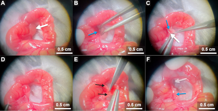
Figure 3: Annotated stills and commentary from mesenteric lymphadenectomy (first station). (A) Proper orientation of the bowel exposes the first mesenteric lymph node station (white arrow). (B) A plane is created between the ileocolic artery (bluearrow) and the node. (C) Suture ligation of feeding vessels entering the apex of the lymph node (blue arrow) is performed while excluding the ileocolic vessels by exiting the space between these structures (white arrow). (D) Suture ligation of the base of the lymph node is performed, again noting and avoiding the distal branches of the ileocolic artery. (E) The node is excised along the previously created tissue plane, staying high towards the lymph node and leaving suture ligations behind (black arrows). (F) Hemostasis is confirmed, distal branches of the ileocolic artery are observed to ensure proper blood flow, and the empty space (faded arrow)where the node previously lived is noted to be free of residual node. Scale bars: 0.5 cm. Please click here to view a larger version of this figure.
- Second lymph node station (mid-distal small intestine drainage)
- Gently eviscerate the small intestine as well. Orient the exteriorized bowel onto moistened drapes with the cecum inferiorly, proximal colon superiorly, and small intestine towards the surgeon's right (animal's left side; Figure 1A).
- Identify the mesenteric lymph node chain, which courses longitudinally along the colon. The second station begins at the confluence of ileal/jejunal vessels (Figure 1B).
- Using angled fine-tipped forceps, bluntly dissect the second lymph node stationaway from the colonic vessels by placing closed forceps in the space between these structures and allowing the forceps to open gently.
- Remove forceps and repeat this maneuver 2-3 times or until a sufficient plane has been created for safe excision. Avoid damage to major colonic blood vessels.Also, take special note of the ileal and jejunal vessels, which would be detrimental if injured.
NOTE: If significant bleeding is encountered, abort the procedure and humanely euthanize the animal.
- OPTIONAL: To decrease a theoretical risk of collaterals to nearby nodes, perform an optional suture ligation with single interrupted sutures along the lymph node chain (one 1 mm distal and one 1 mm proximal to the second lymph node station) using 9-0 black monofilament nylon suture and fine-tipped forceps to drive the needle (Supplementary Figure 2A).
- Gently retract the ligated lymph stationcaudad and, using fine-point scissors, excise the target lymph node station from cephalad to caudad (Supplementary Figure 2B).
NOTE: Often, mice have a small but visible feeding vessel off the ileocolic artery into the posterior aspect of the lymph node. Bleeding from this vessel can be controlled by gentle pressure with a cotton-tipped swab until hemostasis is achieved.
- Second and third lymph node station (prox-mid-distal small intestine drainage; Supplementary Figure 1C,D'
- Gently eviscerate the entirety of the small intestine to the point of posterior tethering (ligament of Treitz). Orient the exteriorized bowel onto moistened drapes with the cecum inferiorly, proximal colon superiorly, and small intestine towards the surgeon's right (animal's left side; Figure 1A).
- Identify the mesenteric lymph node chain, which courses longitudinally along the colon. The second station begins at the confluence of ileal/jejunal vessels (Figure 1B).
- Using angled fine-tipped forceps, bluntly dissect the second and third lymph node station away from the colonic vessels by placing closed forceps in the space between these structures and allowing the forceps to open gently.
- Remove forceps and repeat this maneuver 2-3 times or until a sufficient plane has been created for safe excision. Avoid damage to major colonic blood vessels. Also, take special note of the ileal and jejunal vessels, which would be detrimental if injured.
NOTE: If significant bleeding is encountered, abort the procedure and humanely euthanize the animal.
- OPTIONAL: To decrease a theoretical risk of collaterals to nearby nodes, perform an optional suture ligation with single interrupted sutures along the lymph node chain (usually one 1 mm distal, two 1 mm lateral, and one 1 mm proximal to the second/third lymph node stations) using 9-0 black monofilament nylon suture and fine-tipped forceps to drive the needle (Supplementary Figure 3B,C).
- Note the afferent lymphatic tracts, which lie just superficial to the ileal/jejunal vascular bundles (Supplementary Figure 3B). Take caution to ensure suture ligation courses deep to lymphatics and superficial to major vascular structures, as damage to ileal/jejunal vascular bundles would be detrimental.
- Gently retract the ligated lymph stations cephalad and, using fine or extra-fine point scissors, excise the target lymph node stations from caudad to cephalad.
NOTE: Before proceeding, ensure the small intestine remains pink and well-perfused. Incidental ligation of the ileal/jejunal vessels will result in ischemic small intestine.
- Fourth lymph node station (distal colon drainage; Supplementary Figure 1E,F')
- Gently eviscerate the entirety of the small intestine to the point of posterior tethering (ligament of Treitz). Orient the exteriorized bowel onto moistened drapes with the cecum inferiorly, proximal colon superiorly, and small intestine towards the surgeon's right (animal's left side; Figure 1A).
- Identify the mesenteric lymph node chain, which courses longitudinally along the colon. The fourth station resides just medial to the distal colon and lateral to the third station (Figure 1B, Figure 4A).
- Using angled fine-tipped forceps or extra-fine point scissors, bluntly dissect the fourth lymph node station away from the colonic vessels and surrounding adipose by placing closed forceps/scissors in the space between these structures and allowing the forceps/scissors to open gently.
- Remove forceps/scissors and repeat this maneuver 2-3 times or until a sufficient plane has been created for safe excision. Avoid damage to major colonic blood vessels.
NOTE: If significant bleeding is encountered, abort the procedure and humanely euthanize the animal.
- OPTIONAL: To decrease a theoretical risk of collaterals to nearby nodes, perform an optional suture ligation with single interrupted sutures at each identifiable lymph node (usually one 1 mm from the node) using 9-0 black monofilament nylon and fine-tipped forceps to drive the needle (Figure 4B - D).
- Gently retract the ligated lymph stations cephalad and, using extra-fine point scissors, excise the target lymph node station from caudad to cephalad (Figure 4D,E).
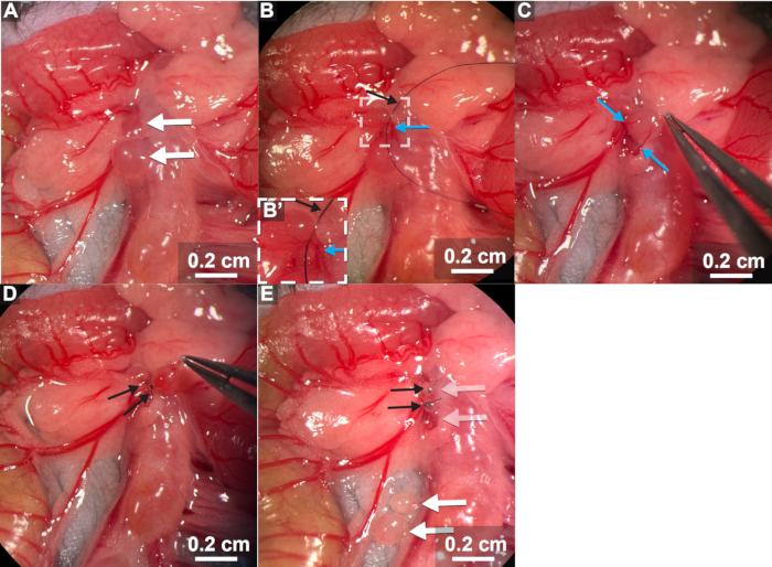
Figure 4: Annotated stills and commentary from mesenteric lymphadenectomy (fourth station). (A) Proper bowel orientation exposes the fourth mesenteric lymph node station (white arrows) medial to the distal colon and lateral to the third station. (B,C) After blunt dissection of the node station, suture ligation (black arrow) is performed at each identifiable node, ensuring to include all lymph node feeding vessels (blue arrows). (D) The node is excised along the previously created tissue plane, staying high towards the lymph nodes and leaving suture ligations behind (black arrows). (E) Following excision of the node station (white arrows), hemostasis is confirmed, suture ligations are visible (black arrows), and the empty space (faded arrows) where the node previously lived is noted to be free of residual node. Scale bars: 0.2 cm. Please click here to view a larger version of this figure.
4. Mesenteric lymphadenectomy completion
- Observe the distal branches of the ileocolic vessels, ileal vessels, and/or jejunal vessels to ensure blood supply is intact. Well-perfused bowel is bright and pink with visible pulsations.
- Using cotton-tipped swabs, return abdominal contents into the peritoneal cavity. Take care to remove any fragments of cotton, lint, drape, or suture from the bowels prior to return, as this can result in additional surgical inflammation.
- Ensure bowel is not twisted on its mesentery by confirming the bowel remains bright/pink (perfused) rather than dull/gray/purple (ischemic).
- Instill 1 mL of body-temperature sterile saline into the abdomen just prior to closure to replete insensible fluid loss/evaporation. Close the abdomen in two layers (abdominal wall and skin) with 6-0 monofilament polypropylene suture in a running fashion.
- Apply sterile surgical glue to the wound.
NOTE: Alternatively, interrupted sutures or wound clips can be used for skin closure.
5. Postoperative care
- Relocate animal to a clean cage, which is half on and half off of a warm-water circulating pad with access to food and water ad lib.
- Allow the animal to recover from anesthesia and monitor closely for signs of distress. If animals do not recover promptly from anesthesia with appropriate behaviors (i.e., increased respiratory rate, self-righting, ambulating, grooming), humanely euthanize the animal.
- When all animals have recovered from anesthesia, return cages to the animal care facility with access to water, food, bedding, and enrichment materials ad libium.
- Assess mice post-operatively every 12 h for 3 days for signs of pain or distress (i.e., hunched posture, decreased activity, decreased engagement, poor grooming). Humanely euthanize mice demonstrating signs of distress.
- If using permanent suture (such as 6-0 monofilament polypropylene), examine the incisions on postoperative day 10 and remove any remaining suture material.
6. Lymph node handling
- Immediately perform a buoyancy test by placing the excised tissue in a 1.5 mL microcentrifuge tube with 500 µL phosphate-buffered saline (PBS). Confirm lymph node excision by observing tissue sinking in PBS versus inadvertently resected adipose, which will float (Figure 5A).
NOTE: The excised lymph node station(s) are immediately ready to enter further downstream processing. - For live cell techniques, place excised lymph node station(s) in cell culture media (e.g., DMEM, RPMI) or PBS on ice. For planned histology, place excised lymph node station(s) in fixative (e.g., 10% neutral buffered formalin, 4% paraformaldehyde).
Access restricted. Please log in or start a trial to view this content.
Results
To confirm the accuracy of this procedure, mesenteric lymphadenectomies were performed on eleven 4-6-week-old, age, sex, and genotype-matched C57BL/6J strain littermates. Procedure times ranged from 5-15 min per mouse. All mice tolerated the procedure well, with 100% perioperative survival. Excised samples were placed in PBS on ice. All samples immediately sunk, confirming low adipose content. Excised samples (and four separate visceral adipose controls) were then crushed through a 100 µm cell strainer and washed with PB...
Access restricted. Please log in or start a trial to view this content.
Discussion
Described here is a survival procedure for safe and well-tolerated murine mesenteric lymphadenectomy at each of four node stations. The major limitation of this procedure is the indirect relationship between exposure and the progression of lymph nodes along the mesenteric chain. The first lymph node station is easily and readily exposed in its entirety. The second lymph node station is often visible, and exposure to the 2nd to 4th stations can be improved by extending the skin/abdominal wall incisio...
Access restricted. Please log in or start a trial to view this content.
Disclosures
R.A.F. is an advisor to GlaxoSmithKline, Plythera Inc., and Ventus Therapeutics. All other authors declare no competing interests.
Acknowledgements
We acknowledge J. Alderman, B. Cadugan, E. Hughes-Picard, and J. Horrocks for administrative assistance, and to C. Hughes for mouse colony management. H.N.B. is a fellow of the Yale Surgeon Scientist Training Program, which partially funded and supported this project. This work was also funded by the Howard Hughes Medical Institute (to R.A.F.) and the American College of Surgeons Resident Research Award (to H.N.B.).
Access restricted. Please log in or start a trial to view this content.
Materials
| Name | Company | Catalog Number | Comments |
|---|---|---|---|
| Anesthesia circuit | Any | Or injectable anesthetic | |
| BD LSRII flow cytometer (or equivalent) | BD Biosciences | Equipped with FACS Diva Software, (for flow cytometry) | |
| Buprenorphine XR | Any | For analgesia | |
| Cell culture media | Any | (for live cell techniques; ie. DMEM or RPMI) | |
| DAPI | Biolegend | 422801 | Working concentration: 15 ug/mL |
| Ethanol (70% in water) | Any | For cleaning | |
| FITC-conjugated anti-mouse CD45.2 (104) | Biolegend | 109806 | Working concentration: 5 ug/mL |
| Forceps: Angled fine-tipped forceps | Roboz | RS-5058 | Material: Dumoxel; Pattern #5, 45 degree angle; tip size 0.1 X 0.06mm |
| Forceps: Fine-tipped forceps | Roboz | RS-4960 | Material: Dumostar; Pattern #1; Tip size: 0.20 X 0.12mm |
| Forceps: Fine-tipped forceps | Roboz | RS-4966 | Material: Dumostar, Biologie tip; Pattern #3; Tip size: 0.08 x0.04mm |
| Lidocaine | Any | For analgesia | |
| Metal gavage needle | Any | 20/22 gauge reusable straight oral gavage needle, stainless steel. | |
| Needle drivers | Roboz | RS-6410 | Castroviejo needle holder; Straight |
| OPTIONAL: Dual/twin gooseneck microscope LED lights | Any | ||
| OPTIONAL: Evans Blue dye | Sigma | E2129-10G | For optional lymph tracing. |
| OPTIONAL: Operating microscope | Leica | MZ9.5 | Leica Mz9.5 stereo head operating microscope |
| Phosphate-buffered saline (PBS) | Any | ||
| Povidone/Iodine solution | Any | For skin preparation/sterilization | |
| Scissors: Extra fine-tipped scissors | Roboz | RS-5605 | McPherson-Vannas; 3mm cutting edge; curved (for station 3/4 nodes) |
| Scissors: Fine-tipped scissors | Roboz | RS-5650SC | Surecut Castroviejo scissors (for station 1/2/3 nodes) |
| Scissors: Medium scissors | Roboz | RS-5676 | Noyes; 13mm cutting edge; straight |
| Surgical glue | Any | 3M Vetbond tissue adhesive | |
| Suture: 6-0 blue monofilament polypropylenex | Any | ||
| Suture: 9-0 black monofilament nylon suture | Any | (optional) Can also use the slightly larger 8-0 size suture. | |
| Tissue fixative | Any | (for planned histology; ie. 10% neutral buffered formalin or 4% paraformaldehyde) | |
| Warm-water circulating heating pad | Any |
References
- Chopp, L., Redmond, C., O'shea, J. J., Schwartz, D. M. From thymus to tissues and tumor: A review of T cell biology. J Allergy Clin Immunol. 151 (1), 81-97 (2023).
- Rouiz-Argüelles, A. Autoimmunity: From bench to bedside. Anaya, J. -M. , El Rosario University Press. Colombia. (2013).
- Crespo, J., Sun, H., Welling, T. H., Tian, Z., Zou, W. T cell anergy, exhaustion, senescence, and stemness in the tumor microenvironment. Curr Opin Immunol. 25 (2), 214-221 (2013).
- Mempel, T. R., Henrickson, S. E., Von Andrian, U. H. T-cell priming by dendritic cells in lymph nodes occurs in three distinct phases. Nature. 427 (6970), 154-159 (2004).
- Divi, V., et al. Lymph node count from neck dissection predicts mortality in head and neck cancer. J Clin Oncol. 34 (32), 3892-3897 (2016).
- Chang, G. J., Rodriguez-Bigas, M. A., Skibber, J. M., Moyer, V. A. Lymph node evaluation and survival after curative resection of colon cancer: Systematic review. JNCI. 99 (6), 433-441 (2007).
- Simoes, P., et al. Lymph node yield in the pathological staging of resected nonmetastatic colon cancer: The more the better. Surg Oncol. 43, 101806(2022).
- Resio, B. J., et al. Commission on cancer standards for lymph node sampling and oncologic outcomes after lung resection. Ann Thorac Surg. 119 (2), 308-315 (2025).
- Connolly, K. A., et al. A reservoir of stem-like CD8 T cells in the tumor-draining lymph node preserves the ongoing antitumor immune response. Sci Immunol. 6 (64), eabg7836(2021).
- Saddawi-Konefka, R., et al. Lymphatic-preserving treatment sequencing with immune checkpoint inhibition unleashes cdc1-dependent antitumor immunity in HNSCC. Nat Commun. 13, 4298(2022).
- Saddawi-Konefka, R., Schokrpur, S., Gutkind, J. S. Let it be: Preserving tumor-draining lymph nodes in the era of immuno-oncology. Cancer Cell. 42 (6), 930-933 (2024).
- Houston, S. A., et al. The lymph nodes draining the small intestine and colon are anatomically separate and immunologically distinct. Mucosal Immunol. 9 (2), 468-478 (2016).
- Brinkmann, V., Pinschewer, D., Chiba, K., Feng, L. Fty720: A novel transplantation drug that modulates lymphocyte traffic rather than activation. Trends Pharmacol Sci. 21 (2), 49-52 (2000).
- Mehandru, S., Colombel, J. -F., Juarez, J., Bugni, J., Lindsay, J. O. Understanding the molecular mechanisms of anti-trafficking therapies and their clinical relevance in inflammatory bowel disease. Mucosal Immunol. 16 (6), 859-870 (2023).
- Mariathasan, S., et al. Absence of lymph nodes in lymphotoxin-alpha (lt alpha)-deficient mice is due to abnormal organ development, not defective lymphocyte migration. J Inflam. 45 (1), 72-78 (1995).
- Kwon, S., Agollah, G. D., Wu, G., Sevick-Muraca, E. M. Spatio-temporal changes of lymphatic contractility and drainage patterns following lymphadenectomy in mice. PLoS One. 9 (8), e106034(2014).
- Blum, K. S., Proulx, S. T., Luciani, P., Leroux, J. -C., Detmar, M. Dynamics of lymphatic regeneration and flow patterns after lymph node dissection. Breast Cancer Res Treat. 139 (1), 81-86 (2013).
- Shaikh, H., et al. Mesenteric lymph node transplantation in mice to study immune responses of the gastrointestinal tract. Front Immunol. 12, 689896(2021).
Access restricted. Please log in or start a trial to view this content.
Reprints and Permissions
