A subscription to JoVE is required to view this content. Sign in or start your free trial.

Method Article

Preparation of Meiotic Chromosome Spreads from Mouse Oocytes for Assessment of Synapsis and Recombination

2.2K views

DOI:

10.3791/68749

July 18th, 2025

In This Article

Summary

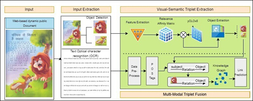
Diagram of OCR method for visual-semantic triplet extraction and knowledge graph linkage.
Figure 1: End-to-end framework. The figure illustrates end-to-end framework for multimodal knowledge Graph VISHAM-KG. Please click here to view a larger version of this figure.

Abstract

Three critical and interdependent processes define meiotic prophase I: homologous chromosomes must pair together, a proteinaceous structure called the synaptonemal complex forms to tether homologs together (synapsis), and homologs undergo recombination, reciprocally exchanging genetic material to form crossovers (COs). Errors in these processes can result in premature ovarian insufficiency, aneuploidy, and ultimately, pregnancy loss and infertility. Meiotic recombination is particularly error-prone in oocytes, with over 7% of human oocytes containing at least one chromosome pair without a crossover, and between 20%-80% of eggs versus 2.5%-7% of sperm are aneuploid. However, it remains unclear why chromosomal errors show such striking sex disparities. The present protocol describes methods for the preparation and analysis of oocyte prophase I and metaphase I chromosome spreads from fetal and juvenile mice, respectively. To trace critical chromosome dynamics throughout meiotic prophase I, this protocol employs immunofluorescence staining of common markers for meiotic recombination (RAD51 for double strand breaks, MSH4 for CO intermediates, and MLH1/MLH3 to identify most COs) and synapsis (SYCP3 for chromosome axes, SYCP1 for synapsed regions, and HORMAD1 for asynapsed regions). Further, this protocol uses in vitro maturation of oocytes collected to assess the number of paired chromosomes (bivalents) and the total number of crossovers (chiasmata) in metaphase I. Together, these techniques provide a comprehensive and quantitative framework to examine mechanisms regulating early chromosome dynamics in female meiosis.

Introduction

Meiosis is a specialized cell division essential to the production of haploid gametes. During prophase I, homologous chromosomes (each consisting of sister chromatids held together by cohesin) must accomplish three critical tasks: 1) recognize and pair with each other, 2) zip together by forming the synaptonemal complex (SC) between their lateral axes, and 3) exchange DNA via homologous recombination, resulting in crossovers (COs) between non-sister chromatids1,2. The physical connection resulting from crossover formation, maintained by cohesin, ensures proper chromosome biorientation and segregation during metaphase I3,4,5.

Meiotic prophase I comprises five stages, each defined by the status of synapsis6. In leptonema (Greek for thin thread), chromosomes condense as the axial element (marked by SYCP3) forms along chromosome axes. During zygonema (joined threads), homologs begin to synapse as the transverse element of the SC (SYCP1) forms between axes7. In pachynema (thick thread), chromosomes are synapsed along their entire length. The SC then disassembles during diplonema (two threads), leaving homologs connected only at crossover sites (chiasmata). Chiasmata are the product of homologous recombination, a process initiated with the programmed induction of DNA double-strand breaks (DSBs). From hundreds of DSBs, only ~10% become COs. In mice, most (90%-95%) form through the class I CO pathway facilitated by MutLγ (MLH1-MLH3), with the remaining COs made by MUS81-EME1 through the class II CO pathway8,9,10,11.

Pairing, synapsis, and recombination are highly regulated and interdependent processes. Errors typically trigger checkpoint mechanisms, leading to cell death. In females, germ cell loss from early prophase I errors can cause premature ovarian insufficiency12,13, while late errors (e.g., homologs failing to form COs, malplacement of COs, or premature cohesin loss) can lead to chromosome missegregation and aneuploidy. In humans, meiotic errors are a leading cause of pregnancy loss and infertility. Female meiosis is particularly error-prone. Around 20%-80% of human eggs (versus 2.5%-7% of sperm) carry abnormal chromosome numbers, most resulting from errors in the first meiotic division14,15,16,17. Although age has been identified as the single greatest cause of maternally aneuploidy, the reason why oocytes are so susceptible to errors remains unclear.

Despite the importance of understanding the factors underlying high error rates in oocytes, most of our knowledge about the regulation of prophase I events comes from studies of male meiosis. Because a small fragment of adult testicular material provides sufficient material to assess all stages of meiotic and post-meiotic germ cells, male meiosis has long been more readily accessible to researchers. In contrast, female meiosis occurs in a discontinuous manner. In mice, prophase I progresses semi-synchronously in the fetal ovary, beginning around 14 days post-coitum (dpc; Figure 1A), shortly after gonadal sex determination18,19,20. Around the time of birth, oocytes enter dictyate arrest until ovulation months (in mice) or decades (in humans) later21,22,23. Consequently, specific prophase I stages are accessible only during limited developmental windows (Figure 1A), with post-prophase I events occurring in only a few oocytes at a time in the juvenile and adult. Though technical advances have improved mammalian oocyte meiosis studies, these methods remain challenging and time-consuming compared to comparable techniques used in male studies.

Female meiotic analysis is further complicated by limitation in sub-staging pachytene oocytes - critical for studying crossover designation and maturation. Male studies benefit from well-characterized sex chromosome pairing dynamics and the presence of the histone variant, H1t, that serve as markers for early, mid, and late pachynema24,25,26. However, no reliable method has been formalized for sub-staging pachytene oocytes.

The protocols presented here facilitate a comprehensive analysis of synapsis and recombination in mouse oocytes from early prophase I through metaphase I. This work builds on classical chromosome spreading techniques developed by Peters et al.27 and Tarkowski28 and prior refinements of those methods by Hunt29 and Sun and Cohen30. This protocol has been developed to emphasize: 1) Enrichment and identification of specific prophase I sub-stages, 2) maximizing the number of applications performed per pair of fetal ovaries, and 3) thorough, accessible metrics for evaluation of synapsis defects and recombination from foci to chiasmata.

Access restricted. Please log in or start a trial to view this content.

Protocol

All animal handling and procedures were performed following approval by the Cornell Institutional Animal Care and Use Committee (IACUC) under protocol 2004-0063.

1. Preparation of oocyte prophase I chromosome spread slide

  1. In late afternoon, set up timed matings of C57BL/6J female mice (2 - 6 months old) with males (2 - 12 months old). Beginning the next day, check females each morning (before 9 AM) for a copulatory plug. Noon on the day of the plug is defined as 0.5 dpc.
  2. Prepare the following prior to sacrificing pregnant mice.
    1. Set up a humidified chamber with room temperature H2O.
    2. Prepare 10 mL of hypotonic extraction buffer (HEB; see Table 1) consisting of 30 mM Tris-HCl, 50 mM sucrose, 17 mM sodium citrate, 5 mM EDTA, 2.5 mM DTT, and 0.5 mM PMSF in Milli-Q water. Prior to adding DTT and PMSF, adjust pH to 8.2 - 8.4 using 50 mM boric acid. Make HEB fresh and use within 2 hours of adding DTT.
    3. Prepare 100 mM sucrose by adding 342 mg sucrose to 10 mL of ultrapure H2O. Store at 4 °C when not in use.
    4. Preheat ultrapure H2O in a standard microwave for 1 min (until 90 - 95 °C). Under a fume hood, add the heated ultrapure H2O to 0.5 g of Paraformaldehyde (PFA). Add two drops of 1 N NaOH, vortex to mix, and let cool to room temperature. Adjust pH to 9.2 - 9.3 using 50 mM boric acid and add Triton X-100 to 0.15 % (v/v). Add ultrapure H2O to a final volume of 50 mL (1% PFA fixative solution) and pour fixative into a 50 mL glass Coplin staining jar.
      NOTE: This protocol was optimized using near-boiling water to prepare PFA; however, heating PFA in solution above 65 °C may cause degradation and release of toxic fumes. Alternatively, preheat ultrapure H2O to 60 °C in a water bath.
    5. Clean glass microscope slides with 70% ethanol. Use a hydrophobic barrier PAP pen to draw up to three squares (approximately 20 x 20 mm; see Figure 1E). A single pair of ovaries will provide sufficient material for six squares (or two slides of 3 squares, each).
  3. Sacrifice pregnant mice 14.5 - 18.5 dpc by CO2 asphyxiation and cervical dislocation according to IACUC guidelines.
    1. Timepoints for enrichment of specific stages of prophase I in C57BL/6J mice are outlined in Figure 1A. For the given developmental timepoints, the approximate percentage of oocytes in each stage of prophase I has been provided using data from Evans et al. (1982)31. For zygotene oocytes, collect on 16.5 dpc; early pachytene on 17.5 dpc; and mid-pachytene - diplotene at 18.5 dpc.
  4. Use dissection scissors to open the abdominal cavity and remove the uterine horns.
  5. Dissect the fetal mice from the uterine horn, decidual tissue, and yolk sac. Place fetal mice in a 100 mm Petri dish with 1x phosphate buffer saline (PBS).
  6. Dissect ovaries from one female fetus at a time while maintaining the rest in 1x PBS. Sacrifice fetal mice by decapitation according to IACUC guidelines. Using stainless steel specimen pins, secure the torso ventral surface up to a silicon pad in a dissection dish.
  7. Use microdissection scissors to make an abdominal incision just below the umbilicus, being careful to avoid nicking arteries. Use fine forceps to displace the intestine until the fetal uterine horns are visible.
  8. To locate the ovaries, follow each uterine horn outward from the bladder towards the kidney. The ovary and oviduct will appear as small globule structures at the termini of the uterine horns, located just below the kidney and near the posterior of the peritoneal cavity (Figure 1B).
  9. Use fine forceps to pinch off the ovaries and collect them in a small watch glass filled with 750 µL of 1x PBS.
  10. Dissect the ovary from the surrounding tissues and place both ovaries in a watch glass containing 750 µL of HEB, taking care to ensure the ovaries are completely submerged (See Figure 1C and 1D). Incubate the ovaries for 15 min at room temperature.
  11. Pipette 35 µL of 100 mM sucrose onto a concavity slide. Use fine forceps to transfer both ovaries from HEB to sucrose.
  12. Use a 25G needle to secure an ovary to the slide while using a second needle, bevel down, to press out the oocytes from the ovary.
    1. When the ovary becomes transparent and jelly-like, remove the ovary from the HEB and repeat the process with the second ovary. Gently pipette the solution to disperse cells.
  13. Dip the slide into the Coplin staining jar of PFA and briefly dab the edge of the slide against a paper towel. Once the PFA has been collected into small drops along the hydrophobic boundary of each square (Figure 1E), pipette 5 µL of the oocyte-sucrose solution into each drop of PFA.
  14. Spread the oocytes over the square sections by gently tilting the slides back and forth. Be careful not to allow the solution from any one square to spill over into adjacent squares.
  15. Place the slide in a humid chamber and continue making slides until the oocyte-sucrose solution is exhausted.
  16. Repeat steps 1.6 - 1.15 until all female fetuses have been used. Incubate slides in a humid chamber at room temperature for 2 h.
  17. Following incubation, open the humid chamber. As most (if not all) the liquid/fixative should still be present on the slide surface (roughly 10 µL in each square section), gently transfer slides to a paper towel, and allow slides to completely air-dry at room temperature on a lab bench (this process typically takes 30 - 60 min, depending on local humidity).
    NOTE: Slides may also be air-dried under a fume hood to mitigate potential exposure to residual PFA fumes. This will also shorten drying to roughly 20 - 30 min.
  18. Wash slides in 0.4% wetting agent in ultrapure H2O for 2 min. Allow slides to completely dry.
    NOTE: At this point, the slides may be stored at -80 °C; however, certain epitopes are unstable (e.g., in our experience, MLH1 stains poorly, and SYCP3 staining has significant artifacts after freezing). It is strongly advised that slides be immunostained immediately.

2. Immunofluorescence staining of prophase I chromosome spreads

  1. Prepare 10x antibody dilution buffer (ADB; see Table 2) containing 10% (v/v) Normal Goat Serum, 3% (w/v) bovine serum albumin (BSA), and 0.05% Triton X-100 in 1x PBS. Filter sterilize using a 0.45 µm filter and store at 4 °C for up to 2 weeks.
  2. Wash slides in a Coplin staining jar of 0.4% wetting agent in 1x PBS for 10 min. Wash slides in a Coplin staining jar of 0.1% Triton X-100 in 1x PBS for 10 min. Block slides in a Coplin staining jar of 1x ADB for 10 min.
  3. Dilute selected primary antibodies in 10x ADB (See Table of Materials for list of antibodies and their respective dilutions).
    1. Prepare four combinations of antibodies for multiplex immunostaining: (1) SYCP332,33, SYCP133, HORMAD133; (2) SYCP332,33, RAD5132,33, MLH332; (3) SYCP332,33, MSH432, MLH332; and (4) SYCP334, MLH132,33,34, CREST35.
  4. Remove slides from ADB, tap off excess liquid, and place slides in a humidified chamber with room temperature water. Apply 30 µL of diluted antibodies per square section of each slide, taking care that antibodies from one square do not spill over into adjacent squares.
  5. Close the humidified chamber and allow slides to incubate overnight at room temperature.
  6. The next morning, open the humid chamber and tap off the diluted antibodies from the slides. Repeat step 2.2.
  7. Dilute selected secondary antibodies in 10x ADB (see Table of Materials).
    NOTE: From this point on, slides and antibodies are photosensitive. Complete all subsequent incubations in the dark.
  8. Remove slides from ADB, tap off excess liquid, and place slides in a humidified chamber of room temperature water. Apply 30 µL of selected diluted antibodies per square section of each slide, taking care so antibodies from one square do not spill over into adjacent squares.
  9. Close the humidified chamber and allow slides to incubate for 2 h at room temperature.
  10. Wash slides 3x for 5 min each in Coplin staining jars of 0.4% wetting agent in 1x PBS. Wash slides for 5 min in a Coplin staining jar of 0.4% wetting agent in ultrapure H2O.
  11. Apply a small drop (approximately 10 µL) of antifade mounting media with 4', 6-diamidino-2-phenylindole (DAPI) or equivalent nucleic acid stain. Place a 60 mm x 24 mm No. 1.5 glass coverslip onto each slide and carefully blot excess liquid.
    NOTE: Slides may be sealed with either clear nail polish or rubber cement. If using a curing mountant, sealing the slides is unnecessary. Slides should be stored in a slide box or folder at 4 °C. Although it is best practice to image slides shortly after staining, in our experience slides stored in the dark at 4 °C can retain their signal intensity for several months to a year. For longer term storage, keep slides can be kept at -20 °C for several years without significant loss of signal quality.
  12. Image cells at 63x using epifluorescence microscopy (see Table of Materials). Use image analysis software (e.g., Zen of ImageJ, see Table of Materials) to measure SC length and fluorescence intensity of all channels along the axis. Trace the length of each chromosome axis (SYCP3), beginning at the centromere (CREST or DAPI dense region).
    1. For analysis of HORMAD1 and SYCP1, record the lengths of fluorescence signal peaks. Calculate the total lengths of HORMAD1 or SYCP1 peaks and divide them by the length of the chromosome axis.
    2. For analysis of MSH4, quantify the number of fluorescence signal peaks for each measurement of SYCP3. Record both the total number of MSH4 peaks per nucleus and the number of MSH4 peaks overlapping MLH3 peaks. Confirm counts of MSH4 by comparing peaks to the corresponding foci count.

3. Diakinesis/prometaphase preparations for observation of chiasmata

  1. To maximize the yield of meiotically competent oocytes, use juvenile female mice (25 - 28 days old).
    NOTE: Stimulation by intraperitoneally injecting female mice with 5 IU pregnant mare's serum gonadotropin (PMSG) 48 h prior to oocyte collection may be performed to enhance yield. However, for the purposes of chiasmata analyses, sufficient germinal vesicle (GV) oocytes can be collected from unstimulated females.
  2. Before sacrificing mice, prepare the following reagents and solutions.
    1. Allow M2 (3 mL per mouse) and KSOM (1 mL per mouse) to equilibrate in a cell culture incubator (37 °C, 5 % CO2) for at least 1 h.
      NOTE: Tubes of frozen media aliquots can be placed in the incubator with their lids loosened the evening prior to oocyte collection, allowing for approximately 14 h of equilibration.
    2. Prepare M2 media with 2.5 µM milrinone (a phosphodiesterase isoenzyme inhibitor that prevents meiotic resumption)36. Pipette five 20 µL drops of M2 + milrinone into a 35 mm Petri dish and overlay with light mineral oil (Figure 2C).
    3. Pipette five 20 µL KSOM drops into a 35 mm Petri dish and overlay with light mineral oil (Figure 2C).
      CAUTION: Control of temperature and pH is critical to oocyte in vitro maturation. Collection of oocytes should be performed on a heated (37 °C) benchtop. All media should be kept in the incubator until it is used, and dishes of media should be removed one at a time to limit disruption caused by exposure to ambient air.
    4. Clean glass microscope slides with 70% ethanol and allow to air-dry. On the back of glass microscope slides, lightly score (with a diamond-tip pen) or draw (with a water-resistant ink) a 5 x 5 grid of ~ 4 mm x 4 mm squares (An example is shown in Figure 2B and can be used to trace the grid).
  3. Collect oocytes from one mouse at a time. Sacrifice a female mouse by CO2 asphyxiation and cervical dislocation according to IACUC guidelines.
  4. Use dissection scissors to open the abdominal cavity. Use fine forceps to displace the intestines.
  5. Follow each uterine horn from the bladder towards the kidney. Ovaries and oviducts will be associated with a small fat pad near the kidney.
  6. Use microdissection scissors to remove the ovaries and place them in gassed M2 media with milrinone.
  7. Use microdissection scissors to remove all fat and other tissues (including the bursa) from the ovaries. Use 25G needles to rupture antral follicles.
  8. Use a transfer pipette to spread the oocytes in M2 + milrinone in a thin layer in a 100 mm Petri dish, taking care not to let the media touch the sides of the dish (Figure 2C).
  9. Use a dissection microscope (magnification 25x - 50x) to visualize GV oocytes (Figure 2F). GV oocytes may be associated with cumulus cells in a cumulus-oocyte complex (COC). Remove cumulus cells with repeated pipetting.
  10. Collect GV oocytes using a mouth-operated glass pipette (100 - 150 µm diameter; Figure 2A). Transfer GV oocytes into a 20 µL drop of M2 + milrinone overlaid with light mineral oil (Figure 2C). Do not put more than 30 oocytes into a single drop.
  11. When all GV oocytes have been collected, pass them through two or three 20 µL drops of M2 + milrinone overlaid with light mineral oil to remove any somatic cells.
  12. Transfer GV oocytes (with as little media as possible) into a 30 µL drop of KSOM. Wash out the milrinone by passing oocytes through five KSOM drops (Figure 2C).
  13. Transfer all GV oocytes into 20 µL drops of KSOM overlaid with light mineral oil, a maximum of 30 oocytes per KSOM drop (Figure 2C). Place oocytes in a cell culture incubator (37 °C, 5 % CO2).
  14. Incubate oocytes for approximately 5 h.
    NOTE: GV breakdown (marking disappearance of the nuclear envelope and meiotic resumption; Figure 2G) typically occurs after 2 h in culture, but the rate of oocyte maturation may vary depending on mouse strain or hormone stimulation. In our experience, a total of 5 h in culture best optimizes for chiasmata spreads in most mouse strains. If left to mature too long, the first meiotic division and polar body extrusion may occur (Figure 2H).
  15. Transfer 5 - 10 oocytes (with as little KSOM as possible) into a 30 µL drop of hypotonic solution (0.9 % sodium citrate in ultrapure H2O). Incubate oocytes in the hypotonic solution for 5 - 10 min.
  16. Pipette a small drop (~ 1 µL) of acidified water (8 drops of glacial acetic acid in 50 mL of ultrapure H2O) onto one of the squares on the prepared slides (Figure 2B).
  17. Use a 100 - 150 µm diameter mouth-operated glass pipette to quickly transfer one oocyte from the to the acidified water drop. Transfer as little hypotonic solution with the oocyte as possible. Use the same pipette to remove some of the liquid until the oocyte sticks to the slide.
    NOTE: The volume of liquid around the oocyte is critical. There should be enough liquid that the oocyte does not dry out before fixation, but not so much that the oocyte cannot adhere to the slide.
  18. Quickly switch to a 150 µm diameter glass pipette tip. Add one drop of fresh Carnoy's fixative (three parts absolute methanol to one-part glacial acetic acid) to the oocyte. Allow the fixative to disperse over the slide. The oocyte should appear to become less distinct and melt as the acid from the fixative dissolves the zona pellucida and the cells burst.
  19. As the front of the fixative begins to contract, quickly add 2 - 3 more drops of fixative. Gently blow on the slide to speed up drying.
  20. Repeat steps 3.16 - 3.19 with additional oocytes from the hypotonic solution.
  21. Repeat steps 3.15 - 3.20 until all oocytes have been fixed. Check the spreading of chromosomes using a phase contrast microscope.
  22. When the slide has fully air dried, place the slide in a Coplin staining jar with 50 mL of 4% Giemsa (v/v) in ddH2O. Stain slides in Giemsa for 6 min at room temperature.
  23. Wash slides 3x for 3 min each in ddH2O at room temperature. Allow slides to completely dry.
  24. Apply a thin layer (~60 µL) of Permount across a 60 mm x 24 mm No. 1.5 glass cover slip. Mount slides by inverting them onto the coverslips and carefully blotting out excess liquid. Allow slides to dry overnight before imaging at 63x on brightfield.

Access restricted. Please log in or start a trial to view this content.

Results

Evaluation of synapsis defects in pachytene-like cells
This experiment evaluates synapsis defects in pachytene oocytes and monitors prophase I progression (Figure 3A). Immunostaining for SYCP3 (axial/lateral SC elements), SYCP1 (central SC element), and HORMAD1 (unsynapsed/desynapsed chromosome axes) enables staging and synapsis assessment. HORMAD1 colocalizes with SYCP3 during zygonema and diplonema (before synapsis and after desynapsis)...

Access restricted. Please log in or start a trial to view this content.

Discussion

Mammalian oocytes are particularly error-prone compared to spermatocytes. While the oocyte's less efficient spindle assembly checkpoint (SAC) contributes to this vulnerability14,52,53, Hassold and Hunt propose that multiple hits lead to aneuploidy, with defective recombination being a primary factor54. Human studies link aneuploidy to insufficient crossover assurance and suboptimal crossover positioni...

Access restricted. Please log in or start a trial to view this content.

Disclosures

The author has no conflicts to disclose.

Acknowledgements

Funding for this project was provided by a K99 award from the Eunice Kennedy Shriver National Institute for Child Health and Development (HD112986 to TH).

Access restricted. Please log in or start a trial to view this content.

Materials

List of materials used in this article
NameCompanyCatalog NumberComments
0.45 μm Syringe FilterMillipore SigmaSLHAR33SBfor making mouth-operated pipette
0.5 M EDTA, pH 8.0Corning46-034-CI for HEB
1 mL Sterile Syringe with 25-gauge, 16 mm needleFisher Scientific14-817-125 for rupturing ovaries
100 x 15 mm Petri dishVWR25384-302 
14.6 cm disposable borosilicate glass Pasteur pipetsFisher Scientific13-678-20B for making mouth-operated pipette
35 x 10 mm Petri dishCorning351008
40 mm x 0.45 mm #3 Stainless Steel Pins for EntomologyAmazonB0CSSPW5S7for pinning fetal mice for dissection
Acetic Acid, GlacialFisher ScientificA38-212for Carnoy's fixative
Aspirator tube assemblies for calibrated microcapillary pipettesMillipore SigmaA5177-5EAfor making mouth-operated pipette
B.P.I. Watch GlassNDS Technologies 1580-00Alternatively use a 30 mm dia x 12 mm deep embryo dish (Electron Microscopy Sciences SKU: 70543-30)
Boric AcidFisher ScientificBP168-1for adjusting pH of HEB, PFA
Bovine Serum AlbuminMillipore SigmaA7906-100Gfor ADB
CO2 Tankfor incubator
Corning Microscope SlidesMillipore SigmaCLS294875X25L x W 75 mm x 25 mm, frosted one side
Corning microscope slides, frosted one side, one endVWR294875X25
Cover glass rectangular. 60 x 24 mm, #1.5VWR48393-251
Dissecting DishElectron Microscopy Sciences70540
Dissection MicroscopeAny standard model
Dissection Scissors, Sharp Tip, 4.5"VWR82027-578
Extra fine, sharp tip forcepsVWR76548-836
Giemsa StainMillipore SigmaGS500-500for staining chiasmata spreads
ImmEdge Hydrophobic Barrier PAP PenVector LaboratoriesH-4000
IncubatorAny standard model with CO2 and water jacketed technology
IVF WorkstationK-SystemsAny model with a heated benchtop
Kodak Photo-Flo 200 SolutionBH Photo & Video1464510for slide washes
MethanolFisher ScientificA412-4for Carnoy's fixative
MP Biomedicals Coplin Staining JarFisher ScientificICN17006201
Normal Goat SerumGibco16210-072for ADB
Paraformaldehyde, EM Grade, Purified PrillElectron Microscopy Sciences19200
PermountFisher ScientificSP15-100for  mounting slides for chiasmata spreads
ProLong Glass Antifade Mountant with NucBlue StainThermo ScientificP36985for mounting immunostained slides
Silicone tube, interior diameter: 0.125", exterior diameter: 0.25"VWR89068-474for making mouth-operated pipette
Sodium Citrate DihydrateFisher ScientificS279-500for HEB, hypotonic solution
StainTray Slide Staining SystemElectron Microscopy Sciences71396-BFor use as a humidified chamber. Must use the black lid for immunofluorescence staining. May alternatively line the bottom of an opaque slide box or tupper ware with damp paper towels for use as a humidified chamber 
Stripper Tips - 100Cooper SurgicalMXL3-100for mouth-operated pipette
Stripper Tips - 150Cooper SurgicalMXL3-150for mouth-operated pipette
SucroseMillipore SigmaS-8501for HEB, Sucrose hypotonic solution
Tris BaseFisher ScientificBP152-5for HEB
Triton X-100Fisher ScientificBP151-100 for PFA, ADB, and Triton-PBS
United Scientific Coplin Staining JarFisher ScientificS17495Aopaque white plastic for washes and blocking slides after addition of photosensitive secondary antibodies
United Scientific Glass Concavity SlidesFisher ScientificS1395331.3 mm thick, 2 well count
Vannas Microdissection Scissors 5mm straightVWR76457-358
Zeiss Axio Imager.Z2, Colibri 7Carl Zeiss000000-2624-307
Zen Blue 3.11 SoftwareCarl ZeissImage acquisition and analysis software
Antibodies
Alexa Fluor 488 AffiniPure F(ab')? Fragment Goat Anti-Mouse IgG, Fc fragment specificJackson ImmunoResearch Laboratories Inc.115-546-071used 1:1,000 dilution
Alexa Fluor 488 AffiniPure F(ab')? Fragment Goat Anti-Rabbit IgG, Fc fragment specificJackson ImmunoResearch Laboratories Inc.111-546-046used 1:1,000 dilution
Alexa Fluor 647 AffiniPure F(ab')? Fragment Goat Anti-Human IgG, Fcγ fragment specificJackson ImmunoResearch Laboratories Inc.109-606-170used 1:1,000 dilution
Alexa Fluor 647 AffiniPure Goat Anti-Guinea Pig IgG, Fc fragment specifiJackson ImmunoResearch Laboratories Inc.106-605-008used 1:1,000 dilution
Guinea Pig anti-MLH3 pAbcustom made with Thermo-Fisher, used 1:500 dilution
Guinea Pig anti-SYCP1 pAbfrom Christer Höög (Kouznetsova et al. 2005), used 1:200 dilution
Human anti-Centromere protein pAbAntibodies Inc.15-234used 1:1,000 dilution
Mouse anti-MLH1 mAbBD Biosciences550838used 1:100 dilution
Mouse anti-SYCP3 mAbAbcamab97672used 1:1,000 dilution
Rabbit anti-HORMAD1 pAbProteintech13917-1-APused 1:500 dilution
Rabbit anti-MSH4 pAbAbclonalA8556used 1:100 dilution
Rabbit anti-RAD51 pAbMilliporePC130used 1:500 dilution
Rabbit anti-SYCP3 pAbAbcamab15093used 1:1,000 dilution
Rhodamine Red-X (RRX) AffiniPure F(ab')? Fragment Goat Anti-Mouse IgG, Fc fragment specificJackson ImmunoResearch Laboratories Inc.115-296-071used 1:1,000 dilution
Rhodamine Red-X (RRX) AffiniPure F(ab')? Fragment Goat Anti-Rabbit IgG, Fc fragment specificJackson ImmunoResearch Laboratories Inc.111-296-046used 1:1,000 dilution

References

  1. Gray, S., Cohen, P. E. Control of Meiotic Crossovers: From Double-Strand Break Formation to Designation. Annu Rev Genet. 50, 175-210 (2016).
  2. Hunter, N. Meiotic Recombination: The Essence of Heredity. Cold Spring Harb Perspect Biol. 7, a016618(2015).
  3. Kudo, N. R., et al. Resolution of Chiasmata in Oocytes Requires Separase-Mediated Proteolysis. Cell. 126, 135-146 (2006).
  4. Murdoch, B., et al. Altered Cohesin Gene Dosage Affects Mammalian Meiotic Chromosome Structure and Behavior. PLoS Genet. 9, 1003241(2013).
  5. Hodges, C. A., Revenkova, E., Jessberger, R., Hassold, T. J., Hunt, P. A. SMC1β-deficient female mice provide evidence that cohesins are a missing link in age-related nondisjunction. Nat Genet. 37, 1351-1355 (2005).
  6. Zickler, D., Kleckner, N. Meiosis: Dances Between Homologs. Ann Rev Genet. 57, 1-63 (2023).
  7. Billmyre, K. K., et al. SYCP1 head-to-head assembly is required for chromosome synapsis in mouse meiosis. Sci Adv. 9, eadi1562(2023).
  8. Kolas, N. K., et al. Localization of MMR proteins on meiotic chromosomes in mice indicates distinct functions during prophase I. J Cell Biol. 171, 447-458 (2005).
  9. Lipkin, S. M., et al. Meiotic arrest and aneuploidy in MLH3-deficient mice. Nat Genet. 31, 385-390 (2002).
  10. Oh, S. D., Lao, J. P., Taylor, A. F., Smith, G. R., Hunter, N. RecQ Helicase, Sgs1, and XPF Family Endonuclease, Mus81-Mms4, Resolve Aberrant Joint Molecules during Meiotic Recombination. Mol Cell. 31, 324-336 (2008).
  11. Holloway, J. K., Booth, J., Edelmann, W., McGowan, C. H., Cohen, P. E. MUS81 generates a subset of MLH1-MLH3-independent crossovers in mammalian meiosis. PLoS Genet. 4, e1000186(2008).
  12. Edelmann, W., et al. Mammalian MutS homologue 5 is required for chromosome pairing in meiosis. Nat Genet. 21, 123-127 (1999).
  13. Kneitz, B., et al. MutS homolog 4 localization to meiotic chromosomes is required for chromosome pairing during meiosis in male and female mice. Genes Dev. 14, 1085-1097 (2000).
  14. Nagaoka, S. I., Hassold, T. J., Hunt, P. A. Human aneuploidy: mechanisms and new insights into an age-old problem. Nat Rev Genet. 13, 493-504 (2012).
  15. Capalbo, A., Hoffmann, E. R., Cimadomo, D., Ubaldi, F. M., Rienzi, L. Human female meiosis revised: new insights into the mechanisms of chromosome segregation and aneuploidies from advanced genomics and time-lapse imaging. Hum Reprod Update. 23, 706-722 (2017).
  16. Gruhn, J. R., et al. Chromosome errors in human eggs shape natural fertility over reproductive life span. Science. 365, 1466-1469 (2019).
  17. Gruhn, J. R., Hoffmann, E. R. Errors of the Egg: The Establishment and Progression of Human Aneuploidy Research in the Maternal Germline. Ann Rev Genet. 56, null(2022).
  18. Soygur, B., et al. Intercellular bridges coordinate the transition from pluripotency to meiosis in mouse fetal oocytes. Sci Adv. 7, eabc6747(2021).
  19. Bowles, J., et al. Retinoid Signaling Determines Germ Cell Fate in Mice. Science. 312, 596-600 (2006).
  20. Bullejos, M., Koopman, P. Germ cells enter meiosis in a rostro-caudal wave during development of the mouse ovary. Mol Reprod Dev. 68, 422-428 (2004).
  21. Borum, K. Oogenesis in the mouse: A study of the meiotic prophase. Exp Cell Res. 24, 495-507 (1961).
  22. Grive, K. J., Freiman, R. N. The developmental origins of the mammalian ovarian reserve. Development. 142, 2554-2563 (2015).
  23. Hu, M., et al. PRC1-mediated epigenetic programming is required to generate the ovarian reserve. Nat Commun. 13, 4510(2022).
  24. Drabent, B., Bode, C., Bramlage, B., Doenecke, D. Expression of the mouse testicular histone gene H1t during spermatogenesis. Histochem Cell Biol. 106, 247-251 (1996).
  25. Goetz, P., Chandley, A. C., Speed, R. M. Morphological and temporal sequence of meiotic prophase development at puberty in the male mouse. J Cell Sci. 65, 249-263 (1984).
  26. Solari, A. J. The spatial relationship of the X and Y chromosomes during meiotic prophase in mouse spermatocytes. Chromosoma. 29, 217-236 (1970).
  27. Peters, A. H., Plug, A. W., van Vugt, M. J., de Boer, P. A drying-down technique for the spreading of mammalian meiocytes from the male and female germline. Chromosome Res. 5, 66-68 (1997).
  28. Tarkowski, A. K. An Air-Drying Method for Chromosome Preparations from Mouse Eggs. Cytogenetics. 5, 394-400 (2008).
  29. Susiarjo, M., Rubio, C., Hunt, P. Analyzing mammalian female meiosis. Methods Mol Biol. 558, 339-354 (2009).
  30. Sun, X., Cohen, P. E. Studying Recombination in Mouse Oocytes. Mammalian Oocyte Reg Meth Protoc. , 1-18 (2013).
  31. Evans, C. W., Robb, D. I., Tuckett, F., Challoner, S. Regulation of meiosis in the foetal mouse gonad. Development. 68, 59-67 (1982).
  32. Horan, T. S., et al. The DNA helicase FANCJ (BRIP1) functions in double strand break repair processing, but not crossover formation during prophase I of meiosis in male mice. PLoS Genet. 20, e1011175(2024).
  33. Wood, A. J., et al. CNTD1 is crucial for crossover formation in female meiosis and for establishing the ovarian reserve. J Cell Biol. 224, e202401021(2025).
  34. Horan, T. S., et al. Replacement Bisphenols Adversely Affect Mouse Gametogenesis with Consequences for Subsequent Generations. Curr Biol. 28, 2948-2954.e3 (2018).
  35. Lee, C., Oh, J. S. Novel BRCA1-PLK1-CIP2A axis orchestrates homologous recombination-mediated DNA repair to maintain chromosome integrity during oocyte meiosis. Nuc Acid Res. 53, gkae1207(2025).
  36. Thomas, R. E., Thompson, J. G., Armstrong, D. T., Gilchrist, R. B. Effect of specific phosphodiesterase isoenzyme inhibitors during in vitro maturation of bovine oocytes on meiotic and developmental capacity. Biol Reprod. 71, 1142-1149 (2004).
  37. Wojtasz, L., et al. Mouse HORMAD1 and HORMAD2, Two Conserved Meiotic Chromosomal Proteins, Are Depleted from Synapsed Chromosome Axes with the Help of TRIP13 AAA-ATPase. PLoS Genet. 5, e1000702(2009).
  38. Kogo, H., et al. Differential phosphorylation of two serine clusters in mouse HORMAD1 during meiotic prophase I progression. Exp Cell Res. 440, 114133(2024).
  39. Jiao, X., et al. Aberrant activation of chromosome asynapsis checkpoint triggers oocyte elimination. Nat Commun. 16, 2260(2025).
  40. Cloud, V., Chan, Y. L., Grubb, J., Budke, B., Bishop, D. K. Rad51 Is an Accessory Factor for Dmc1-Mediated Joint Molecule Formation During Meiosis. Science. 337, 1222-1225 (2012).
  41. Lan, W. H., et al. Rad51 facilitates filament assembly of meiosis-specific Dmc1 recombinase. Proc Natl Acad Sci. 117, 11257-11264 (2020).
  42. Hinch, A. G., et al. The Configuration of RPA, RAD51, and DMC1 Binding in Meiosis Reveals the Nature of Critical Recombination Intermediates. Mol Cell. 79, 689-701.e10 (2020).
  43. Snowden, T., Acharya, S., Butz, C., Berardini, M., Fishel, R. hMSH4-hMSH5 recognizes Holliday Junctions and forms a meiosis-specific sliding clamp that embraces homologous chromosomes. Mol Cell. 15, 437-451 (2004).
  44. Rinaldi, V. D., Bolcun-Filas, E., Kogo, H., Kurahashi, H., Schimenti, J. C. The DNA Damage Checkpoint Eliminates Mouse Oocytes with Chromosome Synapsis Failure. Mol Cell. 67, 1026-1036.e2 (2017).
  45. Premkumar, T., et al. Genetic dissection of crossover mutants defines discrete intermediates in mouse meiosis. Mol Cell. 83, 2941-2958.e7 (2023).
  46. Polani, P. E., Crolla, J. A., Seller, M. J., Moir, F. Meiotic crossing over exchange in the female mouse visualised by BUdR substitution. Nature. 278, 348-349 (1979).
  47. Imai, H. T., Moriwaki, K. A re-examination of chiasma terminalization and chiasma frequency in male mice. Chromosoma. 85, 439-452 (1982).
  48. Kanda, N., Kato, H. Analysis of crossing over in mouse meiotic cells by BrdU labelling technique. Chromosoma. 78, 113-121 (1980).
  49. Hultén, M., Lawrie, N. M., Laurie, D. A. Chiasma-based genetic maps of chromosome 21. Am J Med Genet. 37, 148-154 (1990).
  50. Lawrie, N. M., Tease, C., Hultén, M. A. Chiasma frequency, distribution and interference maps of mouse autosomes. Chromosoma. 104, 308-314 (1995).
  51. Speed, R. M. The effects of ageing on the meiotic chromosomes of male and female mice. Chromosoma. 64, 241-254 (1977).
  52. Gui, L., Homer, H. Spindle assembly checkpoint signalling is uncoupled from chromosomal position in mouse oocytes. Development. 139, 1941-1946 (2012).
  53. Sebestova, J., Danylevska, A., Dobrucka, L., Kubelka, M., Anger, M. Lack of response to unaligned chromosomes in mammalian female gametes. Cell Cycle. 11, 3011-3018 (2012).
  54. Hassold, T. J., Hunt, P. A. Missed connections: recombination and human aneuploidy. Prenatal Diagnos. 41, 584-590 (2021).
  55. Oliver, T. R., et al. New Insights into Human Nondisjunction of Chromosome 21 in Oocytes. PLoS Genet. 4, e1000033(2008).
  56. Ghosh, S., Feingold, E., Dey, S. K. Etiology of Down syndrome: Evidence for consistent association among altered meiotic recombination, nondisjunction, and maternal age across populations. Am J Med Genet A. 149A, 1415-1420 (2009).
  57. Wang, S., et al. Inefficient crossover maturation underlies elevated aneuploidy in human female meiosis. Cell. 168, 977-989.e17 (2017).
  58. Shin, Y. H., McGuire, M. M., Rajkovic, A. Mouse HORMAD1 Is a Meiosis I Checkpoint Protein That Modulates DNA Double-Strand Break Repair During Female Meiosis. Biol Reprod. 89, 29(2013).
  59. Kogo, H., et al. HORMAD1-dependent checkpoint/surveillance mechanism eliminates asynaptic oocytes. Genes Cells. 17, 439-454 (2012).
  60. Ward, J. O., et al. Mutation in Mouse Hei10, an E3 Ubiquitin Ligase, Disrupts Meiotic Crossing Over. PLoS Genet. 3, e139(2007).
  61. Holloway, J. K., Sun, X., Yokoo, R., Villeneuve, A. M., Cohen, P. E. Mammalian CNTD1 is critical for meiotic crossover maturation and deselection of excess precrossover sites. J Cell Biol. 205, 633-641 (2014).
  62. Hodges, C. A., Hunt, P. A. Simultaneous analysis of chromosomes and chromosome-associated proteins in mammalian oocytes and embryos. Chromosoma. 111, 165-169 (2002).
  63. Chambon, J. P., Hached, K., Wassmann, K. Chromosome Spreads with Centromere Staining in Mouse Oocytes. Mammal Oocyte Regul Method Protoc. , 203-212 (2013).
  64. Yun, Y., Ito, M., Sandhu, S., Hunter, N. Cytological Monitoring of Meiotic Crossovers in Spermatocytes and Oocytes. Homologous Recomb Method Protoc. , 267-286 (2021).

Access restricted. Please log in or start a trial to view this content.

Reprints and Permissions

Tags

Synaptonemal ComplexMeiotic RecombinationImmunofluorescence StainingChromosome SynapsisCrossover FormationOocyte MaturationAneuploidy AssessmentChromosome Dynamics