Here, we present a protocol for an optimized blood collection technique in murine subjects through facial vein venipuncture using a lancet. We use this technique alongside flow cytometric assessment to characterize cellular phenotypes of transgenic, congenic, and reporter strains. It is also suitable for serum cytokine analysis and diabetes studies.
Method Article
Facial Vein Venipuncture for Murine Blood Collection
In This Article
Summary
Abstract
This paper presents an optimized protocol for murine blood collection via facial vein venipuncture using a lancet, offering a rapid, minimally invasive, and cost-effective alternative to other common techniques such as tail vein and retro-orbital sampling. This technique is particularly advantageous for studies requiring repeated sampling, such as mouse phenotyping, serum cytokine analysis, and diabetes research. The protocol is efficient, requires minimal specialized equipment, and is well-suited for high-throughput cellular analysis in mice. Our primary application is the phenotyping of transgenic, congenic, and reporter mice using flow cytometry, demonstrated here with antibody staining and tdTomato reporter mice. The paper outlines the complete procedure, from animal preparation to sample analysis. Key steps include blood collection with a 3 mm lancet, cell lysis, staining, and flow cytometric profiling. Facial vein venipuncture enhances collection efficiency while supporting ethical research practices. This practical and adaptable method is suitable for a wide range of experimental protocols involving murine blood sampling and cellular analysis.
Introduction
Efficient, reproducible, and minimally invasive blood collection is essential for many murine studies, particularly those requiring repeated sampling. This protocol provides a streamlined method for collecting small volumes of blood using facial vein venipuncture, followed by red blood cell lysis, antibody staining, and flow cytometric analysis. This workflow is well-suited for high-throughput applications while minimizing technical complexities.
Many blood collection methods, like retro-orbital bleeding or tail vein incision, can require procedures involving heat, anesthesia1, and prolonged handling times, which can cause significant distress to the animal2. In contrast, facial vein venipuncture with a lancet offers a simple and rapid alternative that supports ethical and reproducible experimental design. This method has been validated in prior studies as a humane alternative for small-volume blood sampling in mice3.
We illustrate this method by phenotyping immune cells from transgenic and reporter mice, including TdTomato reporter mice and MD4-IgHEL mice. The latter requires staining with antibodies against CD19 and IgMa, an IgM allotype.
While this protocol is developed for blood cellular profiling, it can be easily adapted for other uses, including serum cytokine analysis, specific antibody detection, or glucose measurements, by omitting the red blood cell lysis step. Overall, this technique enhances data quality and experimental efficiency while causing minimal animal distress, making it broadly useful across diverse research applications.
Protocol
Mice were housed in a specific pathogen-free environment at Dartmouth Hitchcock Medical College, an AAALAC-accredited institution, and used in accordance with an approved protocol under the Institutional Animal Care and Utilization Committee.
1. Materials gathering
- Prepare 5 mL culture tubes, each containing 1.5 mL of 1x Red Blood Cell (RBC) Lysis Buffer (10X RBC Lysis diluted with sterile water). Use one tube per mouse.
- Gather a box of 3 mm lancets (one lancet per mouse).
- Gather a box of gauze sponges (one gauze sponge per mouse).
- Gather transfer pipets and a 50 mL conical tube containing 1x HBSS with 0.5 mM EDTA (sufficient for up to 20 mice).
NOTE: This buffer is used to stop the lysis reaction. If the buffer is only needed for phenotyping and not further downstream analysis, most neutral buffers with pH 7.2- 7.4 can be used in place of HBSS. - Bring an ice bucket, additional 1x RBC lysis buffer, extra 5 mL tubes, and transfer pipets.
2. Mouse and tube labeling
- Label each cage card and mouse with a phenotype identifier that corresponds to labels on each tube to aid in distinguishing which results correlate to which mouse. Commonly used identifiers include different permanent marker colors (e.g., red, blue, green, black) on the tails of mice, which correspond to the markings on the tube.
NOTE: Always include a control mouse for phenotyping. For reporter mice, ensure to have a negative reporter control. For transgenic mice, include a wild-type mouse as a control. - Arrange the labeled tubes in a rack with an ice bucket nearby to store tubes immediately after blood collection.
- Open all the lancets needed for the procedure.
- Open all gauze sponges needed for the procedure.
3. Mouse handling and sample collection
- Use the dominant hand to grasp the mouse by the tail and place it on a wire feeding rack or any flat surface.
- Use the non-dominant index finger and thumb to scruff the mouse by maneuvering from the tail toward the cervical region.
- Use the remaining fingers to secure the tail, minimizing mouse movement.
- Use the dominant hand to retrieve an opened 3 mm lancet.
- Locate the small hairless spot just beyond the mouth and trace a straight line toward the ear. Stop where the line intersects with the outer corner of the eye (Figure 1).
- Insert the lancet at this intersection to ensure an ample blood supply.
- Use the flat lateral edge of the lancet to collect a single drop of blood.
- Deposit the entire lancet, with the blood sample, into the labeled tube containing RBC lysis buffer.
- Gently agitate the tube to ensure the entire sample is mixed into the buffer. Incomplete mixing may cause clotting and poor data quality.
- Use a Gauze sponge to apply gentle pressure to the puncture site for 5-10 s.
- Return the mouse to its cage.
- Maintain a blood-to-lysis buffer ratio greater than 1:10.
- Repeat the steps in this section for each additional mouse.
4. Lysis, preparation, and analysis of the sample
- Wait for 7-10 min after the start of lysis, add approximately 2 mL of 1x HBSS with 0.5 mM EDTA (or other media).
- Centrifuge tubes at 400 g for 5 min. Prepare antibody cocktails during this time.
- Discard the supernatant.
- Remove the lancets from the tubes using forceps.
- Add 30 µL of an antibody cocktail designed to identify the desired cellular populations.
NOTE: To phenotype MD4 IgHEL mice, our cocktail consisted of 1 mL of HBSS, 25 μL of IgMa PerCP cy-5.5, and 25 μL of CD19 APC. - Resuspend cells by vortexing for 2-3 s and incubate for 25-30 min at 4 °C.
- Turn on the flow cytometer and configure the flow plots to the needs of the experiment.
NOTE: There is no need to spin the cells post-staining if the goal is phenotyping. - Add 150-25 µL of HBSS or other media to each sample using a transfer pipette. If needed, DAPI can be added at a final concentration of 1. µg/mL (use 10 µL of DAPI-containing media).
- Vortex each sample for 2-3 s just before acquisition.
- Acquire data 8,000-10,000 events/s, aiming for 100,000 cells if lysis was effective.
- Analyze the acquired data, illustrated in Figure 2 and Figure 3.
Results
Using flow cytometry, Figure 2 shows lysed blood from wild-type (WT) and PF4-Cre TdTomato reporter mice, prepared using the protocol described above. The first set of flow plots displays side scatter (SSC), which reflects cell granularity, versus forward scatter (FSC), which indicates cell size. Cellular debris and platelets are observed near the origin of the axes, while lymphocytes appear smaller compared to the more granular neutrophils. A distinct separation between debris and live cells indicates effective red blood cell lysis and good sample integrity. Reporter mice can be evaluated by plotting fluorescence intensity (from a reporter channel) against forward scatter (FSC). As expected in Figure 2, PF4-Cre mice show TdTomato+ platelets within the cellular debris, in contrast to WT mice.
In this sample, no antibodies were required, further highlighting the clarity and resolution that can be achieved through consistent blood handling and processing.
Figure 3 shows blood samples from WT and MD4 IgHEL transgenic mice. First, samples were plotted as SSC vs. FSC to gate live cells, followed by CD19 versus IgMa staining. CD19 marks B cells, and MD4 IgHEL B cells are IgMa+, whereas WT B cells are not. Minimal smearing and well-defined cell clusters reflect efficient lysis and antibody staining, demonstrating the reproducibility and utility of the protocol for downstream immunophenotyping.

Figure 1: Identifying the site of lancet insertion in a mouse. Scruff the mouse and locate the intersection of the veins, as illustrated in the image. Insert the lancet at a 90˚ angle. A large droplet of blood should appear and can then be transferred into a culture tube containing 1× lysis buffer. Please click here to view a larger version of this figure.
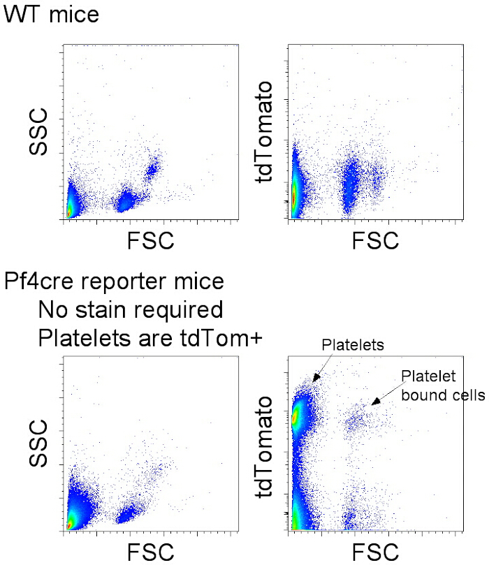
Figure 2: Phenotyping TdTomato reporter mice. Top plots, wild-type control, TdTomato-negative mice. Bottom plots, Pf4cre crossed with RosaLSLtdTom mice. Left plots illustrate lysed blood cells with side scatter versus forward scatter. Centered cells: smaller cells are lymphocytes; larger cells are neutrophils and eosinophils (not readily visible in this plot), debris, and platelets in the lower corner. The right plots are tdTomato reporting cells versus forward side scatter. Please click here to view a larger version of this figure.
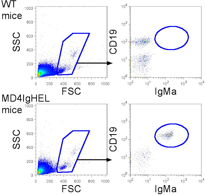
Figure 3: Phenotyping MD4 IgHEL mice. Top plots, wild-type control mice. Bottom plots, MD4IgHEL mice. Left flow plots illustrate lysed blood cells with side scatter versus forward scatter. Smaller cells are lymphocytes; larger cells are neutrophils and eosinophils (not readily visible in this plot). The right plots show gated live cells, CD19 versus allotype IgMa. Please click here to view a larger version of this figure.
Discussion
This optimized facial vein blood collection protocol provides an efficient and simple alternative to other commonly used methods, such as tail vein or retro-orbital bleeding. While the protocol is simple and straightforward, a few critical steps must be carefully followed. The most important step to execute properly is the correct placement of the puncture site4. Targeting the appropriate location on the cheek ensures sufficient blood flow with minimal distress to the mouse. Immediately depositing the blood sample into pre-chilled lysis buffer is also essential, as this prevents clotting and preserves cellular integrity. Centrifuging the lysed samples at 400 x g for 5 min is critical for proper separation and recovery of viable cells for analysis.
Over the years of use, several refinements have been introduced to improve data quality and reproducibility. Keeping all media and samples on ice throughout the procedure significantly enhances cell preservation and reduces smearing during flow cytometric analysis5. Timing is also essential. If lysis is stopped in under seven minutes, red blood cells may not fully lyse. On the other hand, if lysis exceeds 10 min, it may result in excessive cell death. Antibody staining protocols may also require slight adjustments depending on the specific antigens or fluorophores used. Despite these variables, the core blood collection and preparation steps remain consistent across diverse experimental needs. If phenotyping is performed regularly, antibody cocktails can be prepared in advance, although they should not be stored for more than a few weeks.
One large drawback of retro-orbital sampling is the use of anesthesia6. When mice are placed under anesthesia, they must then be monitored for recovery, and this adds an additional layer of complexity and time demand that facial vein bleeding does not require7. Facial vein sampling requires fewer materials and less technical training. It can be done quickly without anesthetics to provide rapid results, making it ideal for high-volume mouse phenotyping. Tail vein sampling, while common, can be time-consuming. The facial vein method is well-suited for repeat sampling, which is particularly useful in longitudinal studies such as cytokine monitoring, diabetes research, and immune phenotyping in lineage tracing and bone marrow chimeric mouse models.
The limitations to facial vein bleeding lie in the possibility of over-collection and potential for tissue trauma. While overcollection is certainly a concern, the chances of overcollection can be minimized with proper hemostasis. It is acceptable to take 7.5% of the total circulating blood volume once per week8. For adult mice, the accepted circulating blood volume is 72 mL/kg-1 of the mouse's body weight8. Female adult C57BL/6 mice weigh on average 18-22 g. Based on these numbers, an acceptable volume of blood to remove from an adult C57BL/6 female mouse is between 97 µL and 118 µL. Based on our experience, the average drop of blood that comes from the facial vein bleeding is approximately 30-50 µL, and others have documented even lower average volumes9. Approximately, one drop of blood is collected per mouse, so even taking excess bleeding into the gauze into consideration, it is unlikely that one would exceed the limit of drawing more than 7.5% of the circulating blood volume.
When it comes to tissue trauma and animal distress, facial vein venipuncture is considered to cause some level of distress and discomfort in mice. While this method may cause some distress, it is very unlikely to be fatal when bleeding is controlled after venipuncture10. Additionally, physical and lasting trauma is minimal when using the facial vein for blood collection, with minimal lesions and distress after 24 h8,9,10,11. Finally, while stress can cause variation in hormone levels and other changes in blood makeup, these effects do not take place until at least 3 minutes after the stressor occurs12 . Since the protocol described above only requires handling of the mouse for about 30-60 s, it is unlikely that this would affect the data produced by using this method.
Overall, this protocol promotes an ethical research method and refines blood collection practices to reduce the time spent on collection as well as the time during which the animals are restrained8. Its low cost, flexibility, and compatibility with flow cytometry make it broadly applicable across fields ranging from immunology to pharmacology. The ability to generate high-quality cellular data from small blood volumes makes this protocol a valuable resource for researchers seeking both experimental precision and rapid results in animal studies.
Disclosures
The authors declare no competing interests.
Acknowledgements
This work was funded by the National Institutes of Health Grant: R35 HL155458 (C.V.J.)
Materials
| Name | Company | Catalog Number | Comments |
|---|---|---|---|
| APC anti-mouse CD19 Antibody | Biolegend | 152410 | CD19 APC |
| Centrifuge 5810 R, without rotor, 120 V, 50/60 Hz | Eppendorf | 22625501 | Centrifuge |
| Dermacea Gauze sponges 8 ply 2" x 2" | Covidien | 441211 | Guaze Ponges |
| Disposable Graduated Transfer Pipettes | Fisherbrand | 13-711-91am | Transfer pipet |
| ErgoOne Single Channel Pipette (200 µL) | USA Scientific | 7100-2200 | Pipette |
| Ethylenediaminetetraacetic Acid (0.5 M Solution/pH 8.0) | Fisherbrand | BP2482-100 | EDTA 5 M |
| FACSymphony A3 Cell Analyzer | BD Biosciences | 660937 | Flow Cytometer |
| Fisherbrand Analog Vortex Mixer | Fisherbrand | 02-215-414 | Vortex |
| Goldenrod Animal Lancet 3 mm Sterile 5 trays of 200 | Goldenrod | GR 3MM | Lancet 3 mm |
| Graefe Forceps | Fine Science Tools | 11051-10 | Forceps |
| Hank’s Balanced Salt Solution, 1X without calcium and magnesium | Corning | 21-021-CVR | HBSS |
| PerCP/Cyanine5.5 anti-mouse IgMa Antibody | Biolegend | 408612 | IgMa PerCP cy-5.5 |
| RBC Lysis Buffer (10x) | Biolegend | 420301 | RBC Lysis Buffer |
| Round-Bottom Polystyrene Test Tubes Without Cap | Fisherbrand | FB1496110 | Culture tubes |
| Sharpie Permanent Markers, Chisel Tip | Sharpie | 38254PP | Sharpies |
| Tested for USP Sterile Water | Corning | 25-055-CI | Cell Culture Grade Water |
| TipOne Pipette Tip Refills (200 µL Natural Beveled) | USA Scientific | 1111-1730 | Pipette tips |
References
- Harikrishnan, V. S., Hansen, A. K., Abelson, K. S., Sorensen, D. B. A comparison of various methods of blood sampling in mice and rats: Effects on animal welfare. Lab Anim. 52 (3), 253-264 (2018).
- Jo, E. J., Bae, E., Yoon, J. H., Kim, J. Y., Han, J. S. Comparison of murine retroorbital plexus and facial vein blood collection to mitigate animal ethics issues. Lab Anim Res. 37 (1), 12(2021).
- Golde, W. T., Gollobin, P., Rodriguez, L. L. A rapid, simple, and humane method for submandibular bleeding of mice using a lancet. Lab Anim (NY). 34 (9), 39-43 (2005).
- Francisco, C. C., Howarth, G. S., Whittaker, A. L. Effects on animal wellbeing and sample quality of 2 techniques for collecting blood from the facial vein of mice. J Am Assoc Lab Anim Sci. 54 (1), 76-80 (2015).
- Ng, M. S., Ng, A. S., Chan, J., Tung, J. P., Fraser, J. F. Effects of packed red blood cell storage duration on post-transfusion clinical outcomes: a meta-analysis and systematic review. Intensive Care Med. 41 (12), 2087-2097 (2015).
- Hoggatt, J., Hoggatt, A. F., Tate, T. A., Fortman, J., Pelus, L. M. Bleeding the laboratory mouse: Not all methods are equal. Exp Hematol. 44 (2), 132-137.e1 (2016).
- Navarro, K. L., et al. Mouse anesthesia: The art and science. ILAR J. 62 (1-2), 238-273 (2021).
- Ahrens Kress, A. P., Zhang, Y., Kaiser-Vry, A. R., Sauer, M. B. A comparison of blood collection techniques in mice and their effects on welfare. J Am Assoc Lab Anim Sci. 61 (3), 287-295 (2022).
- Lavin, E. S., Feldman, E. R., Soprano, S. M., Moore, E. S. Venipuncture site influences blood-drop volume in C57BL/6 mice. J Am Assoc Lab Anim Sci. 63 (3), 325-332 (2024).
- Forbes, N., et al. Morbidity and mortality rates associated with serial bleeding from the superficial temporal vein in mice. Lab Anim (NY). 39 (8), 236-240 (2010).
- Moore, E. S., et al. Comparing phlebotomy by tail tip amputation, facial vein puncture, and tail vein incision in C57BL/6 mice by using physiologic and behavioral metrics of pain and distress. J Am Assoc Lab Anim Sci. 56 (3), 307-317 (2017).
- Gong, S., et al. Dynamics and correlation of serum cortisol and corticosterone under different physiological or stressful conditions in mice. PLoS One. 10 (2), e0117503(2015).
Reprints and Permissions
