Here, we describe a simple and quick procedure for detecting DNA from bee pathogens, such as Lotmaria passim and Nosema ceranae, using an amplification-ready cell lysis, recombinase polymerase amplification, and CRISPR/Cas12a assays.
Method Article
On-site DNA Detection of Trypanosomatid Parasites and Nosema ceranae Through Alkaline Lysis Coupled to RPA/CRISPR/Cas12a System
In This Article
Erratum Notice
Important: There has been an erratum issued for this article. View Erratum Notice
Summary
Abstract
Early on-site detection of microscopic pathogens is essential for the effective management and health of honeybee colonies. Currently, the gold standard for molecular detection of bee pathogens is qPCR or RT-qPCR. Here, we present a rapid, sensitive, and cost-effective alternative for field-deployable DNA pathogen detection. This method combines direct, amplification-ready cell lysis of worker bee abdomens with recombinase polymerase amplification (RPA), CRISPR/Cas12a-mediated trans-cleavage of reporter probes, and detection via lateral flow assays. We successfully validated this protocol in honeybees infected with Nosema ceranae and Lotmaria passim. The described protocol can be performed using a simple heat block or at room temperature and is potentially applicable to any DNA-based pathogen or gut microbiota of the honeybee. The entire process takes approximately 180 min and achieves a sensitivity comparable to qPCR, detecting as few as 96 parasite copies/µL. In conclusion, this approach offers a promising tool for reliable and rapid field diagnosis of honeybee infections without the need for complex laboratory equipment, making it ideal for on-site colony surveillance.
Introduction
Bees are key species implicated in the pollination of plants and are essential for plant reproduction, biodiversity, and food security1. Their pollination services benefit a wide range of crops with high economic value, contributing an estimated $235–577 billion annually to global agriculture. However, the worldwide decline in bee populations has raised serious concerns due to its potential consequences for ecological stability and crop productivity, which have resulted in an estimated loss of $4.8–16.3 billion in heavily indebted poor countries2. The mass of bee disappearance involves multiple interactions between several factors: pesticide application, habitat loss, climate change, and infections by pathogens, including viruses, bacteria, fungi, and protists.
Among these microorganisms, Nosema spp., a microsporidian fungus that disturbs the functioning of the gut, and trypanosomatid parasites — particularly Lotmaria passim — are widely distributed in honeybee colonies worldwide3,4. Recent findings show that L. passim infections, even when asymptomatic, can downregulate immune-related genes and alter energy metabolism in honeybees, potentially compromising their resilience under additional stress5. Similarly, Nosema spp. infections reduce bee longevity, suppress immune function, and disrupt the colony's foraging dynamics, leading to reduced honey production and increased colony mortality. Moreover, coinfections involving both Nosema spp. and trypanosomatids may have synergistic effects, exacerbating physiological stress and weakening host defenses. These findings underscore the importance of monitoring multiple pathogens simultaneously.
Addressing these threats requires the development of rapid and accurate diagnostic tools, which in turn depend on a deeper understanding of the biology, transmission, and survival strategies of these pathogens6. The detection of these pathogens through traditional methods, including light microscopy and molecular amplification, remains reliable but faces certain limitations. Examples of qPCR, RTqPCR (requires cycling at different temperatures), and isothermal methods, such as Loop Mediated Amplification (LAMP, operates at constant temperature), have been developed for fungal, viral, bacterial, or protozoan parasites6,7,8,9,10. However, the implementation of these methods needs specialized equipment, together with trained personnel and well-equipped laboratory facilities, which restricts their practicality for field applications and large-scale monitoring. To address these issues, new molecular techniques have been developed — the amplification of DNA targets with recombinase polymerase amplification (RPA), together with CRISPR/Cas12a-based detection — a fast, specific, and sensitive approach. The method works isothermally and produces visual results that make it suitable for on-site or point-of-care diagnostics, according to Shao et al.11 and Xiao et al.12.
RPA is a simple and rapid DNA amplification method that operates at a constant temperature, typically between 37 °C and 42 °C. It relies on recombinase enzymes, single-stranded DNA-binding proteins, and a strand-displacing polymerase to amplify DNA without the need for thermal cycling. RPA is especially suitable for on-site testing and field diagnostics due to its speed, minimal equipment requirements, and compatibility with crude samples11,12,13. To enhance specificity, the RPA product can be detected using the Cas12a endonuclease, which also functions within the same temperature range. Cas12a binds to a CRISPR guide RNA (gRNA), a short RNA that guides Cas12a towards the amplified target sequence and binds to the complementary sequence in the DNA. Upon binding, Cas12a becomes activated and exhibits collateral (trans) cleavage activity, specifically against single-stranded DNA (ssDNA). This enables the use of ssDNA reporter molecules labeled with fluorophores or biotin, which can generate fluorescence or visual signals, including lateral flow strip readouts. Together, RPA and CRISPR/Cas12a form a powerful molecular diagnostic platform that is rapid, sensitive, and suitable for use in field or resource-limited settings12.
To further enhance the practicality of this molecular diagnostic approach, we also implemented a simplified amplification-ready cell lysis (ARCL) method based on sodium hydroxide (NaOH) lysis of bee abdomens11,14, eliminating the need for extensive sample preparation and laboratory reagents. This rapid nucleic-acid extraction technique, combined with the isothermal amplification and CRISPR/Cas12a detection system, enables a truly field-deployable workflow that significantly reduces both time and cost. Importantly, the assay maintains the quality, high sensitivity, and specificity comparable to qPCR, the current gold standard for pathogen detection in bees.
Here, we successfully applied this methodology for the detection of Nosema ceranae and L. passim in honey bees (Figure 1). We compared this method to standard qPCR assays and validated its performance using naturally infected samples. Our findings show that this approach is a practical and reliable alternative for field-based monitoring of honeybee health, with strong potential for large-scale surveillance and early intervention efforts in beekeeping.
Protocol
All procedures were conducted in accordance with institutional and national guidelines for working with invertebrates and using the lowest number of bees needed for proof of concept. Honeybees were transported in specialized hive boxes with ventilation and appropriate temperature and stored in the lab at -20 °C until analysis.
1. HotSHOT alkaline cell lysis
- Solution preparation
- HotSHOT Lysis buffer-preparation of 100 mL
- Weigh 99.993 mg of NaOH and 8.32 mg of EDTA, add 90 mL of distilled water, and mix. Adjust the pH to 12 and the volume to 100 mL; autoclave.
- 40 mM Tris-HCl, pH 5
- To 484.544 mg of Tris, add 90 mL of deionized water, adjust the pH to 5 with HCl, and bring the volume to 100 mL.
- HotSHOT Lysis buffer-preparation of 100 mL
- Lysis protocol
- Dissect the abdomen of the bee using a sterile scalpel and transfer the dissected tissue to a 1.5 mL microcentrifuge tube.
NOTE: For pathogen detection, only the abdomens were used, and the remaining body parts were discarded. - Add 400 µL of HotSHOT buffer, macerate the tissue using a disposable pestle, and further homogenize it by vortexing.
NOTE: The pestle can be reused if decontaminated by incubating the material overnight in 0.1 N NaOH solution, then rinsed with double deionized water, followed by one autoclave cycle and subsequent drying in an incubator. - Incubate the homogenate at 95 °C for 10 min to lyse the cells. After incubation, immediately place the tube on ice to cool.
- To neutralize the reaction, add an equal volume (400 µL) of 40 mM Tris-HCl, resulting in a final concentration of 20 mM Tris-HCl in the solution.
NOTE: This lysis procedure is illustrated in Figure 2.
- Dissect the abdomen of the bee using a sterile scalpel and transfer the dissected tissue to a 1.5 mL microcentrifuge tube.
2. Isothermal amplification by RPA
NOTE: The design of the primers was made with the help of the eprimer3 software tool (https://www.bioinformatics.nl/cgi-bin/emboss/eprimer3), although other primer designing tools could be used.
- Preparation of working solutions
- RPA primer solutions
- Prepare stock solutions at a concentration of 100 µM according to the manufacturer's instructions. For example, reconstitute 10.0 nmol of lyophilized primer in 100 µL of Diethyl Pyrocarbonate (DEPC)-treated water and store at -20 °C. Prepare working solutions at a 10 µM concentration by diluting the stock 1:10; mix 90 µL of DEPC-treated water with 10 µL of the 100 µM stock solution in a 0.2 mL tube. Store these solutions at -20 °C.
- RPA primer solutions
- RPA protocol
- Prepare the master mix according to the number of reactions to be performed. Include both a negative control (substituting ARCL with DEPC-treated water) and a positive control using pathogen DNA (in this example, DNA from cell culture L. passim promastigotes and bees positive for Nosema spp. (spores detected by microscopy)) (Table 1).
NOTE: Always prepare the RPA master mix on ice and use filter tips to avoid contamination. - Transfer an aliquot of 46.5 µL of the master mix into each 0.2 mL tube.
- Add magnesium acetate (MgOAc) and template DNA to the caps of the tubes, following the volumes specified in Table 2. Briefly centrifuge the tubes to mix the contents and initiate the RPA reaction.
NOTE: For better reproducibility, briefly vortex and centrifuge all components before use. Positive and negative controls must be included in each batch to validate the reaction. - Incubate the samples at 39 °C for 40 min for the RPA amplification reaction.
NOTE: After incubation, RPA reactions can be directly used for downstream detection (e.g., CRISPR/Cas12a-based assays), described in section 3.
- Prepare the master mix according to the number of reactions to be performed. Include both a negative control (substituting ARCL with DEPC-treated water) and a positive control using pathogen DNA (in this example, DNA from cell culture L. passim promastigotes and bees positive for Nosema spp. (spores detected by microscopy)) (Table 1).
3. Pathogen DNA detection by CRISPR/Cas12a
NOTE: Detection after RPA can be performed in different ways. In this protocol, we will detail detection by fluorescence (FAM-TTTTTTTT-MGB) and immunochromatographic strips (Biotin-TTTTTTTT-FITC).
Perform all procedures in a clean, DNA-free area (e.g., dedicated PCR space). Clean work surfaces and pipettes with 0.5% bleach, followed by 70% ethanol. Always use filter tips and wear gloves. Handle DNA or amplified material in a separate area from the reagent preparation zone. Thaw all the reagents on ice and maintain cold conditions throughout preparation.
- Preparation of working solutions
- crRNA
NOTE: The designing of Nosema ceranae and Lotmaria passim crRNA was performed using the Eukaryotic Pathogen CRISPR guide RNA/DNA Design Tool (EuPaGDT) http://grna.ctegd.uga.edu/. For customized crRNA design, other software such as GPP sgRNA Designer (https://portals.broadinstitute.org/gpp/public/analysis-tools/sgrna-design) or CHOPCHOP (https://chopchop.cbu.uib.no/) may be used.- To prepare a 100 µM crRNA stock solution, reconstitute 10 nmol of each lyophilized crRNA with 100 µL of DEPC-treated water and store at -20 °C. To prepare a 1 µM working solution, perform a 1:100 dilution by mixing 1 µL of the 100 µM stock solution with 99 µL of DEPC-treated water in a 0.2 mL tube (Figure 3).
NOTE: Both solutions should be stored at -20 °C.
- To prepare a 100 µM crRNA stock solution, reconstitute 10 nmol of each lyophilized crRNA with 100 µL of DEPC-treated water and store at -20 °C. To prepare a 1 µM working solution, perform a 1:100 dilution by mixing 1 µL of the 100 µM stock solution with 99 µL of DEPC-treated water in a 0.2 mL tube (Figure 3).
- Cas12a preparation
NOTE: Cas12a can be provided at concentrations of either 1 µM or 100 µM.- If supplied at 100 µM, dilute it to a 1 µM working solution before use by mixing 1 µL of the 100 µM Cas12a enzyme with 99 µL of the diluent provided in the kit (1:100 dilution, Figure 3).
NOTE: The Cas12a enzyme concentration can be adjusted depending on the sample type. As the enzyme is stored in a glycerol-based diluent, it is important to ensure that the volume of enzyme added does not exceed 20% of the total reaction volume to avoid potential inhibitory effects.
- If supplied at 100 µM, dilute it to a 1 µM working solution before use by mixing 1 µL of the 100 µM Cas12a enzyme with 99 µL of the diluent provided in the kit (1:100 dilution, Figure 3).
- Preparation of FAM and Biotin probe solutions
NOTE: The FAM probe is provided in lyophilized form.- To prepare a 100 µM stock solution, resuspend the probe by adding 1 µL of DEPC-treated water per 1 nmol of probe (e.g., for 102 nmol, add 102 µL of DEPC-treated water). Store the stock solution at -20 °C. For use in the detection mastermix, dilute the stock 1:10 (90 µL of DEPC-treated water with 10 µL of 100 µM FAM) to obtain a 10 µM working solution (Figure 3).
- crRNA
- CRISPR/Cas12a detection protocol
- Fluorescence-based detection (FAM probe)
- In a 1.5 mL microcentrifuge tube, prepare the reaction mix as described in Table 3, excluding the amplicons.
- Mix thoroughly using a vortex and distribute the mixture into PCR tubes.
- Then, add 4 µL of RPA amplicon (described in step 2.2) to each tube.
- Incubate the reactions at 37 °C for 120 min in a thermocycler with fluorescence reading capability, recording fluorescence every minute.
- Optional: At the end of the incubation, transfer the reactions to 0.2 mL tubes and visualize using a gel documentation system for imaging.
- Detection using a Biotin probe in a lateral flow test
- In a 1.5 mL microcentrifuge tube, add the components shown in Table 4.
- Make the mix as described in section 3.2.1, mix, and distribute it into a PCR plate. Finally, add 4 µL of amplicon to the respective wells.
- Incubate for 120 min in a thermocycler (without fluorescence reading) or thermal block at 37 °C.
- Transfer the samples to 0.2 mL tubes and perform the detection with immunostrips. To do this, add 50 µL of running buffer and 10 µL of the reaction mixture into each tube and insert the strip directly into the tube. Incubate for 15 min at room temperature and read the results after 10 min of immersion, as illustrated in Figure 3.
- Fluorescence-based detection (FAM probe)
Results
Here we have described a fast on-site method using alkaline lysis of worker bee abdomens coupled to RPA amplification and CRISPR/Cas12a-based detection by fluorescence and/or lateral flow approaches. The schema of the full workflow is represented in Figure 3, and the schema of the HotSHOT alkaline lysis of worker bee abdomens is in Figure 4. Representative results of the RPA/CRISPR/Cas12a assay are shown in Figure 5. To detect L. passim and N. ceranae, a thermocycler was used to quantify the relative fluorescence units (RFUs) using a FAM-labeled probe (Figure 4A). The RPA reactions were performed using 10 ng of DNA, and the fluorescence data were plotted in a graph (Figure 4B). At the final stage of the assay, samples in conical tubes were photographed using a gel imaging system (Figure 4C). For detection with a biotin-labeled probe, the samples were incubated in a thermoblock (Figure 4A); the detection solution was prepared according to the manufacturer's instructions and applied onto a lateral flow strip (Figure 4B, C). The method resulted in a successful detection of both honeybee pathogens.
To determine the efficiency of RPA reactions, we compared this method with standard qPCR using DNA purified from honeybees using the referenced kit (see the Table of Materials). A total of 32 bee samples were tested with primers targeting the β-tubulin gene of L. passim for both methods and then detected by CRISPR/Cas12a trans-cleavage of fluorescent probes. Notably, as a result, 12 samples were found to be positive by qPCR, while RPA/CRISPR/Cas12a detected L. passim in 16 samples (Figure 5A). This indicates that RPA reaction performance is more sensitive than PCR, as the gold-standard DNA amplification method.
Likewise, we determined the detection limit (LOD) for qPCR and RPA coupled to CRISPR/Cas12a detection for L. passim DNA as the pathogen model using serial dilutions of L. passim DNA ranging from 66 ng to 6 fg. qPCR detected as little as 6 pg, corresponding to approximately 96 parasites (Figure 5B). In comparison, the LOD for RPA corresponded to 96 parasites (Figure 5C, D).
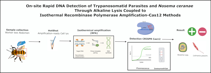
Figure 1: General workflow of the HotSHOT alkaline lysis coupled with RPA amplification and Cas12 detection methods in honeybees. Abbreviations: RPA = recombinase polymerase amplification; CRISPR = Clustered Regularly Interspaced Short Palindromic Repeats; Cas = CRISPR-associated protein. Please click here to view a larger version of this figure.
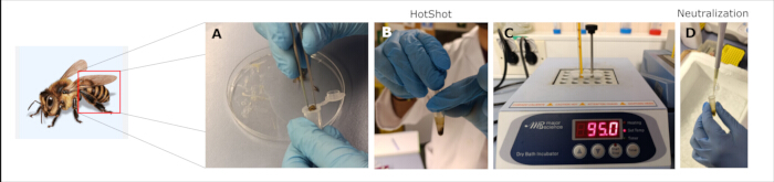
Figure 2: Processing of amplification-ready cell lysis with HotSHOT buffer. (A) Cut the honeybee abdomen with a scalpel and put it in a centrifuge tube; (B) with a pestle, make a maceration with 400 µL of HotShot; (C) incubate on a thermoblock at 95 °C for 10 min; and (D) neutralize with 400 µL of 40 mM Tris-HCl. Please click here to view a larger version of this figure.
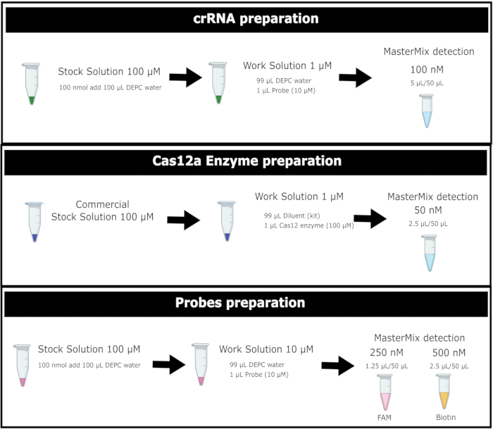
Figure 3: General workflow for the preparation of Cas12a enzyme and FAM- and Biotin-labeled reporter probe solutions. Abbreviations: DEPC-water = Diethyl Pyrocarbonate-treated water; crRNA= RNA CRISPR; Cas12 = type of CRISPR-associated protein. Please click here to view a larger version of this figure.
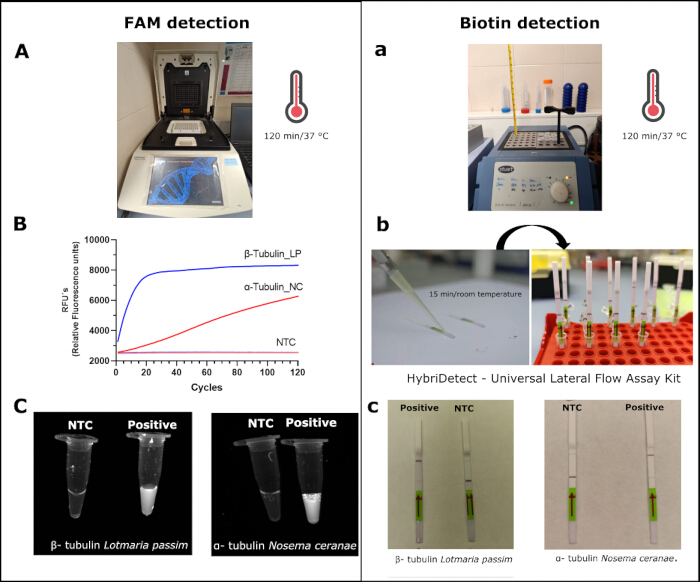
Figure 4: Detection of Lotmaria passim and Nosema ceranae. by the CRISPR/Cas12a method with FAM and biotin probes. For FAM detection, (A) samples are being incubated in a thermocycler; (B) the relative fluorescence units (RFU´s) of L. passim and N.ceranae; (C) visualization of the same samples under a photodocumentation system. In biotin tests, (a) a sample incubating in a thermo block; (b) samples were added in an immunostrip from the kit (see the Table of Materials); (c) the results after incubation. Abbreviations: LP= Lotmaria passim; NC= Nosema ceranae; NTC= no template control; RFU: relative fluorescence units Please click here to view a larger version of this figure.
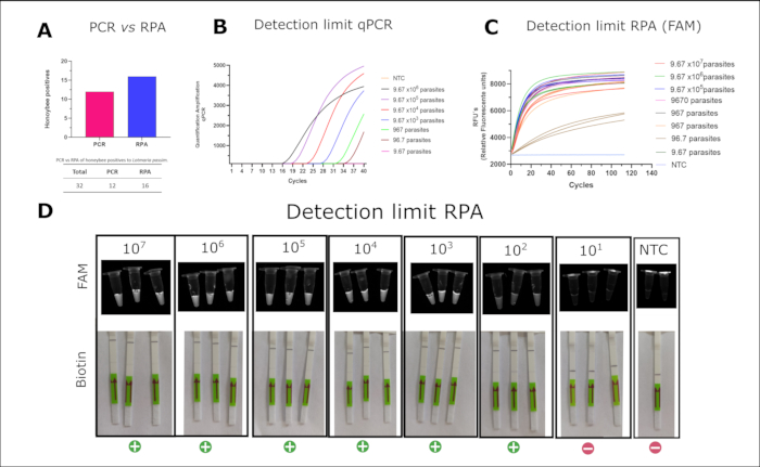
Figure 5: Comparison between qPCR and RPA, and detection limit of Lotmaria passim. (A) Detection of L. passim in honeybee samples using conventional qPCR and RPA. (B) qPCR detection limit of L. passim using SsoFast EvaGreen. (C) Detection limit of L. passim using CRISPR/Cas12a with a FAM-labeled probe; (D) visualization of the same CRISPR/Cas12a detection (from C) under a gel imaging system (top row), and corresponding lateral flow results using CRISPR/Cas12a with a biotin-labeled probe (bottom row). Abbreviations: RPA = recombinase polymerase amplification; qPCR= quantitative polymerase chain reaction. Please click here to view a larger version of this figure.
| Reagent | Final Concentration | 10 µL | 50 µL |
| 2x Reaction Buffer | 1x | 5 µL | 25 µL |
| dNTPs (10 mM) | 1.8 mM | 1.8 µL | 9 µL |
| 10x Basic E-Mix | 1x | 1 µL | 5 µL |
| Forward Primer (10 µM) | 480 nM | 0.5 µL | 2.4 µL |
| Reverse Primer (10 µM) | 480 nM | 0.5 µL | 2.4 µL |
| 20x Core Reaction Mix | 1x | 0.5 µL | 2.5 µL |
| DEPC Water | -- | 0.7 µL | 3.7 µL |
Table 1: Mastermix components for RPA with final volumes of 10 µL and 50 µL, and corresponding working concentrations:
| Reagent | Final concentration | 10 µL reaction | 50 µL reaction |
| 280 mM MgOAc | 1.8 mM | 0.5 µL | 2.5 µL |
| DNA | 10-0.1 ng | 1 µL | 2 µL |
Table 2: Volume of reagents to be added per sample in a reaction of RPA
| Reagent | [stock] | [final] | 1x (µL) |
| Cas12a enzyme | 1 µM | 50 nM | 2.5 |
| cRNA | 1 µM | 100 nM | 5 |
| FAM probe | 10 µM | 250 nM | 1.25 |
| NEB buffer 10x | 10x | 1x | 5 |
| RPA amplicon | - | - | 4 |
| Water | - | - | 32.25 |
| Total | 50 |
Table 3: Concentrations of reagents to make a mix of FAM detection by the method CRISPR/Cas 12a.
| Reagent | [stock] | [final] | 1x (µL) |
| Cas12a enzyme | 1 µM | 50 nM | 2.5 |
| cRNA | 1 µM | 100 nM | 5 |
| Biotin probe | 10 µM | 500 nM | 2.5 |
| NEB buffer 10x | 10X | 1X | 5 |
| RPA amplicon | - | - | 4 |
| Water | - | - | 31 |
| Total | 50 |
Table 4: Concentrations of reagents to make a mix of biotin detection by the CRISPR/Cas 12a method.
Discussion
In this study, we developed and validated a practical molecular diagnostic protocol for detecting honeybee pathogens. The workflow integrates amplification-ready cell lysis, isothermal amplification using RPA, and highly specific detection via CRISPR/Cas12a. Among the chemical DNA extraction methods evaluated, the HotSHOT alkaline lysis protocol offers significant advantages in cost, speed, and simplicity. It efficiently produces amplification-ready lysates without the need for commercial kits. This method has proven reliable in various applications, including pathogen detection in honeybees14,15,16,17,18. The high-temperature alkaline conditions disrupt cell membranes and hydrogen bonding between DNA strands, releasing single-stranded DNA suitable for downstream amplification. Although potential limitations include the presence of PCR inhibitors or suboptimal neutralization, alkaline lysis has been shown to work effectively in diverse organisms such as archaea19, bacteria20, and fungi14, and is compatible with PCR, qPCR, and isothermal methods like LAMP21 and RPA12.
In our hands, the RPA/CRISPR/Cas12a system showed strong specificity and satisfactory sensitivity. The use of crRNAs targeting conserved regions in each pathogen allowed for accurate identification with no cross-reactivity to host DNA or unrelated microorganisms. Previous applications of this system in honeybees have achieved detection limits of approximately 6.5 × 10² and 6.2 × 101 copies/µL for Deformed Wing Virus types A and B8,12. However, these assays have not been optimized for in-field conditions with rapid RNA and/or DNA extraction. In this work, we adapted the method specifically for DNA pathogens -- N. ceranae and L. passim -- enabling field-compatible detection.
Our results show that RPA/CRISPR/Cas12a outperformed conventional PCR in identifying positive samples (16 vs. 12), likely due to the improved signal using the CRISPR/Cas12a collateral cleavage activity, which remains effective even in crude lysates. The improvement in sensitivity over PCR-based methods has been previously reported in different pathogen models such as Yersinia enterocolitica22, Diaporthe aspalathi23, or Vibrio vulnificus24. Furthermore, quantitative analysis revealed an equivalent limit of detection of RPA/CRISPR/Cas12a and qPCR under laboratory conditions (~96 parasite copies/µL). This implies a good performance of the method with comparable specificity to TaqMan qPCR, due to the added sequence recognition provided by the crRNA. Furthermore, the assay requires only minimal equipment, does not depend on a thermocycler or fluorescence reader, and can be visualized using lateral flow strips as previously developed for other pathogens25,26. This simplifies interpretation and makes the test highly accessible for field technicians and beekeepers.
The current protocol yields robust results, but further improvements are possible. Adapting the assay to target other DNA-based bee pathogens would only require redesigning RPA primers and crRNAs. For RNA viruses, the protocol can be expanded to include a reverse transcription step (RT-RPA) as previously demonstrated for several viral infectious diseases26,27,28, including DWV-A and DWV-B in honeybees15. Additionally, the total assay time -- currently around 180 minutes -- can likely be reduced through protocol optimization for decreasing the timing of each step. Another alternative would be one-pot RPA/CRISPR/Cas12a that combines all RPA/CRISPR/Cas12a reagents in a single reaction. This all-in-one methodology has been successfully applied for the detection of a wide range of viral and bacterial pathogens25,26,29. Besides and despite that ssDNA probes have been extended and popularized for Cas12a detection, some studies have reported the capacity of Cas12a of trans-cleavage double-stranded DNA (dsDNA) probes that may be useful to be tested due to the stability of dsDNA30. This would lead to a time-to-result under 30 minutes, further enhancing field applicability. Lyophilizing RPA and CRISPR reagents would also improve reagent stability and eliminate the need for a cold chain, increasing suitability for remote or low-resource environments. The successful performance of RPA/CRISPR/Cas12a using lyophilized reagents has been successfully determined for the detection of Actinobacillus pleuropneumonia31 and respiratory infectious viruses28,32, including SARS-Cov226.
In summary, this study presents a fully in-field, cost-effective, and portable molecular diagnostic platform for honeybee pathogen detection. This methodology, as it stands, would last 180 min approximately, but its total timing could still be subjected to further improvements. While we focused on L. passim and Nosema spp. as proof-of-concept targets, the system is modular and adaptable to a wide range of DNA-based bee pathogens (i.e., Paenibacillus larvae or Melisococcus plutonious) as well as for monitoring commensal symbionts of interest. This approach expands access to molecular diagnostics beyond traditional laboratory settings and supports early intervention strategies in apiculture. More broadly, our results demonstrate the viability of CRISPR-based technologies in field-ready diagnostic applications.
Disclosures
The authors have no conflicts of interest to disclose.
Acknowledgements
This work was supported by the grant "New molecular tools for rapid monitoring of honeybee diseases in apiaries in the province of Granada" (GOPG-GR-23-0002) under the call Aid to operational groups of the European Innovation Association (AEI) in agricultural productivity and sustainability of the Andalusian government. We also acknowledge the support by the Spanish Programme for Knowledge Generation and Scientific and Technological Strengthening of the R+D+I System, grant PID2021-126938OB-I00 funded by MCIN/AEI/10.13039/501100011033 and by "ERDF/EU". We strongly appreciated the help and resources provided by beekeepers of the Asociación de Pequeños Apicultores de Granada (APAG) and Cooperativas Agroalimentarias de Granada (FAECA). The study is part of the PhD thesis of D. Pedro García Olmedo. Doctoral Program in Fundamental and Systems Biology (University of Granada, UGR).
Materials
| Name | Company | Catalog Number | Comments |
|---|---|---|---|
| 0.2 mL DNAse- and RNAse-free Eppendorf tubes | Axygen | MTC-02-C | |
| 1.5 mL DNAse- and RNAse-free Eppendorf tubes | Axygen | MTC-150-C | |
| 10 μL low retention filtered tips | LabClinics | 5-063-P5-0 | |
| 100 μL low retention filtered tips | LabClinics | 5-120-P5-0 | |
| 1000 μL low retention filtered tips | LabClinics | 5-203-P4-0 | |
| BH-200 Series Stuart Dry Block Heater | Cole-Parmer | EW-36610-00 | |
| DEPC-treated water | Ambicon | AM9906 | |
| EDTA | Sigma-Aldrich | 798681 | |
| EnGen Lba Cas12a (Cpf1) | New England Biolabs | M0653T | |
| Sigma-Aldrich | Sigma-Aldrich | 64-17-5 | |
| HCl | Sigma-Aldrich | 7647-01-0 | |
| Microtube pestles | Fisherbrand | 12141364 | |
| Milenia GenLine HybriDetect | Milenia | MGHD 1 | |
| NaOH | Sigma-Aldrich | 221465 | |
| RPA kit TwistAmp Liquid Basic | TwistAmp | TALQBAS01 | |
| Scalpel blades | Swann-Morton | 201 | |
| Single Chanel Pipete PIPETMAN P1000L Micropipette Metal eject | Gilson | F144059M | |
| Single Chanel Pipete PIPETMAN P100L Micropipette Metal eject | Gilson | F144057M | |
| Single Chanel Pipete PIPETMAN P10L Micropipette Metal eject | Gilson | F144055M | |
| Sodium hypochlorite | |||
| Touch Real-Time PCR Detection System | BioRad | CFX96 | |
| Tris-HCl | Sigma-Aldrich | T1503 | |
| TwistAmp Liquid Basic | Twist | TALQBAS01 |
References
- Goulson, D., Nicholls, E., Botías, C., Rotheray, E. L. Bee declines driven by combined stress from parasites, pesticides, and lack of flowers. Science. 347 (6229), 1255957(2015).
- Murphy, J. T., Breeze, T. D., Willcox, B., Kavanagh, S., Stout, J. C. Globalisation and pollinators: pollinator declines are an economic threat to global food systems. People and Nature. 4 (3), 773-785 (2022).
- Zattara, E. E., Aizen, M. A. Worldwide occurrence records suggest a global decline in bee species richness. One Earth. 4 (1), 114-123 (2021).
- Carreira de Paula, J., et al. Promastigote EPS secretion and haptomonad biofilm formation as evolutionary adaptations of trypanosomatid parasites for colonizing honeybee hosts. NPJ Biofilms Microbiomes. 10 (1), 27(2024).
- Requier, F., et al. First large-scale study reveals important losses of managed honeybee and stingless bee colonies inLatin America. Sci Rep. 14 (1), 10079(2024).
- Babin, A., Schurr, F., Rivière, M. P., Chauzat, M. P., Dubois, E. Specific detection and quantification of three microsporidia infecting bees, Nosema apis, Nosema ceranae, and Nosema bombi, using probe-based real-time PCR. Eur J Protistol. 86, 125935(2022).
- Cameron, T. C., Wiles, D., Beddoe, T. Current status of loop-mediated isothermal amplification technologies for the detection of honeybee pathogens. Front Vet Sci. 8, 659683(2021).
- Schurr, F., et al. Validation of quantitative real-time RT-PCR assays for the detection of six honeybee viruses. J Virol Methods. 270, 70-78 (2019).
- Xu, G., et al. Triplex real-time PCR for detection of Crithidia mellificae and Lotmaria passim in honeybees. Parasitol Res. 117 (2), 623-628 (2018).
- D'Alvise, P., Seeburger, V., Gihring, K., Kieboom, M., Hasselmann, M. Seasonal dynamics and co-occurrence patterns of honeybee pathogens revealed by high-throughput RT-qPCR analysis. Ecol Evol. 9 (18), 10241-10252 (2019).
- Shao, G., Zhu, X., Hua, R., Chen, Y., Yang, G. Development of a copro-RPA-CRISPR/Cas12a assay to detect Echinococcus granulosus nucleic acids isolated from canine feces using NaOH-based DNA extraction method. PLoS Negl Trop Dis. 18 (12), e0012753(2024).
- Xiao, Y., Fei, D., Li, M., Ma, Y., Ma, M. Establishment and application of CRISPR-Cas12a-based recombinase polymerase amplification and a lateral flow dipstick and fluorescence for the detection and distinction of deformed wing virus types A and B. Viruses. 15 (10), 2041(2023).
- Li, Z., et al. Cas-OPRAD: a one-pot RPA/PCR CRISPR/Cas12 assay for on-site Phytophthora root rot detection. Front Microbiol. 15, 1390422(2024).
- Osmundson, T. W., Eyre, C. A., Hayden, K. M., Dhillon, J., Garbelotto, M. M. Back to basics: an evaluation of NaOH and alternative rapid DNA extraction protocols for DNA barcoding, genotyping, and disease diagnostics from fungal and oomycete samples. Mol Ecol Resour. 13 (1), 66-74 (2013).
- Truett, G. E., et al. Preparation of PCR-quality mouse genomic DNA with hot sodium hydroxide and Tris (HotSHOT). BioTechniques. 29 (1), 52-54 (2000).
- Klintschar, M., Neuhuber, F. Evaluation of an alkaline lysis method for the extraction of DNA from whole blood and forensic stains for STR analysis. J Forensic Sci. 45 (3), 669-673 (2000).
- Galanis, A., et al. Bee foraging preferences, microbiota and pathogens revealed by direct shotgun metagenomics of honey. Mol Ecol Resour. 22 (7), 2506-2523 (2022).
- Xiang, Z., et al. A simple alkali lysis method for Plasmodium falciparum DNA extraction from filter paper blood samples. Mol Biochem Parasitol. , 254(2023).
- Shwani, A., Zuo, B., Alrubaye, A., Zhao, J., Rhoads, D. D. A simple, inexpensive alkaline method for bacterial DNA extraction from environmental samples for PCR surveillance and microbiome analyses. Appl Sci. 14 (1), 141(2024).
- Fowler, E. V., et al. Development of a cost-effective, morphology-preserving method for DNA isolation from bulk invertebrate trap catches: tephritid fruit flies as an exemplar. PLoS One. 18 (2), e0281759(2023).
- Yuan, J., et al. CRISPR-Cas12a-mediated hue-recognition lateral flow assay for point-of-need detection of Salmonella. Anal Chem. 96 (1), 220-228 (2024).
- Xiao, Y., et al. Ultra-sensitive and rapid detection of pathogenic Yersinia enterocolitica based on the CRISPR/Cas12a nucleic acid identification platform. Foods. 11 (14), 2160(2022).
- Dong, J., et al. Comparative evaluation of PCR-based, LAMP and RPA-CRISPR/Cas12a assays for the rapid detection of Diaporthe aspalathi. Int J Mol Sci. 25 (11), 5773(2024).
- Zhang, X., et al. CRISPR/Cas12a combined with recombinase polymerase amplification for rapid and sensitive detection of Vibrio vulnificus in one tube. Acta Biochim Biophys Sin. 55 (2), 322-326 (2023).
- Li, P. R., et al. An RPA-assisted CRISPR/Cas12a assay combining fluorescence and lateral flow strips for the rapid detection of enterotoxigenic Bacillus cereus. J Agric Food Chem. 72 (24), 14000-14010 (2024).
- Sun, Y., et al. One-tube SARS-CoV-2 detection platform based on RT-RPA and CRISPR/Cas12a. J Transl Med. 19 (1), 74(2021).
- Aman, R., et al. iSCAN-V2: a one-pot RT-RPA-CRISPR/Cas12b assay for point-of-care SARS-CoV-2 detection. Front Bioeng Biotechnol. 9, 800104(2022).
- Peng, R., Lu, Z., Liu, M., Hu, F. RT-RPA-assisted CRISPR/Cas12a for rapid and multiplex detection of respiratory infectious viruses based on centrifugal microfluidics. Sens Actuators B Chem. 399, 134838(2024).
- Liu, Y., et al. One-tube RPA-CRISPR Cas12a/Cas13a rapid detection of methicillin-resistant Staphylococcus aureus. Anal Chim Acta. 1278, 341757(2023).
- Nguyen, L. T., Smith, B. M., Jain, P. K. Enhancement of trans-cleavage activity of Cas12a with engineered crRNA enables amplified nucleic acid detection. Biochemistry. 59 (17), 1474-1481 (2020).
- Hao, J., et al. Application of a lyophilized CRISPR/Cas12a and RPA assay for rapid detection of Actinobacillus pleuropneumoniae. Microchem J. 206, 111443(2024).
- Lin, C., et al. A universal all-in-one RPA-Cas12a strategy with de novo autodesigner and its application in on-site ultrasensitive detection of DNA and RNA viruses. Biosens Bioelectron. 239, 115609(2023).
Reprints and Permissions
Erratum
Formal Correction: Erratum: On-site DNA Detection of Trypanosomatid Parasites and Nosema ceranae Through Alkaline Lysis Coupled to RPA/CRISPR/Cas12a System
Posted by JoVE Editors on 8/14/2025. Citeable Link.
This corrects the article 10.3791/68874
