This article demonstrates CRISPR-Cas9-mediated genome engineering in Steinernema hermaphroditum, an entomopathogenic (EPN: insect-parasitic) nematode and an emerging genetic model. The described technology is useful for creating mutants, allowing for the elucidation of gene functions in nematode biology that are relevant to mutualistic and parasitic symbiosis.
Method Article
CRISPR-Cas9-based Mutagenesis in the Entomopathogenic Nematode Steinernema hermaphroditum and the Maintenance of Mutant Lines
In This Article
Summary
Abstract
Entomopathogenic nematodes (EPNs) in the genus Steinernema and Heterorhabditis maintain mutualistic interactions with Xenorhabdus and Photorhabdus symbiotic bacteria, respectively. Together, these nematode-bacterium pairs infect and kill insect hosts that are primarily larvae from the orders of Lepidoptera and Coleoptera, forming a tractable tripartite system for dissecting the molecular basis of mutualism and parasitism. A key step towards fully utilizing this model is the development of stable and transgenerational genetic tools in EPNs. Here, we demonstrate a reliable CRISPR–Cas9 genome editing platform in the emerging model Steinernema hermaphroditum, a species that is readily maintained in vivo and in vitro, and is highly amenable to gonadal microinjection. Importantly, its hermaphroditic reproduction greatly streamlines the generation and maintenance of homozygous mutant lines. We provide a detailed protocol for efficient, targeted gene disruption using microinjection-based delivery of Cas9 ribonucleoprotein complexes. As a proof of concept, we modified the conserved muscle-associated gene unc-22, generating a characteristic twitching phenotype that validates targeted mutagenesis in this system. This CRISPR–Cas9 platform opens the door to stable genetic manipulation in S. hermaphroditum, such as transgene expression, and provides a framework that can be extended to additional EPN species of agricultural and ecological importance.
Introduction
Entomopathogenic nematodes (EPNs) are insect-killing parasites that form species-specific mutualistic partnerships with their bacterial symbionts1. EPNs consist of two families: Steinermatidae and Heterorhabditidae, which associate with Gram-negative bacteria of Photorhabdus and Xenorhabdus spp, respectively2,3. During the free-living infective juvenile stage (IJ) of Steinernema nematodes, Xenorhabdus bacteria are housed within an intestinal pocket of the nematode known as the receptacle, and upon penetration of the insect host into the hemocoel, the bacteria are released, after which they multiply, and kill the insect by septicaemia3,4. In this mutualistic relationship, the bacteria produce antimicrobial and insecticidal compounds, which protect against the host immune response and provide nutritional support to the nematode5. In return, the bacteria benefit from the protection provided by the nematode host against environmental microbes and are facilitated in their dispersal to new insect prey6,7.
Over the past few decades, the Steinernema-Xenorhabdus partnership has been established as a powerful experimental system to study various aspects of parasitic and mutualistic interactions, such as insect host-seeking behaviors3, bacterial colonization in the nematode host8, and adaptive behaviors of symbiotic bacteria that facilitate their transitions between nematodes and insect host animals9. EPNs are also important species in soil sustainability and are used as organic pest control agents to promote agricultural productivity10,11.
Despite the rapid development of genetic tools in the symbiotic bacteria Xenorhabdus and Photorhabdus12, consistent genetic tools in EPNs have been scarce. Gonadal microinjection has been successfully established in both Heterorhabditis and Steinernema nematodes13,14. Previously, gonadal microinjection in Heterorhabditis bacteriophora has been used to consistently deliver double-stranded RNA for gene knockdown among the first generation of progeny13,15. Recently, CRISPR-Cas9 genome editing in Steinernema has been developed, delivered via gonadal microinjection, and resulted in precise, stable, and heritable mutations14.
One Steinernema spp, S. hermaphroditum, has been demonstrated to be highly tractable and has great potential as an emerging genetic model organism4,14. Originally isolated from the soil in Indonesia16 and re-isolated in India17, S. hermaphroditum is consistently hermaphroditic, with a small number of males in each generation, two features that facilitate the adaptation of genetic tools from the classical model nematode C. elegans4. In addition, the genome of S. hermaphroditum has been sequenced and assembled into chromosomes, simplifying the task of identifying potential Cas9 target sites and designing primers18.
During microinjection of EPN, an injection mix containing appropriate reagents, such as dsRNAs, Cas9 protein, or guide RNAs, is delivered to the syncytial nematode gonad via a needle pulled from a quartz capillary. The appropriate volume and pressure of injection result in a visible 'flow' of liquid throughout the gonadal arm. Since these nematodes have a pair of syncytial gonads composed of germline nuclei that share the same cytosol19, this microinjection approach allows multiple germ cells to be accessible for editing, before cytokinesis occurs, separating the germline nuclei into developing oocytes20. Although gonadal microinjection serves as a consistent technique for genetic modulation in several species of non-model nematodes14,21,22,23,24,25, it is crucial to adapt the injection techniques to the appropriate gonadal structures, life stage, and physiological condition of each nematode species to ensure success and high efficiency in delivery. Recovery techniques post-injection and maintenance of mutant lines are both important to ensure this technique is cost-efficient.
This study demonstrates a CRISPR-Cas9 genome editing protocol in Steinernema hermaphroditum. The gonadal microinjection is described in the young adult, a stage that is resilient to high-pressure microinjection and is receptive to gene editing14. In addition, detailed methods describing the maintenance of heritable and stable mutant lines are provided, both via cryopreservation and propagation through insects.
Protocol
The reagents and the equipment used are listed in the Table of Materials.
1. Nematode preparation prior to microinjection
- Prepare Nematode Growth Media (NGM) plates seeded with bacteria Xenorhabdus griffiniae and Comamonas aquaticus.
NOTE: Grow S. hermaphroditum nematodes on their native symbiotic bacterium Xenorhabdus griffiniae before microinjection to maximize nematode growth and recover injected animals on bacterium Comamonas aquaticus to increase post-injection survival14.- Streak bacterium X. griffiniae on LB-pyruvate agar plates (2.5 g yeast extract, 5 g tryptone, 2.5 g sodium chloride, 0.5 g sodium pyruvate, 7.5 g agar in 500 mL water) and incubate overnight at 30 °C.
- Streak bacterium C. aquaticus on LB agar plates and incubate overnight at 37 °C.
- Pick a single colony of bacteria to inoculate dark LB liquid media (5 g yeast extract, 10 g tryptone, 5 g sodium chloride in 1 L of water). Grow bacterial culture overnight (16 h maximum) with aeration (X. griffiniae at 30 °C and C. aquaticus at 37 °C).
- Prepare NGM agar: (3 g NaCl, 2.5 g peptone, and 20 g agar added to 975 mL water, with 1 mL cholesterol, 1 mL 1 M CaCl2, 1 mL 1 M MgSO4, 25 mL 1 M KPO4 added after autoclaving) 10 mL per 60 mm x 15 mm Petri dish.
- Seed bacteria: Add a few droplets of the overnight bacterial culture onto each NGM agar plate, gently shake to spread the bacterial droplets into a patch.
- Incubate bacteria-seeded NGM plates at room temperature to allow agar to dry and bacteria to grow.
NOTE: X. griffiniae-seeded plates should be used as soon as possible; C. aquaticus plates can be stored at room temperature for 1-2 weeks.
- Nematode staging
NOTE: Pick the J3 stage of the hermaphrodite the night before injection and young adults on the day of injection.- Pick or chunk S. hermaphroditum nematodes onto the prepared NGM plates seeded with X. griffiniae. Incubate at a temperature of 25-28 °C for optimal growth.
- The night before microinjection, pick J3 stage of hermaphrodites onto NGM agar seeded with X. griffiniae. As shown in Figure 1A, the J3 stage is estimated by the size of the nematode and the morphology of the gonads and vulva.
- On the day of microinjection, pick young adult hermaphrodites (Figure 1B).
2. CRISPR-Cas9 preparation for microinjection
- Selecting the CRISPR crRNAs and reagent storage
- Extract the target gene sequence from the reference Steinernema hermaphroditum genome using BlastP, PRJNA982879.
- Select an early coding exon sequence, if possible, preferably the first exon, and input the sequence into CRISPRScan using default settings26 to generate a list of possible CRISPR RNAs (crRNAs).
- Blast the top crRNAs against the S. hermaphroditum genome to ensure only unique crRNAs are chosen.
NOTE: Optional: Confirm the sequence of the target region of the genome by PCR amplification and Sanger sequencing before crRNA design, to ensure efficient binding of crRNAs. - Store the CRISPR RNAs (crRNAs) and universal trans-activating CRISPR RNAs (tracrRNAs) at 100 µM each in duplex buffer at -80 °C.
- Store Cas9 protein at 10 mg/mL at -20 °C or -80 °C. Store 1 M KCl at 4 °C.
- Making the 2% agarose pad for injection (adapted from Berkowitz et al.27)
- Melt 2% agarose in water using a microwave oven.
- Transfer 50 µL of melted agarose onto a cover glass (48 x 60 mm no.1). Immediately cover the agarose droplet with another cover glass.
- Let the agarose dry at room temperature for 20 min. Remove the top cover glass. Label the front side of the agarose pad.
NOTE: Optional: Bake the agarose pad at 37 °C overnight to further dry the agarose pad. - Store the 2% agarose pads at room temperature for months to years.
- Needle preparation
- Manufacture quartz needles using the following parameters on a laser filament puller. With the needle puller used in this study, the suggested program is as follows: Heat: 700, Filament: 4, Velocity: 60, Delay: 145, Pull: 175.
- Pull the quartz needles (OD: 1.0 mm, ID: 0.7 mm) immediately before injection, or ahead of time, and store them in a pipette storage box.
NOTE: Alternatively, we also have success injecting with borosilicate needles (OD: 1.2 mm, ID: 0.68 mm). Note that different sizes of needles will match different sizes of needle holders.
3. CRISPR-Cas9 microinjection and recovery of the nematodes after microinjection
- Prepare the CRISPR-Cas9 microinjection mixture (adapted from Wang et al.28)
- Prepare fresh guide RNA duplexes by mixing the chosen crRNAs and universal tracrRNA in a 1:1 ratio, i.e., total crRNA to tracrRNA in a PCR tube:
crRNA #1 (100 µM) - 1.5 µL
crRNA #2 (100 µM) - 1.5 µL
tracrRNA (100 µM) - 3.0 μL
NOTE: If using co-CRISPR, crRNA #1 is targeting the unc-22 marker gene, while crRNA #2 can be replaced with another sequence targeting a second gene of interest. - Incubate the PCR tube in a thermocycler at 94 °C for 2 min and then cool to room temperature.
- Assemble the injection mix by combining the following, then incubate for 5 min at room temperature:
Cas9 (10 mg/mL) - 2 µL
1M KCl - 0.58 µL
gRNA duplexes (from step 3.1.1) - 2.7 µL - Mix three times by flicking the PCR tube, followed by a brief spin (10-15 s) in a mini centrifuge to collect the contents of the tube. After the final mix, spin down for longer, for up to 1 min.
- Prepare fresh guide RNA duplexes by mixing the chosen crRNAs and universal tracrRNA in a 1:1 ratio, i.e., total crRNA to tracrRNA in a PCR tube:
- Load and break the needle
- Load the needle with 1-2 µL of injection mixture using a microloader tip.
- Assemble the needle to the pipette (needle) holder, then break open the tip by dragging the needle across a piece of double-sided tape covered in halocarbon oil. Test the opening of the needle using the CLEAN function of the microinjector .
- Microinjection into the syncytial gonads of a young adult nematode
- Transfer a small droplet of Halocarbon oil onto 2% agarose pad.
- Under the dissecting stereoscope, use a thin (soft) platinum wire to pick up a young adult hermaphrodite nematode and wash it in an M9 buffer (per liter: 3 g KH2PO4, 11.3 g Na2HPO4, 5 g NaCl, with 1 ml 1 M MgSO4 added after autoclaving) to remove bacteria (see young adult nematode in Figure 1).
- Transfer the washed nematode onto an agarose pad and place it in the droplet of halocarbon oil. Use a soft platinum wire to gently brush the nematode until the animal is immobilized.
- Carefully place the agarose pad on the microinjection stage. The orientation of the gonadal syncytium should be pointing to the needle at about a 45° angle. Start from 5x magnification, then switch to 20x and 40x magnification for the injections.
- Ensure the needle penetrates the syncytium, then use the CLEAN function of the microinjector to introduce the injection mix at approximately 99 psi until a clear gonadal flow of injected liquid is observed to ensure reagents access germ cells in the gonadal syncytium.
- If the second gonadal arm is accessible, move the stage to position the needle to inject into the second arm.
- Nematode recovery post-injection
- Immediately after injection, resuspend the injected animal in 1-2 µL of M9 buffer on the agarose pad.
- Using a thin platinum wire, pick up the injected nematode off the agarose pad and wash off the oil using M9 buffer.
- Recover the nematodes on NGM plates with a lawn of C. aquatica bacteria (NGM plate preparation: see step 1.1), which increases the survival rate of the nematodes post-injection14. Incubate at 25 °C overnight.
- The next day, isolate each injected nematode (P0) onto an individual NGM plate seeded with X. griffiniae. Let P0 animals produce F1 progeny overnight (either by laying eggs or bagging) for phenotyping and genotyping (see step 4).
4. Screening to confirm CRISPR-Cas9-mediated knockouts
- Nicotine-based (CAUTION) screen of F1 for twitching phenotype when using unc-22 as a co-CRISPR marker
- Transfer F1 progeny from injected P0 animals into a spot of 2% Nicotine solution.
- Pick an individual twitching animal from the nicotine spot and immediately recover the animal on NGM agar seeded with X. griffiniae. Isolate each twitching F1 onto an individual plate and let it produce F2 animals for genotyping (see step 4.2).
- Genotyping
- Pick eight F2 or F3 progeny from each F1 twitching line for genotyping. These animals are likely to include both +/mut heterozygous and mut/mut homozygous animals.
- Prepare fresh DNA extraction buffer by adding 20 µL Proteinase K (10 mg/mL) to 180 µL worm lysis buffer (50 mM KCl, 10 mM Tris pH 8.0, 2.5 mM MgCl2, 0.45% NP-40, 0.45% Tween 20, 0.01% gelatin).
- Add nematodes to 10 µL of the DNA extraction buffer in PCR tubes. Incubate at 65 °C for 10 min, then 95 °C for 15 min to extract genomic DNA.
- Genotype the nematodes by PCR-amplifying the Cas9 target region following the manufacturer's instructions, using 1-2 µL of lysate from step 4.2.2. It is important to allow for ample space, if possible (at least 300 bp), between the Cas9 cut sites and the primer binding sites in case large deletions occur.
NOTE: Agarose gel electrophoresis can be used to confirm successful amplification before sequencing. - Clean up the PCR product following the manufacturer's instructions to ensure accurate Sanger sequencing trace analysis.
- Sequence the PCR amplicons generated in step 4.2.4 using Sanger sequencing and analyze the chromatograms using the ICE program29.
- If mutant alleles are confirmed in step 4.2.6, go back to the original F1 twitching plate and isolate at least eight hermaphrodites onto individual plates. Allow each single nematode to produce progeny, then mass genotype the progeny to confirm homozygosity. Homozygous lines can be maintained in the laboratory as outlined in step 5.
5. CRISPR line maintenance
NOTE: There are three main methods of line maintenance. These include, trehalose-DMSO based cryopreservation (step 5.1), in vivo passaging through waxworms (step 5.2), and in vitro mainenance on NGM plates (step 5.3).
- Trehalose-DMSO cryopreservation of wild-type and mutant lines
NOTE: Using a trehalose-DMSO cryopreservation method4, S. hermaphroditum can be stably stored long term at -80 °C and recovered on NGM medium. Nematodes are frozen in 2 mL cryogenic vials. Optimal freezing is achieved through gradual cooling using a polystyrene foam container.- Grow S. hermaphroditum on NGM seeded with X. griffiniae (see step 1.1) until the majority of the population is J1 or bagging hermaphrodites.
NOTE: Avoid infective juvenile (IJ) stages since they do not freeze well using this method. - Wash nematode worms off from NGM agar plates in M9 buffer and collect in 15 mL conical tubes up to the 5 mL mark. Concentrate by centrifugation (1372 x g for 1-2:30 min is sufficient).
- Remove the supernatant and wash once in 5 mL room temperature Trehalose-DMSO freezing buffer by centrifugation following the parameters provided in step 5.1.2.
- Resuspend the worms in 5 mL room temperature Trehalose-DMSO freezing buffer. Incubate at room temperature for 30 min with the tube on its side. This increases the rate of survival.
- Dispense 1 mL per tube in 4 cryogenic vials.
- Place vials in polystyrene foam boxes in the -80 °C freezer.
- One week later, test thaw one of the vials.
- Transfer the rest of the vials into -80°C freezer boxes and store permanently.
- The test thaw or recovery of S. hermaphroditum from frozen stocks
- Remove a vial from the freezer and allow it to thaw completely at room temperature.
- Pour the liquid contents onto a fresh NGM plate. Note that the addition of a symbiotic bacterium is not necessary since the frozen stock usually contains sufficient symbiotic bacteria. Worms should start recovering within minutes to hours.
- Observe the nematode movement and reproduction. After a day or two, transfer several nematodes to new NGM plates seeded with X. griffiniae.
- Grow S. hermaphroditum on NGM seeded with X. griffiniae (see step 1.1) until the majority of the population is J1 or bagging hermaphrodites.
- In vivo maintenance through Galleria mellonella
NOTE: For in vivo growth, propagate S. hermaphroditum mutant lines through Galleria mellonella (waxworm) 5th instar larvae. This method is especially useful for lines that are not viable after cryopreservation. A detailed and similar protocol of entomopathogenic nematode natural propagation through insects, and other media for EPN growth can be found in (McMullen et al.30). In brief:- Allow nematodes to starve and become IJs on NGM plates.
- Use M9 buffer to remove IJs from the NGM plates.
- Transfer IJs to infect Galleria insects using the White trap.
- For in vitro maintenance on NGM plates, follow the steps:
- Seed NGM plates with approximately 100 µL of fresh liquid culture of the S. hermaphroditum symbiont, X. griffiniae.
- Allow the bacterial patch to fully dry before transferring the nematodes onto the plate.
- Store the plates at 25 °C and transfer the nematodes to freshly seeded plates approximately once a month.
Results
Two CRISPR RNAs were designed (identified by the Pam site number - Pam 3 and Pam 5) targeting the S. hermaphroditum homolog of unc-22 at previously published Pam sites14 (Figure 2A). Each crRNA complexed with tracrRNA to form a single guide RNA (sgRNA) and was incorporated into Cas9 to form the ribonucleoprotein as described in step 3.1. In this protocol, two crRNAs were designed to target the same gene; however, if using the co-CRISPR method developed for use in C. elegans31, crRNA #2 can be replaced by the gene of interest crRNA. Alternatively, the unc-22 crRNA can be added to the two targeting the gene of interest, making sure to preserve the 1:1 ratio between total crRNA and tracrRNA.
The day prior to microinjections, well-fed early J3 nematodes with visible gonad arms (Figure 1A) were picked onto a fresh plate seeded with the symbiotic bacterium Xenorhabdus griffiniae and allowed to grow into young adults overnight at room temperature (Figure 1B). On the day of injections, young adults (Figure 1B) carrying few to no fertilized embryos were isolated onto a new Petri dish for injection. Based on observation, younger nematodes had a relatively more penetrable cuticle; however, they were less likely to recover after injection. The needle was made (see step 2.3), loaded with the injection mix, and broken open as described in step 3.2. The nematode was washed in M9 buffer and immobilized in a droplet of halocarbon oil on the agarose pad (step 2.2). At the injection site (Figure 1C), external pressure from the microinjector was used to create a visible liquid flow in the gonadal syncytia, ensuring the germline nuclei were bathed in the injection mix (step 3.3). Where possible, both gonads were injected to increase the probability of obtaining edited F1 progeny.
The injected nematode (P0) was quickly recovered by washing in M9 buffer and incubated overnight on NGM plates seeded with Comamonas aquaticus bacteria for overnight recovery (step 3.4). Approximately 40%-90% of injected P0 animals recovered soon after injection, and 20%-90% of injected P0 nematodes survived overnight on C. aquatica. (Table 1). Individual P0 animals that survived were then isolated onto NGM Petri-dishes seeded with X. griffiniae with a transfer rate of 20%-90%. Nicotine screening for the unc-22 deletion phenotype was performed on F1, F2, and F3 animals to determine the heritability of the deletion (step 4.1). S. hermaphroditum nematodes with the unc-22 gene deletion twitch when exposed to 2% nicotine, while wild-type nematodes become paralyzed, which allows for screening and selection of animals with putative deletions28. In this experiment, up to 64% of F1 nematodes isolated from injected P0 produce consistently twitching F2 progeny (Table 1). For the ease of screening co-CRISPR injections, individual twitching nematodes can be monitored and quantified using imaging and tracking systems, with one example shown in Figure 2B.
To confirm the genotype of twitching progeny, genomic DNA was extracted from F3 nematodes to genotype the unc-22 locus (step 4.2). By PCR-amplifying the genomic region surrounding targeted sites, multiple faint bands were observed, indicating small and large insertions and deletions (indels) (Figure 2C,D), some of which were confirmed by sequencing analysis (Figure 2E). These data suggest CRISPR-Cas9 created double-stranded breaks at the genomic target sites, which were repaired via the non-homologous end-joining (NHEJ). To confirm the specific modification of the genome at the nucleotide level, PCR-amplified products from the target sites were analyzed using Sanger sequencing: alignment of the chromatograms from both the twitching and wild-type nematodes allowed for identification of insertions or deletions at the target sites (Figure 2E). Once confirmed by phenotyping and genotyping, the CRISPR mutant lines can be maintained in the laboratory by passing through the insect Galleria mellonella (waxworm) or preserved by cryopreservation (step 5). If using unc-22 as a co-CRISPR marker, see Figure 3 for an illustration of a suggested strategy to isolate F2 and F3 progeny carrying mutations in the target gene.
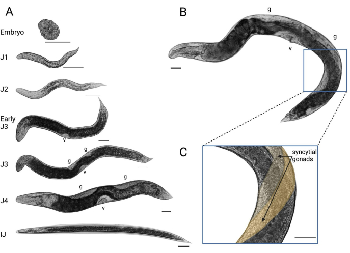
Figure 1: The development of S. hermaphroditum in a laboratory setting. (A) Estimated developmental stages of hermaphrodites grown on NGM agar feeding on its symbiotic bacterium X. griffiniae. Scale bar = 50 µM. v = vulva; g = gonad. (B) A young adult hermaphrodite at the appropriate developmental stage for gonadal microinjection. Blue box: location of gonadal injection. (C) The syncytial gonad of the hermaphrodite targeted for microinjection. The two gonads are colored in different shades of yellow to facilitate visualization. Please click here to view a larger version of this figure.
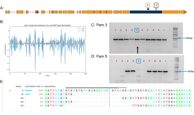
Figure 2: Representative results of CRISPR-Cas9 genome editing in S. hermaphroditum. (A) The gene structure of S. hermaphroditum unc-22 homolog. Blue box: the targeted exon. Black star: Pam 3 target site. Red star: Pam 5 target site. (B) Phenotypic analysis of a representative twitching progeny based on jerk movement when treated with 2% nicotine. Blue: a twitching adult from an unc-22 mutant line. Orange: a wild-type adult paralyzed by 2% nicotine. (C,D) Genotyping of nine independent F3 twitching lines (1-9) from two injected P0 animals. Each sample contains four F3 progeny from the same F1 line. Primers were used to amplify the genomic region around Pam 3 (C) and Pam 5 (D). '+': wild-type control; "-": a negative control with no DNA loaded. Blue boxes: example of a confirmed heterozygous sample amplicon on an agarose gel. Black arrow in (C), the amplicon analyzed in (E). (E) ICE analysis of the unc-22 mutant at Pam 3. PCR sample 5 from (C) (black arrow) was sequenced and analyzed using ICE, showing 50% of the alleles being wild-type, while various deletion mutations (ranging from 20 bp to 31 bp) were detected in the genomic DNA. Please click here to view a larger version of this figure.
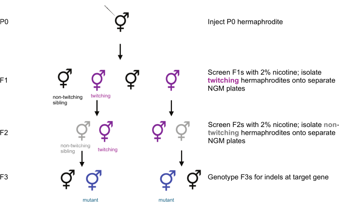
Figure 3: Co-CRISPR screening workflow for isolating mutants in the target gene. When using unc-22 as a co-CRISPR marker, F1 nematodes are screened with 2% nicotine, and twitching F1s are isolated onto separate NGM plates seeded with the symbiotic bacterium X. griffiniae. The F2 progeny of twitching F1s are then screened, and non-twitching F2s are isolated onto individual plates. After allowing F2s to produce progeny, F3s are genotyped to determine whether the targeted gene has been knocked out. Non-twitching siblings of twitching nematodes may also carry indels in the gene of interest and should be included in genotyping. Please click here to view a larger version of this figure.
| Replicate | No. injected P0 | No. P0 that survived injections* | No. P0 that survived overnight | No. P0 transferred to X. griffiniae patches | No. P0 with twitching F2 | % injected P0 that produced twitching F2 |
| 1 | 8 | 4 | 4 | 3 | 0 | 0 |
| 2 | 9 | 4 | 4 | 4 | 3 | 33.33 |
| 3 | 11 | 10 | 10 | 10 | 7 | 63.64 |
| 4 | 9 | 4 | 2 | 2 | 2 | 22.22 |
Table 1: Representative results of the S. hermaphroditum unc-22 homolog modification via microinjection and rate of success as evidenced by twitching progeny. *Survival was determined by the movement of the pharyngeal pump approximately 30 min after microinjection.
Discussion
Unconventional genetic model animal systems provide valuable opportunities to study eukaryotic gene function within the ecological and physiological contexts in which those traits naturally occur, for example, in the presence of a parasitic host or an organism’s native microbiome. In entomopathogenic nematodes (EPNs), genetic manipulation has relied on some cases of RNAi-based approaches13,32. Although RNAi enables functional genetic studies in some species, it is limited by the transient nature of dsRNA, and gene silencing may not be reliably heritable. These constraints underscore the need for stable, transgenerational genetic tools33,34. The present work describes the first protocol for precise genome editing in Steinernematids, one of the two EPN families within the order Rhabditida, as demonstrated in Cao (2023)14.
Successful CRISPR–Cas9 mutagenesis using this protocol depends on the careful handling of the nematodes. Poorly fed individuals often exhibit collapsed gonads that are difficult to penetrate with the injection needle. Injection volume must be adjusted, either by slightly enlarging the needle opening or by fine-tuning the injection pressure to ensure the gonad is properly flushed with the injection mix. Rapid transfer of injected animals from the agarose pad to the C. aquatica recovery plate is essential for post-injection survival, and execution speed improves substantially with experience. Post-injection, it is important that wounded nematodes not be exposed to their symbiotic bacterium, as recovery on X. griffiniae is low14.
Needle clogging is a common technical challenge encountered during microinjection. Clogs can sometimes be cleared by gently dragging the needle across double-sided tape submerged in halocarbon oil. Thoroughly rinsing nematodes in M9 prior to microinjection also prevents symbiotic bacteria from entering and obstructing the needle. If blockage persists, carefully enlarging the needle tip opening may restore flow, though doing so excessively can result in uncontrolled expulsion of injection mix. Transitioning from borosilicate14 to quartz capillaries for needle fabrication markedly improved injection performance. Quartz needles yield sharper tips which facilitate successful injections into slightly older adults with thicker cuticles, thereby increasing survival rates.
A current limitation of this protocol is that CRISPR-based transgene insertion remains challenging and requires further optimization. Homology-directed repair (HDR) of Cas9-induced double-stranded breaks has been difficult to achieve in S. hermaphroditum using short ssDNA donors14,35. Precise transgene insertion is a key capability in any genetic model, enabling high-fidelity modifications such as single amino acid substitutions or transcriptional reporters36. Thus, developing a reliable CRISPR-Cas9-mediated knock-in protocol would significantly advance the genetic toolkit for EPNs and benefit the broader research communities in parasitology, microbial symbiosis, and beyond.
Disclosures
The authors declare that there is no conflict of interest.
Acknowledgements
We thank Margaret McFall-Ngai and Edward Ruby for the use of their confocal microscope. We thank Alexis (Cody) Hargadon and Grischa Chen for training on confocal microscopy, and Carly R. Myers for help with obtaining the pictures and analysis of twitching videos. We also thank Stephanie Hampton for allocating funds for the purchase of the microinjection system. The Carnegie Institution for Science supported this work.
Materials
| Name | Company | Catalog Number | Comments |
|---|---|---|---|
| Agar (80-100 Mesh) | Fisher Scientific | BP26411 | Also used in LB Pyruvate plates |
| Agarose | Fisher Scientific | 16500500 | Also used in genotyping |
| Alt-R CRISPR-Cas9 tracrRNA | IDT | 1072532 | |
| Alt-R S.p. Cas9 Nuclease V3 | IDT | 1081058 | |
| Borosilicate needles | World Precision Instrument | 1B120F-4 | |
| Calcium chloride (CaCl2) | Sigma Aldrich | C4901 | |
| Cholesterol | VWR | 433 | 5 mg/mL in ethanol |
| Comamonas aquatica (DA1877) | |||
| Cover glass for agarose pad | Brain research laboratories | #4860-1D | 48 x 60 mm no1 thickness |
| CRISPRScan | https://www.crisprscan.org/ | ||
| Dimethyl Sulfoxide (DMSO) | Sigma-Aldrich | 472301 | 0.5 M |
| DNA Clean and Concentrator-5 | Zymo Research | D4014 | |
| FemtoJet 4x Microinjector with Grip head set 4 size 0 needle holder | Eppendorf/Calibre | 5253000017 | |
| Galleria melonella 5th instar larvae | GrubCo | https://www.grubco.com/index.cfm | |
| Gel imaging system | Azure400 | AZI400-01 | |
| Gel loading dye | Thermo Fisher Scientific | B72 | |
| Gelatin (2%) | Sigma Aldrich | G1393 | |
| GelRed | Biotium | #41003 | |
| Genotyping primers: DNA oligo, 25 nmol | IDT | N/A | |
| Gibco Bacto Peptone | Fisher Scientific | DF0118-17-0 | |
| Gibco Bacto Tryptone | Fisher Scientific | DF0123-17-3 | Also used in Dark LB, LB Pyruvate plates |
| Gibco Bacto Yeast Extract | Fisher Scientific | DF0127-17-9 | Also used in Dark LB, LB Pyruvate plates |
| Grip head set 4 size 1 needle holder | Calibre | EPE-5196083008 | (paired with borosilicate needle 1B120F-4) |
| Halocarbon oil 700 | Sigma Aldrich | H8898 | |
| Hard platinum wire worm pick for daily maintenance | Tritech Research | PT-9901 | |
| Inference of CRISPR edits (ICE) | Synthego | N/A | Software used to analyze Sanger sequences to identify indels |
| KCl | Sigma Aldrich | P9541 | |
| Laser needle puller P-2000 | Sutter | P-2000G | |
| Magnesium chloride (MgCl2) | Sigma Aldrich | M8266 | |
| Magnesium sulfate (MgSO4) | Sigma Aldrich | M7506 | For M9 buffer, Also used in NGM |
| Microloader tip 2x96ST | Calibre | 930001007 | |
| Micromanipulator Mounting Adapter for Zeiss Axio Observer and Axiovert 200 | Tritech Research | NZ-19-2 | |
| Microscope | Tritech | SMT1 | |
| Nicotine | Sigma Aldrich | N3876 | |
| NP-40 | Thermo Fisher Scientific | 85124 | |
| Nuclease Free Duplex Buffer | IDT | 11-01-03-01 | |
| OneTaq MasterMix | NEB | M0486 | |
| Pipette storage box | Sutter Instruments | BX10 | |
| Potassium chloride (KCl) | Sigma Aldrich | 793590 | |
| Potassium phosphate, dibasic (K2HPO4) | Sigma Aldrich | P3786 | KOH for NGM |
| Potassium phosphate, monobasic (KH2PO4) | Sigma Aldrich | P5655 | For M9 buffer, Also used in NGM |
| Proteinase K | Sigma Aldrich | P6556 | |
| Quartz needles | Sutter Instruments | QF100-70-10 | |
| S. hermaphroditium genome | NCBI | https://www.ncbi.nlm.nih.gov/datasets/genome/GCA_030435675.1/ | |
| sh-unc-22 Alt-R CRISPR-Cas9 crRNAs | IDT | N/A | |
| Sodium chloride (NaCl) | VWR | SS0430 | Also used in M9, Dark LB, LB Pyruvate plates |
| Sodium phosphate, dibasic (Na2HPO4.7H20) | Sigma Aldrich | S9390 | For M9 buffer |
| Sodium pyruvate | Sigma Aldrich | P2256 | Also used in LB Pyruvate plates |
| Soft platinum wire worm pick for microinjection | Surepure Chemetals | 6824 | Platinum 90%, Iridium 10% Alloy Wire, 0.002 inch diameter x 5 ft long |
| Standard Needle Holder | Tritech Research | HI-7 | |
| Steinernema hermaphroditum | |||
| Stereo microscope | Leica | Inveta 3 | |
| Trehalose | Cole-Parmer | EW-88195-99 | 0.08 M |
| Tris | Fisher Scientific | AM9855G | pH 8.0 |
| Tween 20 | Sigma Aldrich | P1379 | |
| Vertical Micromanipulator Mounting Adapter | Tritech Research | NR2 | |
| Xenorhabdus griffiniae (HGB2511) |
References
- Stock, S., Blair, H. G. Entomopathogenic nematodes and their bacterial symbionts: The inside out of a mutualistic association. Symbiosis. 46, 65-75 (2008).
- Murfin, K. E., et al. Nematode-bacterium symbioses--cooperation and conflict revealed in the "omics" age. Biological Bull. 223 (1), 85-102 (2012).
- Dillman, A. R., et al. An entomopathogenic nematode by any other name. PLoS Pathog. 8 (3), e1002527(2012).
- Cao, M., Schwartz, H. T., Tan, C. -H., Sternberg, P. W. The entomopathogenic nematode Steinernema hermaphroditum is a self-fertilizing hermaphrodite and a genetically tractable system for the study of parasitic and mutualistic symbiosis. Genetics. 220 (1), iyab170(2022).
- Sergeant, M., et al. Identification, typing, and insecticidal activity of Xenorhabdus isolates from entomopathogenic nematodes in United Kingdom soil and characterization of the xpt toxin loci. Appl Environ Microbiol. 72 (9), 5895-5907 (2006).
- Walsh, K. T., Webster, J. M. Interaction of microbial populations in Steinernema (Steinernematidae, Nematoda) infected Galleria mellonella larvae. J Invertebr Pathol. 83 (2), 118-126 (2003).
- Herbert, E. E., Goodrich-Blair, H. Friend and foe: The two faces of Xenorhabdus nematophila. Nat Rev Microbiol. 5 (8), 634-646 (2007).
- Flores-Lara, Y., Renneckar, D., Forst, S., Goodrich-Blair, H., Stock, P. Influence of nematode age and culture conditions on morphological and physiological parameters in the bacterial vesicle of Steinernema carpocapsae (Nematoda: Steinernematidae). J Invertebr Pathol. 95 (2), 110-118 (2007).
- Cao, M., Goodrich-Blair, H. Ready or Not: Microbial adaptive responses in dynamic symbiosis environments. J Bacteriol. 199 (15), e00883-e00916 (2017).
- Lacey, L. A., et al. Insect pathogens as biological control agents: Back to the future. J Invertebr Pathol. 132 (C), 1-41 (2015).
- Campos-Herrera, R., et al. Intraspecific virulence of entomopathogenic nematodes against the pests Frankliniella occidentalis (Thysanoptera: Thripidae) and Tuta absoluta (Lepidoptera: Gelechiidae). J Nematol. 53 (1), 1-14 (2021).
- Rill, A., Zhao, L., Bode, H. B. Genetic toolbox for Photorhabdus and Xenorhabdus: pSEVA based heterologous expression systems and CRISPR/Cpf1 based genome editing for rapid natural product profiling. Microb Cell Fact. 23 (1), 98(2024).
- Ratnappan, R., et al. RNAi-mediated gene knockdown by microinjection in the model entomopathogenic nematode Heterorhabditis bacteriophora. Parasit Vectors. 9 (1), 160(2016).
- Cao, M. CRISPR-Cas9 genome editing in Steinernema entomopathogenic nematodes. bioRxiv. , (2023).
- Heryanto, C., Ratnappan, R., O'Halloran, D. M., Hawdon, J. M., Eleftherianos, I. Culturing and genetically manipulating entomopathogenic nematodes. J Vis Exp. (181), e63885(2022).
- Stock, S. P., Griffin, C. T., Chaerani, R. Morphological and molecular characterisation of Steinernema hermaphroditum n. sp. (Nematoda: Steinernematidae), an entomopathogenic nematode from Indonesia, and its phylogenetic relationships with other members of the genus. Nematology. 6 (3), 401-412 (2004).
- Bhat, A. H., Chaubey, A. K., Shokoohi, E., William Mashela, P. Study of Steinernema hermaphroditum (Nematoda, Rhabditida), from West Uttar Pradesh, India. Acta Parasitol. 64 (4), 720-737 (2019).
- Schwarz, E. M., et al. Genomes of the entomopathogenic nematode Steinernema hermaphroditum and its associated bacteria. bioRxiv. , (2025).
- Haglund, K., Nezis, I. P., Stenmark, H. Structure and functions of stable intercellular bridges formed by incomplete cytokinesis during development. Commun Integr Biol. 4 (1), 1-9 (2011).
- Ghanta, K. S., Ishidate, T., Mello, C. C. Microinjection for precision genome editing in Caenorhabditis elegans. STAR Protoc. 2 (3), 100748(2021).
- Gang, S. S., et al. Targeted mutagenesis in a human-parasitic nematode. PLoS Pathog. 13 (10), e1006675(2017).
- Adams, S., Pathak, P., Shao, H., Lok, J. B., Pires-daSilva, A. Liposome-based transfection enhances RNAi and CRISPR-mediated mutagenesis in non-model nematode systems. Sci Rep. 9 (1), 483(2019).
- Culp, E., et al. Genome editing in the nematode Caenorhabditis briggsae using the CRISPR/Cas9 system. Biol Methods Protoc. 5 (1), bpaa003(2020).
- Hellekes, V., et al. CRISPR/Cas9 mediated gene editing in non-model nematode Panagrolaimus sp. PS1159. Front Genome Ed. 5, 1078359(2023).
- Patel, R., et al. The generation of stable transgenic lines in the human-infective nematode Strongyloides stercoralis. G3 (Bethesda). 14 (8), jkae122(2024).
- Moreno-Mateos, M. A., et al. CRISPRscan: Designing highly efficient sgRNAs for CRISPR-Cas9 targeting in vivo. Nat Methods. 12 (10), 982-988 (2015).
- Berkowitz, L. A., Knight, A. L., Caldwell, G. A., Caldwell, K. A. Generation of stable transgenic C. elegans using microinjection. J Vis Exp. (18), e833(2008).
- Wang, H., Park, H., Liu, J., Sternberg, P. W. An efficient genome editing strategy to generate putative null mutants in Caenorhabditis elegans using CRISPR/Cas9. G3 (Bethesda). 8 (11), 3607-3616 (2018).
- Conant, D., et al. Inference of CRISPR Edits from Sanger Trace Data. CRISPR J. 5 (1), 123-130 (2022).
- McMullen, J. G. 2nd, Stock, S. P. In vivo and in vitro rearing of entomopathogenic nematodes (Steinernematidae and Heterorhabditidae). J Vis Exp. (91), e52096(2014).
- Kim, H., et al. A co-CRISPR strategy for efficient genome editing in Caenorhabditis elegans. Genetics. 197 (4), 1069-1080 (2014).
- Ciche, T. A., Sternberg, P. W. Postembryonic RNAi in Heterorhabditis bacteriophora: A nematode insect parasite and host for insect pathogenic symbionts. BMC Dev Biol. 7 (1), 101(2007).
- Du, X., McManus, D. P., French, J. D., Jones, M. K., You, H. CRISPR/Cas9: A new tool for the study and control of helminth parasites. Bioessays. 43 (1), e2000185(2021).
- Quinzo, M. J., Perteguer, M. J., Brindley, P. J., Loukas, A., Sotillo, J. Transgenesis in parasitic helminths: A brief history and prospects for the future. Parasit Vectors. 15 (1), 110(2022).
- Schwartz, H. T., Tan, C. -H., Peraza, J., Raymundo, K. L. T., Sternberg, P. W. Molecular identification of a peroxidase gene controlling body size in the entomopathogenic nematode Steinernema hermaphroditum. Genetics. 226 (2), (2024).
- Dokshin, G. A., Ghanta, K. S., Piscopo, K. M., Mello, C. C. Robust genome editing with short single-stranded and long, partially single-stranded DNA donors in Caenorhabditis elegans. Genetics. 210 (3), 781-787 (2018).
Reprints and Permissions
