Herein, we describe the protocols to measure the amount of fluorescent phospholipid specifically removed from lipid donor particles by high-density lipoprotein (HDL) present in whole human plasma or serum. We have shown that this metric of HDL functionality predicts incident cardiovascular disease.
A subscription to JoVE is required to view this content. Sign in or start your free trial.
Method Article
High-Density Lipoprotein-Specific Phospholipid Efflux Assay
* These authors contributed equally
In This Article
Summary
Abstract
Plasma levels of high-density lipoprotein (HDL)-cholesterol (HDL-C) is currently a key metric for the clinical assessment of cardiovascular disease (CVD) risk. HDL-mediated removal of plaque lipids from cells and extracellular deposits in the arterial wall is one of several anti-atherogenic functions of HDL that may account for its inverse association with CVD risk. The HDL-mediated removal of cellular cholesterol in vitro has recently proven to be an even better predictor of CVD risk than HDL-C.
HDL is composed of a heterogeneous population of particles, which perform different functions. HDL particles involved in the removal of atherosclerotic plaque lipids contain exchangeable apolipoproteins, mostly apolipoprotein A-I, that dissociate from the particle and then remove plaque lipids. Nascent HDL is formed by the solubilization and removal of both phospholipids and cholesterol from ABCA1-generated cellular plasma membrane domains by HDL-derived exchangeable apolipoproteins. This process plays a critical role in both the prevention and regression of atherosclerotic plaque.
Herein, we describe the protocol for the cell-free, HDL-specific phospholipid efflux (HDL-SPE) assay, which we have previously shown can predict incident CVD risk. This assay specifically measures HDL apolipoprotein-mediated removal of a non-exchangeable fluorescent phospholipid from a lipid donor particle. Our case-control clinical studies have established that the HDL-SPE assay performs as well as and potentially even better than the "gold-standard" cell-based cholesterol efflux capacity assay, a laborious and technically complex assay that often utilizes radioactive cholesterol.
The goal of this protocol is to enable basic and clinical researchers alike to assess the functionality of HDL in lipid mobilization, using a simple, standardized, cell-free, high-throughput assay. This assay can be used by basic researchers to gain insights into mechanisms underlying HDL-mediated lipid efflux and for screening new therapeutic agents that enhance HDL functionality. HDL-SPE can also serve as a clinical laboratory diagnostic assay for CVD risk assessment.
Introduction
The high-density lipoprotein-specific phospholipid efflux (HDL-SPE) assay measures HDL functionality and is intended for use in biomedical research and clinical diagnostic laboratories to (i) gain further insights into the mechanisms underlying HDL-mediated removal of lipids from atherosclerotic lesions, (ii) serve as a new platform to improve clinical CVD risk assessment and, (iii) develop new therapeutics to treat cardiovascular disease.
HDL-cholesterol (HDL-C) is an important determinant of CVD risk1, but its usefulness is compromised insofar as elevated levels of HDL-C (>80 mg/dL) are associated with greater CVD risk2,3,4,5. Elevated levels of HDL-C may represent the accumulation of dysfunctional HDL that has a reduced capacity to remove arterial plaque lipids6.
The cellular cholesterol efflux capacity (CEC) assay was the first in vitro assay developed to measure HDL functionality. In this assay, cultured macrophages (mouse or human), typically labeled with 3H- or fluorescent Bodipy-cholesterol, are incubated with apoB-depleted plasma or serum. The % cholesterol efflux is calculated based on the amount of cell- and medium-associated fluorescence/radioactivity7,8,9. The CEC assay has been shown to be a better predictor of incident CVD than HDL-C7. The CEC assay is not amenable to conversion to a routine clinical diagnostic assay mainly due to its requirements for cell culture and long duration (e.g., days). Furthermore, the CEC assay is limited by its inherent variability (e.g., different types of cells, labeling techniques, and PEG-depletion of β-lipoproteins, which alters HDL composition10).
Several research groups have developed cell-free HDL cholesterol efflux assays for use as tools to gain new insights into HDL function and for potential translation into a clinical diagnostic assay to better assess CVD risk11,12. These efflux assays typically involve multiple steps and use a lipid donor particle labeled with a fluorescent cholesterol, and like the CEC assay, they either require depletion of β-lipoproteins or, alternatively, the capture of apoA-I-containing particles using anti-apoA-I antibodies, to measure plasma HDL cholesterol efflux. Although these alternative assays have been validated to be associated to varying degrees with CVD risk, to date, only one13, which assesses apoA-I exchange rate, has been shown to predict incident CVD risk14. Interestingly, the kinetics of the exchange of apoA-I on and off plasma HDL has been shown to be independently associated with CEC15 and to be markedly reduced in patients with acute coronary syndrome16. In addition, several HDL exchangeable apolipoproteins have been shown to mediate both cholesterol and phospholipid from ABCA1-expressing cells in vitro17.
We have developed an alternative approach to assess HDL functionality18. The simple, rapid, and reliable assay described here requires only two components, namely whole human (or animal) plasma or serum samples (fresh or freeze-thawed), and a fluorescent lipid-coated donor particle. The assay measures the ability of apoA-I and other HDL-associated exchangeable apolipoproteins in whole plasma/serum to remove a non-exchangeable, head group-tagged fluorescent phosphatidylethanolamine (PE) from a lipid-coated donor particle18 (Figure 1).
The donor particles in this assay consist of calcium silicate hydrate crystals (CSH) coated with cholesterol, the phospholipid phosphatidylcholine (PC), and a fluorescent PE. Exchangeable HDL-associated apolipoproteins, mostly apoA-I, released from HDL particles bind to the donor particles and then dissociate from them along with donor particle lipids, including fluorescent PE. In this manner, HDL, but not the other lipoproteins present in whole plasma/serum, specifically acquires fluorescent PE from the donor particles18,19. Whole plasma/serum is incubated with the fluorescent lipid-coated calcium silicate hydrate (LC-CSH) donor particles and saline at 37 °C, with shaking for 1 h. Since LC-CSH is about twice the density of water, the donor particles are readily separated from plasma by low-speed centrifugation.
The % PE efflux measured in clinical studies is typically normalized by dividing by the value of the PE fluorescence acquired by HDL in the supernatant of the reaction mixture of a clinical sample by the value obtained by HDL in a reference control human plasma. Normalization to a reference plasma is performed to correct for variations in reagent preparation. For research applications, % PE efflux can be determined by calculating the supernatant PE fluorescence divided by the total fluorescence (supernatant PE fluorescence + LC-CSH fluorescence). Supernatant fluorescent measurement values are pasted into a template in spreadsheet files to calculate %PE Efflux (Supplementary Table 1 for triplicate measurements and Supplementary Table 2 for duplicate measurements). The instructions for performing these calculations in the spreadsheet are provided as supplementary files (Supplementary File 1, Supplementary File 2, Supplementary File 3, Supplementary File 4, Supplementary File 5, Supplementary File 6, Supplementary File 7, and Supplementary File 8), and an example calculation is provided as Supplementary Table 3.
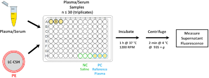
Figure 1: HDL-SPE Assay. HDL-specific phospholipid efflux is assayed in 96-well plates. Saline, calcium silicate hydrate crystals (gray) coated with lipids (LC-CSH), including non-exchangeable lissaminerhodamine-labeled phosphatidylethanolamine (LRh-PE; red membrane layers), and up to 30 whole plasma/serum samples (yellow; in triplicate) are loaded onto 96-well plates. After mixing for 1 h at 37 °C in a thermomixer, plates are briefly centrifuged to pellet the LC-CSH donor particles. Aliquots of the supernatants, containing HDL specifically labeled with LRh-PE, are transferred to a black plate containing detergent (to solubilize Lh-Rh-PE) and saline. PE fluorescence is then measured with a fluorimeter. Saline (Negative Control; NC), for background correction, and pooled healthy human plasma (Positive Control; PC) for plate normalization, are loaded instead of sample plasma/serum in the last set of wells. Please click here to view a larger version of this figure.
Access restricted. Please log in or start a trial to view this content.
Protocol
Blood collection is carried out following the rules of the Declaration of Helsinki of 1975 (https://www.wma.net/what-we-do/medical-ethics/declaration-of-helsinki/), revised in 2013, and should conform to institutional guidelines regarding informed consent.
1. Preparation of fluorescent phospholipid-labeled donor lipid particles
- Formation of lipid film
CAUTION: Prepare and dispense stock solutions in a chemical hood. Wear a lab coat, gloves, and protective eyewear. Dispose of chloroform solutions in organic chemical waste, Triton X-100 solutions (without plasma) in chemical waste, and plasma-containing solutions in biohazard/medical waste.- Purchase lipid stock solutions in chloroform from a vendor or make them in the lab:
25 mg/mL: 1,2-Dimyristoyl-sn-glycero-3-phosphocholine (DMPC):
10 mg/mL: Cholesterol
1 mg/mL: 1,2-dioleoyl-sn-glycero-3-phosphoethanolamine-N-(lissaminerhodamine B sulfonyl) [LRh-PE]
NOTE: Use screw top air-tight glass tubes. Seal cap with Teflon tape.
Store at -20 oC. - Determine the amount LC-CSH that will be needed and prepare the appropriate number of glass tubes.
In a chemical fume hood, add the following to each 10 mm 150 mm borosilicate glass tube: 472 µL of DMPC stock solution (17.7 µmol, 11.8 mg), 339 µL of cholesterol stock solution (8.8 µmol, 3.39 mg), and 305 µL of LRh-PE (200 nmol, 0.305 mg). Vortex briefly to mix. - In a chemical fume hood, dry the lipid mixture in a glass tube under a gentle stream of N2 for 1 h, to form a dry lipid film at the bottom of the tube.
NOTE: The flow of N2 should just cause a slight rippling on the fluid surface. - For assays that require several 96-well plates over days or weeks, make a large batch of LC-CSH that can be aliquoted into tubes, or used to make ready-to-use LC-CSH plates, which are stored at -20 °C or -80 °C. See steps 2.2.1-2.2.2 below.
- Purchase lipid stock solutions in chloroform from a vendor or make them in the lab:
- Coating calcium silicate hydrate with fluorescent lipid film
CAUTION: Wear a lab coat, gloves, a face mask, and protective eyewear while handling calcium silicate hydrate.- Insert a weighing paper cone into the glass tube containing the dried lipids. Add 80 mg of calcium silicate hydrate (CSH) powder (lipid removal reagent) to the glass tube using the weighing paper cone to direct powder to the base of the tube. Immediately add 2 mL of normal saline to the glass tube containing the dried lipids and CSH. Cover the top of the tube with parafilm.
- Vortex by hand to dislodge the bulk of the lipid from the bottom of the glass tube. Insert the glass tube into a hole made in a Styrofoam platform attached to the top of a vortex. Secure the tube to the vortex platform using reinforced tape.
NOTE: Pre-adjust vortex speed to allow mixture to ride up about halfway up the glass tube. - Vortex for 10 min. After vortexing, confirm that no lipid remains on the wall of the tube. If necessary, manually vortex as needed until no lipid remains. Transfer the total volume in the glass tube to a 15 mL conical plastic tube, using a 1 mL pipette. Wash the walls of the glass tube with 3 mL of additional saline and transfer to the same 15 mL conical plastic tube.
- Washing LC-CSH
- Centrifuge the 15 mL plastic tube containing the lipid-coated CSH (LC-CSH) for 2 min at 935 × g at 4 °C to pellet the donor particles. Carefully remove the supernatant by vacuum aspiration using a long, very fine pipette tip.
- Start at the top of the supernatant and while slowly gliding the pipette tip along the side of the tube, tip the tube as the supernatant is being removed. Leave a small amount (≈ 200 µL) of saline to prevent accidental aspiration of the LC-CSH from the pellet. Add enough saline to bring the total volume to 5 mL.
- Repeat steps 1.3.1-1.3.2 four times. After the final wash, add enough saline to bring the final volume to 2.5 mL.
NOTE: Store at LC-CSH preparations at 4 °C for 2 weeks or at -20 °C or -80 °C for up to 6 months.
- Large batch preparation for ready-to-use LC-CSH plates
NOTE: This approach enables the preparation of measurement plates with uniform reagent composition from the same LC-CSH batch, thereby minimizing inter-plate measurement variability.- Prepare 12 LC-CSH tubes (2.5 mL x 12 tubes) as described in steps 1.1.1-1.3.2 above. Combine the entire content of all 12 tubes into a 50 mL conical plastic tube (30 mL total volume). Carefully vortex to obtain a homogenous distribution of LC-CSH particles, and dispense 6 mL into a 15 mL plastic conical tube. Repeat four times to yield a total of 5 plastic conical tubes, each containing 6 mL of LC-CSH.
- Each tube will provide enough LC-CSH to make 5 Ready-to-Use LC-CSH plates (containing 50 µL per well for all 96 wells in a single plate). See steps 2.2.1-2.2.2, below.
2. Incubation of plasma/serum samples with fluorescent phospholipid-labeled donor lipid particles (LC-CSH)
- Individual plates for immediate use.
- Pipette 75 µL of saline into each well of a 0.3 mL 96-well plate using as many wells as needed for the sample set to be assayed. Pipette 100 µL of saline into triplicate negative control (NC) wells, and 75 µL of saline into duplicate positive control (PC) wells (Figure 2). Use a multi-well pipette to dispense.
- Prior to dispensing LC-CSH, vortex the 15 mL plastic stock tube 3 times, 10 s each. Using a single well pipette, dispense 50 µL of LC-CSH along the right side of the saline-containing wells. Rapidly dispense 50 µL each to only 3 wells. Repeat the vortex protocol prior to dispensing LC-CSH into the next 3 wells.
NOTE: This procedure is critical to ensure that the heavy LC-CSH, which spontaneously sediments, is homogeneously suspended, hence allowing equal amounts to be dispensed to each well. - Rotate the 96-well plate by 180° so that the left side of the wells is now to the right. Pipette 25 µL of plasma/serum samples along the right side of the well (Figure 2). For the PC wells, add 25 µL of reference standard human normolipidemic plasma/serum. The total volume per well for the assay is 150 µL.
NOTE: This procedure prevents cross-contamination of plasma/serum with LC-CSH, since they are dispensed along opposite sides of the wells. - Tightly seal the plate with adhesive film. Incubate the sealed 96-well plate in the dark for 1 h at 37 °C at 1200 rpm in a thermomixer. After incubation is complete, remove the plate from the thermomixer and put it on ice. Centrifuge the plate for 2 min at 4 °C (to stop the transfer reaction) at 935 g to pellet the donor particles.
- Ready-to-use LC-CSH plates
- For use, thaw frozen Ready-To-Use plates (see steps 1.4.1-1.4.2 for plate preparation) in the cold (4 °C to 10 °C) for 2 h (or, overnight if needed). Centrifuge for 2 min at 4 °C at 935 × g to collect all the material (125 µL) together. Carefully remove the adhesive film so as not to splash the contents of the wells.
- Dispense 25 µL of plasma/serum samples, saline, and reference human plasma, in triplicate, to the appropriate wells (Figure 1) of the Ready-To-Use plate, on ice, and seal with adhesive film. Incubate the sealed 96-well in the dark for 1 h at 37 °C at 1200 rpm in a thermomixer. Remove the plate from the thermomixer and put it on ice. Centrifuge the plate for 2 min at 4 °C (to stop the transfer reaction) at 935 × g (to pellet the donor particles).
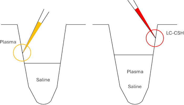
Figure 2: Dispensing reagents into wells without cross-contamination. Dispense plasma/serum and LC-CSH on opposite walls of the wells. Cross-contamination is minimized if the plate is rotated by 180° after first dispensing LC-CSH. Please click here to view a larger version of this figure.
3. Measurement of fluorescent phospholipid efflux to plasma/serum HDL
- Transfer to a black plate and fluorescence measurement
- Carefully remove the adhesive film so as not to disturb the LC-CSH pellet.
NOTE: If pellet disruption occurs, centrifuge again. - Transfer 50 µL of supernatant from the 96-well reaction plate into the wells of a 96-well black polystyrene flat-bottom plate for fluorescence measurements. Use a multi-well pipette.
NOTE: Do not aspirate any LC-CSH. If this should happen, inject back into the well and centrifuge again. - Fill a reservoir with normal saline. Add 50 µL of saline to each well using a multi-well pipette.
- Prepare 1% TX-100 by adding 90 mL of distilled water to 10 mL of 10% Triton X-100 stock solution in a 125 mL plastic Erlenmeyer flask. Fill a reservoir with 1% Triton-X 100.
- Add 100 µL of 1% Triton X-100 at room temperature to each well and mix by gently pipetting up and down 2-3 times using a multi-well pipette. Avoid forming bubbles. Pop any bubbles with air from a 3 mL transfer pipette.
- Measure Lissaminerhodamine fluorescence with fluorimeter (540 nm excitation/ 600 nm emission).
- Carefully remove the adhesive film so as not to disturb the LC-CSH pellet.
- Calculation of %PE efflux
- PE efflux calculation
- %PE efflux = ([Total Supernatant Plasma or Serum FLU - Supernatant NC FLU]/Total FLU)) × 100
FLU = fluorescence units; NC = negative control. Since only 50 µL of the 150 µL of the supernatant reaction mixture is used to measure the fluorescence per well, it is necessary to correct as follows: - Total supernatant FLU/well = Measured FLU of 50 µL (black plate) × 3
- Total FLU per 150 µL reaction mixture/well = Total FLU emitted by 50 µL of LC-CSH.
NOTE: LC-CSH fluorescence is calculated as described below (step 4.2.5).
- %PE efflux = ([Total Supernatant Plasma or Serum FLU - Supernatant NC FLU]/Total FLU)) × 100
- Normalized %PE efflux calculation
- Use the normalized %PE efflux to correct for variation between assays performed under different conditions (i.e., experiment performed on different days, batch differences).
- Use this formula to calculate normalized %PE efflux:
Normalized %PE Efflux = Sample supernatant %PE efflux value (FLU)/positive reference control supernatant %PE efflux FLU value. - Use identical pooled human plasma from healthy donors with a normal lipid profile as a reference control. There is no need to calculate the Total supernatant FLU/well since this value is the same for both the sample and the reference.
- For triplicate samples, paste the raw data obtained in step 3.1.6 into the spreadsheet (Supplementary Table 1). For duplicate samples, paste the raw data obtained in step 3.1.6 into the spreadsheet (Supplementary Table 2).
- PE efflux calculation
4. LC-CSH standard curve
NOTE: The LC-CSH standard curve is used to calculate the amount of PE fluorescent signal present in the aliquot of LC-CSH per well. Typically, 50 µL of LC-CSH/well is used. This value can be used to calculate the %PE efflux to HDL in a sample normalized to the Total FLU (see step 3.2.1) present in the reaction mixture.
%PE efflux = ((supernatant sample fluorescence units [FLU] - supernatant saline FLU)/(total LC-CSH FLU)) x 100.
- Serial dilution of LC-CSH
- Prepare 10-fold and 100-fold dilutions of stock LC-CSH (as prepared in steps 1.1.1-1.3.3):
10-fold dilution: 100 µL of LC-CSH + 900 µL of 1% Triton X-100
100-fold dilution: 100 µL of 10-fold diluted LC-CSH + 900 µL of 1% Triton X-100 - Dispense triplicate samples of 10- and 100-fold diluted LC-CSH into a 96-well incubation plate for the standard curve (see Figure 3).
- Add sufficient volume of 1% Triton X-100 to the wells containing LC-CSH to bring to a final volume of 200 µL (see Table 1).
- Prepare 10-fold and 100-fold dilutions of stock LC-CSH (as prepared in steps 1.1.1-1.3.3):
- Solubilization of LC-CSH for fluorescence measurement
- Seal the plate with adhesive film and incubate for 1 h at 24 °C with shaking at 1200 rpm. Centrifuge plate for 2 min at 4 °C at 935 × g. Carefully remove the adhesive film.
- Using a multi-well pipette, mix samples by pipetting up and down 2-3x. Transfer 100 µL of LC-CSH per well to a 96-well black polystyrene flat-bottom plate for fluorescence measurements, using a multi-well pipette.
- Add 100 µL per well of 1% Triton-X at room temperature to each well, using a multi-well pipette. Mix by pipetting up and down 2-3x.
- Measure fluorescence with a fluorimeter (540 nm excitation/600 nm emission).
- Paste the fluorescence values measured using the fluorimeter into the Linear Regression Analysis template in Supplementary Table 1 as delineated in the Supplementary File 3. Final concentrations of LC-CSH in the standard curve measurement plate are shown in Table 2.
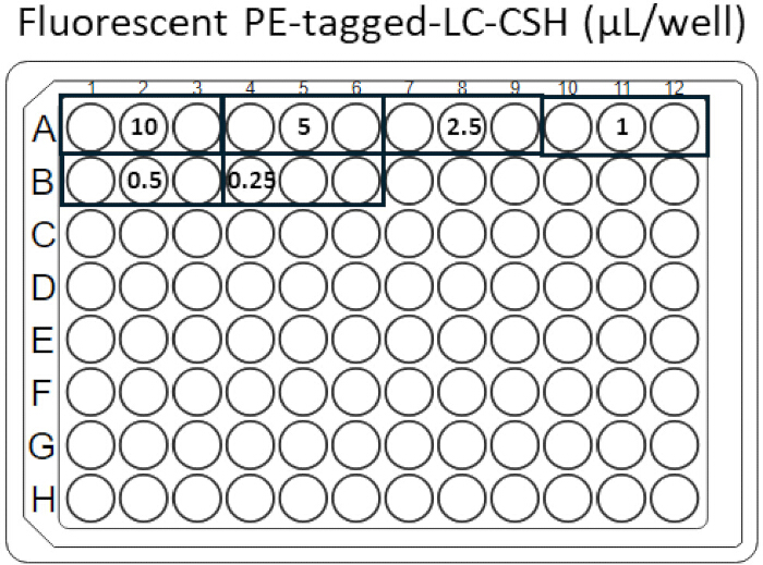
Figure 3: Plate for LC-CSH standard curve. Serially-diluted LRh-PE-labeled-LC-CSH is dispensed into a 96-well plate, as shown. Please click here to view a larger version of this figure.
| Final Concentration | 10-fold Diluted | 100-fold Diluted | 1% Triton X-100 | Total |
| LC-CSH | LC-CSH | LC-CSH | Detergent | Volume |
| (μL/well) | (μL) | (μL) | (μL) | (μL) |
| 10 | 100 | 0 | 100 | 200 |
| 5 | 50 | 0 | 150 | 200 |
| 2.5 | 25 | 0 | 175 | 200 |
| 1 | 0 | 100 | 100 | 200 |
| 0.5 | 0 | 50 | 150 | 200 |
| 0.25 | 0 | 25 | 175 | 200 |
Table 1: Volumes of reagents added to wells of incubation plate for LC-CSH standard curve.
| Solubilized | Saline | Total | Added | Measured |
| LC-CSH | Volume | Volume | LC-CSH | LC-CSH |
| (μL) | (μL) | (μL) | (μL/well) | (μL/well) |
| 100 | 100 | 200 | 10 | 5 |
| 100 | 100 | 200 | 5 | 2.5 |
| 100 | 100 | 200 | 2.5 | 1 |
| 100 | 100 | 200 | 1 | 0.5 |
| 100 | 100 | 200 | 0.5 | 0.25 |
| 100 | 100 | 200 | 0.25 | 0.125 |
Table 2: Volumes of reagents added to wells of the measurement plate for the LC-CSH standard curve.
Access restricted. Please log in or start a trial to view this content.
Results
Analytical methods
As noted above, HDL-SPE sample analyses can be modified for different applications, as demonstrated below. Prior to conducting studies with research samples, LC-CSH standard curve validation, reproducibility, and plasma concentration assays are first performed in order to master the required techniques. First, perform the LC-CSH standard curve analysis to assess the quality of LC-CSH preparations. Next, perform the reproducibility assay. Finally, the plasma ...
Access restricted. Please log in or start a trial to view this content.
Discussion
HDL-C is a key determinant used to clinically assess cardiovascular disease (CVD) risk and thus the need for lipid-lowering or other potential preventative type treatments1. The function of HDL in cellular cholesterol efflux in vitro has been shown to be an even better predictor of incident cardiovascular disease than HDL-C7. Unfortunately, this cellular assay is not amenable to conversion to a high-throughput clinical laboratory assay for routine diagnostic testin...
Access restricted. Please log in or start a trial to view this content.
Disclosures
PATENTS: US20220178953A1; WO2020185598A1 (E.B.N., M.S., A.T.R.)
Masaki Sato is employed by Eiken Chemical Co., Ltd.
Acknowledgements
This research was supported [in part] by the Intramural Research Program of the National Institutes of Health (NIH). The contributions of the NIH author(s) are considered Works of the United States Government. The findings and conclusions presented in this paper are those of the author(s) and do not necessarily reflect the views of the NIH or the U.S. Department of Health and Human Services.
Access restricted. Please log in or start a trial to view this content.
Materials
| Name | Company | Catalog Number | Comments |
|---|---|---|---|
| 1,2-dimyristoyl-sn-glycero-3-phosphocholine | Avanti Research | 850345 | 14:0 PC (DMPC) Chloroform Soluiton |
| 1,2-dioleoyl-sn-glycero-3-phosphoethanolamine-N-(lissamine rhodamine B sulfonyl) (ammonium salt) | Avanti Research | 810150-C | Chloroform Solution LRh-PE |
| Adhesive Film | Applied Biosystems | 4306311 | MicroAmp Clear Adhesive Film |
| Bay Corporation High Density Teflon Tape 1/2 inch x 520 | Medical Testing Solutions | 86732 | |
| Borosilicate glass tubes 20 mm ´ 150 mm | Fisher Scientific | 14-961-33 | |
| Centrifuge | Thermo Scientific | Sorvall X Pro Series | |
| Chloroform | Sigma-Aldrich | 366927 | suitable for HPLC, ≥99.8% |
| Cholesterol Powder | Sigma-Aldrich | C8667 | 3β-Hydroxy-5-cholestene, 5-Cholesten-3β-ol |
| Glassine Weighing Paper | Cole-Palmer | UX-01338-02 | For handling LC-CSH |
| Incubation plate | USA Scientific | 1402-9700 | TempPlate 96 Well Semi Skirt 0.2mL PCR Plates |
| Lipid Removal Agent (LRA) | Supelco | 13358-U | LC-CSH |
| Measurement plate | Greiner bio-one | 655076 | Microplate, PS, 96-well, F-BOTTOM(CHIMNEY WELL), BLACK; FLUOTRAC, MED BINDING |
| Microsoft Excel | Microsoft 365 MSO | Version 2507 | |
| MultiFlex Pipet Tips | Fisher Scientific | 50-550-206 | For aspiraitng supernatant during LC-CSH washes |
| Multilabel plate reader | Perkin Elmer | 1420-050 | Victor3 Wallac1420 Multilabel Counter |
| Normal Saline | Quality Biological | 114-055-101 | For multiple uses |
| Parafilm | Cole-Palmer | HS23452 | To cover glass tube top during vortexing |
| Polycabonate Erlenmeyer Flasks | Fisher Scientific | 10-041-8 | For 10% Triton-X solution |
| Pooled human plasma (blood derived) Li Heparin) | Innovative Research Inc | IPLAWBLIH | Reference plasma, positive control (PC) |
| Surfact-Amps X 100 | Thermo Scientific | 28314 | 10% Triton X-100, dilute 10x with distilled water |
| Thermomixer | Eppendorf | 5382000023 | Thermomixer C with 96-well thermoblock |
| Thermomixer Smartblock | Eppendorf | 5306000006 | 96-well SmartBlock |
| Transfer pipettes (3 mL, Falcon) | Fisher Scientific | 13-711-9CM | For removing bubbles from top of well |
| Vortex mixer | Daigger | 3030A | Affix holder for glass tube on top |
References
- Grundy, S. M., et al. 2018 AHA/ACC/AACVPR/AAPA/ABC/ACPM/ADA/AGS/APHA/ASPC/NLA/PCNA guideline on the management of blood cholesterol: A report of the american college of cardiology/american heart association task force on clinical practice guidelines. Circulation. 139 (25), e1082-e1143 (2019).
- Zhong, S., et al. Increased coronary heart disease in Japanese-American men with mutation in the cholesteryl ester transfer protein gene despite increased HDL levels. J Clin Invest. 97 (12), 2917-2923 (1996).
- Madsen, C. M., Varbo, A., Nordestgaard, B. G. Extreme high high-density lipoprotein cholesterol is paradoxically associated with high mortality in men and women: Two prospective cohort studies. Eur Heart J. 38 (32), 2478-2486 (2017).
- Ko, D. T., et al. High-density lipoprotein cholesterol and cause-specific mortality in individuals without previous cardiovascular conditions: The CANHEART study. J Am Coll Cardiol. 68 (19), 2073-2083 (2016).
- Liu, C., et al. Cholesterol efflux capacity is an independent predictor of all-cause and cardiovascular mortality in patients with coronary artery disease: A prospective cohort study. Atherosclerosis. 249, 116-124 (2016).
- Rosenson, R. S., et al. Dysfunctional HDL and atherosclerotic cardiovascular disease. Nat Rev Cardiol. 13 (1), 48-60 (2016).
- Khera, A. V., et al. Cholesterol efflux capacity, high-density lipoprotein function, and atherosclerosis. N Engl J Med. 364 (2), 127-135 (2011).
- Rohatgi, A., et al. HDL cholesterol efflux capacity and incident cardiovascular events. N Engl J Med. 371 (25), 2383-2393 (2014).
- Sankaranarayanan, S., et al. A sensitive assay for ABCA1-mediated cholesterol efflux using bodipy-cholesterol. Journal of Lipid Research. 52 (12), 2332-2340 (2011).
- Schachtl-Riess, J. F., et al. Lysis reagents, cell numbers, and calculation method influence high-throughput measurement of HDL-mediated cholesterol efflux capacity. J Lipid Res. 62, 100125(2021).
- Harada, A., et al. Cholesterol uptake capacity: A new measure of HDL functionality for coronary risk assessment. J Appl Lab Med. 2 (2), 186-200 (2017).
- Horiuchi, Y., et al. Validation and application of a novel cholesterol efflux assay using immobilized liposomes as a substitute for cultured cells. Biosci Rep. 38 (2), BSR20180144(2018).
- Lorkowski, S. W., et al. A novel cell-free fluorescent assay for HDL function: Low apolipoprotein a1 exchange rate associated with increased incident cardiovascular events. J Appl Lab Med. 5 (3), 544-557 (2020).
- Denimal, D. Rethinking 'good cholesterol' for cardiovascular risk stratification. QJM: An Int J Med. 117 (4), 243-245 (2023).
- Borja, M. S., et al. HDL-apolipoprotein A-I exchange is independently associated with cholesterol efflux capacity. J Lipid Res. 56 (10), 2002-2009 (2015).
- Borja, M. S., et al. HDL-apoa-I exchange: Rapid detection and association with atherosclerosis. PLoS One. 8 (8), e71541(2013).
- Remaley, A. T., et al. Apolipoprotein specificity for lipid efflux by the human ABCAI transporter. Biochem Biophys Res Commun. 280 (3), 818-823 (2001).
- Sato, M., et al. Cell-free, high-density lipoprotein-specific phospholipid efflux assay predicts incident cardiovascular disease. J Clin Invest. 133 (18), e165370(2023).
- Neufeld, E. B., et al. ApoA-I-mediated lipoprotein remodeling monitored with a fluorescent phospholipid. Biology (Basel). 8 (3), 53(2019).
- Davidson, W. S., et al. The effects of apolipoprotein B depletion on HDL subspecies composition and function. J Lipid Res. 57 (4), 674-686 (2016).
- Phillips, M. C. Molecular mechanisms of cellular cholesterol efflux. J Biol Chem. 289 (35), 24020-24029 (2014).
- Oram, J. F., Yokoyama, S. Apolipoprotein-mediated removal of cellular cholesterol and phospholipids. J Lipid Res. 37 (12), 2473-2491 (1996).
- Phillips, M. C. Is ABCA1 a lipid transfer protein. Journal of Lipid Research. 59 (5), 749-763 (2018).
- Smith, J. D., et al. Abca1 mediates concurrent cholesterol and phospholipid efflux to apolipoprotein A-I. J Lipid Res. 45 (4), 635-644 (2004).
- Ossoli, A., et al. Lipoprotein X causes renal disease in LCAT deficiency. PLoS One. 11 (2), e0150083(2016).
- Martínez-Beamonte, R., et al. Effect of extra virgin olive oil high in bioactive compounds on atherosclerosis in apoe-deficient mice. Mol Nutr Food Res. , (2025).
Access restricted. Please log in or start a trial to view this content.
Reprints and Permissions
