This study optimizes a Hot Phenol–based RNA extraction method for Mycobacterium, yielding high-quality RNA suitable for transcriptomic analyses. When benchmarked against TRIzol and RNeasy under identical conditions, the method provided higher yield, comparable integrity, and improved cost-efficiency, offering a practical alternative for large-scale gene expression studies.
Method Article
Optimized Hot Phenol–Based RNA Extraction from Mycobacteria: A Robust Approach for Reliable Gene Expression Analysis
507 views
⸱
September 11th, 2026
In This Article
Summary
Abstract
Mycobacterium tuberculosis (Mtb) remains a major global health threat, underscoring the need for reliable transcriptomic studies to understand its biology and drug resistance mechanisms. Such analyses depend on obtaining high-quality, high-yield RNA. Although several RNA extraction methods are available, many require expensive reagents, large culture volumes, or specialized equipment, limiting their suitability for large-scale studies, particularly in resource-constrained settings. Here, an optimized Hot Phenol based RNA extraction method specifically tailored for mycobacteria is presented. The method uses minimal culture volume and commonly available reagents to consistently yield high-quality RNA suitable for high-throughput transcriptomic applications. RNA quantity and integrity were assessed by gel electrophoresis and RNA integrity analysis (RIN), and its suitability for downstream applications was confirmed by qPCR and Qubit 4. To benchmark the performance of the optimized method, a parallel RNA extraction using TRIzol and RNeasy under identical experimental conditions was carried out, including the same Mycobacterium species, culture volume, growth phase (logarithmic and stationary), and lysis conditions. This allowed a direct comparison of yield, quality, feasibility, and cost. The optimized Hot Phenol method demonstrated comparable or improved RNA yield and quality while significantly reducing reagent cost and dependence on specialized equipment. Owing to its efficiency, reproducibility, and affordability, this protocol provides a practical alternative for large-scale gene expression and transcriptomic studies in Mtb and other mycobacterial species.
Introduction
M. tuberculosis (Mtb), the causative agent of tuberculosis (TB), remains the leading cause of death from a single infectious pathogen, surpassing HIV/AIDS. In 2024, TB accounted for approximately 1.3 million deaths worldwide1. This burden is compounded by the rising prevalence of multidrug-resistant (MDR) and extensively drug-resistant (XDR) strains. Therefore, there is an urgent need to understand the molecular basis of TB pathogenesis and identify new therapeutic targets2.
Transcriptional profiling of Mtb has become an important tool for elucidating functional elements involved in host-pathogen interactions3, adaptation to environmental stress4, and mechanisms underlying drug resistance5. Advanced methodologies such as real-time PCR, microarrays, and RNA sequencing (RNA-seq) have significantly enhanced the ability to analyze the mycobacterial transcriptome6. However, the success of these methods heavily depends on the availability of high-quality RNA, which in turn relies on efficient cell lysis during extraction. The complex mycolyl–arabinogalactan–peptidoglycan cell wall of Mtb makes RNA extraction particularly difficult7. Existing lysis techniques, including enzymatic hydrolysis8, sonication9, and bead-beating10, often require costly equipment and large culture volumes, limiting their practicality in many BSL-3 laboratories. Additionally, Mtb’s slow growth rate necessitates prolonged culture times to obtain sufficient biomass11.
To overcome these limitations, an optimized Hot Phenol–based RNA extraction method that is rapid and economical was developed. Using only 3 mL of mid-log phase culture, the method yields 45–60 µg of high-quality RNA suitable for downstream transcriptomic analyses. Cell lysis is achieved using a compact hand-held tissue grinder and readily available reagents, eliminating the need for expensive instruments. Although the Hot Phenol method has been reported previously11,12, the optimized Hot Phenol method improves convenience, cost-effectiveness, and RNA yield. Optimization was performed using M. smegmatis mc2155 (Msmeg) and successfully validated with Mtb H37Rv.
Protocol
The reagents and the equipment used are listed in the Table of Materials.
1. Culturing of Mycobacterium
- Inoculate 1mL from the glycerol stock of Msmeg or Mtb in 10 mL of Middlebrook 7H9 medium supplemented with 0.2% glycerol, 0.05% Tween 80, and 10% ADC at 37 °C with shaking at 150 rpm until the OD reaches 1–1.5.
- Grow the secondary culture by adding 1% from the primary culture in 10 mL of 7H9 media until mid-log phase, OD600nm = 0.6–0.7.
- Split into 3 mL aliquots and centrifuge the cultures at 2000 x g for 5 min at room temperature (23–25 °C).
- Store the pellets at -80 °C until RNA extraction.
CAUTION: For Mtb cultures, follow proper BSL3 guidelines and heat-inactivate the pellet at 95 ˚C for 20–25 min.
NOTE: Figure 1 shows the flowchart of the basic steps followed during RNA extraction by the Hot Phenol method.
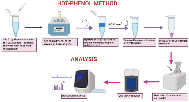
Figure 1: A schematic representation of the Hot-Phenol method and analysis. Please click here to view a larger version of this figure.
2. RNA extraction
- Hot-Phenol method (HP method)
- Add 200 µL of TES buffer (100 mM Tris-HCl, pH 8.0; 5 mM EDTA, pH 8.0; 0.1 M NaCl; % SDS; 1% β-mercaptoethanol) to the cell pellet and resuspend the pellet by vortexing.
- Add approximately 0.1 g of 0.1 mm silica beads to the sample. Use a hand-held tissue grinder for 10–15 s until the pellet is completely thawed on ice. Repeat this step for 2 more cycles, followed by snap freezing in liquid nitrogen after each repeat.
- OR use a high-speed tabletop bead ruptor for 1 min at the speed setting of 5 (5.0 m/s) at room temperature. Repeat this step for 3 cycles and incubate the sample on ice to prevent heating after each cycle.
NOTE: Repeat Mtb cell lysis for 6–8 cycles (each cycle for 1 min) with either a hand-held tissue grinder or a bead ruptor, and intermittently cool on ice after each cycle. Use DEPC (Diethylpyrocarbonate) treated water for the preparation of all the buffers and reagents needed for RNA extraction. - Add an additional 200 µL of TES buffer (pre-warm to 65 °C) to each sample to make up the volume to 400 µL. To each sample, add 200 µL of acidic phenol (phenol equilibrated with 100 mM citrate buffer, pH 4.5) and 200 µL of chloroform (mix the acid phenol and chloroform in an equal ratio before adding it to the sample).
- Incubate the sample at 65 °C on a heat block with shaking at 900 rpm for 30 min and centrifuge at 12000 x g for 15 min at room temperature. Ensure a proper phase separation after centrifugation (a clear upper aqueous phase containing RNA, a distinct interphase of DNA, and a lower organic phase containing proteins and lipids will be observed).
- Remove the upper layer of aqueous phase and transfer it into a new 1.5 mL microcentrifuge tube. Add an equal volume of acidic phenol: chloroform again. Centrifuge at 12,000 x g for 15 min at room temperature. Ensure a proper phase separation.
- Remove the upper aqueous layer into a fresh 1.5 mL microcentrifuge tube, then add 30 µL of 3 M sodium acetate buffer, pH 5.3, and 240 µL of chilled isopropanol. Incubate at −80 °C for 30 min to 1 h.
NOTE: The microcentrifuge tube can be stored overnight at -80 °C at this step. Seal the microcentrifuge tube with multiple layers of parafilm to prevent it from opening at 65 °C during incubation.
CAUTION: Phenol and chloroform are hazardous chemicals; therefore, proper personal protective equipment (PPE), engineering controls like fume hoods, and specific waste disposal procedures should be followed. - Centrifuge at 12,000 x g for 1 h at room temperature. Ensure pellet formation and carefully remove the supernatant without disturbing the pellet.
- Resuspend the pellet in 1 mL of 75% ethanol and centrifuge at 12,000 x g for 1 min at room temperature. Perform this step twice and air-dry the pellet.
- Resuspend the pellet in 50 µL of nuclease-free water.
NOTE: To dissolve RNA, incubate it in RNase-free water for 10–30 min at room temperature.
- TRIzol method
- Add 200 µL of TRIzol reagent to the cell pellet.
CAUTION: TRIzol is a hazardous chemical; therefore, proper personal protective equipment (PPE), engineering controls like fume hoods, and specific waste disposal procedures should be followed. - Follow the cell lysis method as in steps 2.1.2 - 2.1.3..
- Add an additional 200 µL of TRIzol reagent, followed by 200 µL of chloroform, and vortex for 1 min. Centrifuge at 13,400 x g for 15 min at 4 °C. Ensure the proper phase separation after centrifugation.
- Transfer the aqueous phase carefully into a new 1.5 mL microcentrifuge tube.
- Precipitate the RNA by adding 500 µL of isopropanol to the aqueous phase and incubating at −80 °C for 30 min to 1 h.
- Pellet down the RNA by centrifugation at 15,000 x g for 20 min at 4 °C. Carefully observe the pellet and remove the supernatant without disturbing the pellet.
- Resuspend the pellet with 1 mL of 75% ethanol and centrifuge at 13,400 x g for 1 min at 4 °C. Repeat this step twice, air-dry the pellet for 30 min at room temperature, and resuspend in 50 µL of nuclease-free water.
- Add 200 µL of TRIzol reagent to the cell pellet.
- Commercially available RNA extraction kit method
- Resuspend the cell pellet in 200 µL of Resuspension buffer (provided in the kit).
- Follow the cell lysis method as in steps 2.1.2 - 2.1.3.
- Carry out the subsequent experiment as per the manufacturer's instructions.
3. DNase treatment, quantification, and visualization
- To remove genomic DNA contamination, add 1 µL of DNase I (10units) and 5 µL of RDD buffer (1x) to each RNA sample and incubate at room temperature for 30 min.
- Quantify the RNA sample spectrophotometrically using Nanodrop and RNA integrity using a Qubit4. Accepted value of RIN is between 7.5 -10. Accepted values of A260/A280 and A260/A230 in the range 1.8–2.0 and 2.0–2.2, respectively.
- Visualize RNA on 1% agarose gel using 1X RNA loading dye.
4. cDNA synthesis and quantitative real-time PCR
- Set up a 20 µL reaction for cDNA preparation using the cDNA Reverse Transcription kit and 1 µg RNA, following the manufacturer’s instructions in a thermal cycler.
- Set up a 10 µL real-time PCR reaction using 50 ng of cDNA, 0.4 µM of each primer, and SYBR according to the manufacturer’s instructions. Run the sample on the Real-Time PCR Thermal cycler.
NOTE: The following primers were used for qPCR analysis:
Mtb sigA Fw 5' cctcaaacagatcggcaagg 3', Mtb sigA Rv 5' cagatccacatcatgtcgcg 3'
Mtb hsp70 Fw 5’ gctggtggacaagttcaagg 3', Mtb hsp70 Rv 5' ggtcagctgctcgtctaaga 3'
Mtb16srRNA Fw 5' gcgatacgggcagactagag 3', Mtb16srRNA Rv 5' aaggaaggaaacccacacct 3'
Ms. sigA Fw 5' gaagacaccgacctggaact 3', Ms.sigA ms Rv. 5' gactcttcctcgtcccacac 3'
Ms. sigH Fw 5' gaagggttcccgtacaagg 3', Ms sigH Rv. 5' tcatgacgtcacctcctcg 3'
Ms. sigE Fw 5' caccaccaaccttttcctcg 3', Ms. sigE Rv. 5' accctcgatgtcacacagg 3'
5. Statistical analysis
- Perform Student’s t-test with all the biological replicates using Graph Pad Prism, P ≤ 0.05 was considered statistically significant. Error bars represent standard error from at least three biological replicates.
Results
RNA quality and yield
RNA quality and quantity were assessed by agarose gel electrophoresis (Figure 2). Distinct, sharp bands corresponding to 23S and 16S rRNA were observed for all extraction methods, confirming successful isolation of total RNA. The Hot Phenol and TRIzol methods showed three clear bands representing 23S (~2900 bp), 16S (~1500 bp), and 5S rRNA (~120 bp), indicating intact, high-quality RNA. In contrast, the column-based kit method showed only the 23S and 16S bands, with the 5S rRNA band absent or below detection, likely due to preferential loss of small RNAs during column purification. Quantitatively, the Hot Phenol method yielded 1.5–2-fold more RNA than TRIzol and RNeasy respectively (Figure 2A,C). Average yields were ~60 µg for Hot Phenol, ~45 µg for TRIzol, and ~30 µg for RNeasy, the latter representing ~50% lower yield than Hot Phenol. Despite yield differences, RNA integrity and purity were comparable across methods (Figure 2D,E).
To evaluate the efficiency of the lysis approach, RNA was also extracted using a bead ruptor. Comparable yields, quality, and integrity were obtained, demonstrating that effective lysis can be achieved using a simple hand-held tissue grinder without reliance on specialized equipment such as a bead ruptor/beater (Figure 2B,F–H).
Since Mtb is more resistant to lysis than Msmeg, we further validated the optimized Hot Phenol method using the Mtb H37Rv strain. Cell lysis using both the tissue grinder and bead ruptor resulted in RNA of good yield, quality, and integrity (Figure 3A–D), confirming the robustness of the method. Though RNA extraction using a hand-held tissue grinder had a lower yield (~35–45 µg/3 mL culture) when compared to bead ruptor (50–60 µg/3 mL), the difference was found to be not statistically significant, and also the quantity was in the acceptable range enough for large-scale gene expression studies. All the RNA Extractions were repeated multiple times, and all the independent biological replicates have been presented. Densitometry analysis has been performed on the gel images, which further validates the statistical differences in the yield of RNA across different methods, with Hot Phenol giving the highest yield (Supplementary Figure 1A–D).
RNA purity and integrity
All methods yielded RNA with acceptable A260/A280 ratios around 2.0, indicating minimal protein contamination (Figure 2D). RNA integrity (RIN) was comparable across all methods, with the value ranging from 7.5 to 10 (Figure 2D–H, Supplementary Figure 2, lower panel). This indicates good quality, integrity, and minimal degradation.
Quantitative PCR analysis to check RNA quality
To evaluate the suitability of RNA extracted by each method for downstream gene expression studies, qRT-PCR was performed. We studied the gene expression of Msmeg housekeeping gene, mysA ( MSMEG 2758), and two transcriptional regulators, sigE (MSMEG 5072) and sigH (MSMEG 1914), using equivalent input RNA amounts. The Ct (cycle threshold) values obtained reflect the relative abundance of intact, amplifiable RNA in each sample. As shown in (Figure 2I), RNA extracted using Hot Phenol and TRIzol gave comparable Ct values in the range of approximately 22–25, reflecting higher RNA integrity and compatibility with reverse transcription and amplification. While the kit method exhibited slightly higher Ct values (~27–30), indicating lower levels of detectable transcript. Melt curve analysis showed specific amplification of mysA, sigE, and sigH genes, showing qPCR specificity and efficiency (Supplementary Figure 2, upper panel). qPCR analysis using Mtb genes also showed acceptable Ct values (~20–25), confirming suitability for downstream gene expression studies (Figure 3E).
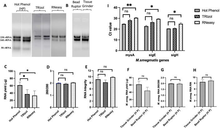
Figure 2: Comparison of RNA extraction methods for Mycobacterium. (A) Agarose gel electrophoresis showing total RNA extracted using Hot Phenol, TRIzol, and RNeasy methods. Distinct 23S and 16S rRNA bands are visible in all samples, indicating intact RNA. (B) Agarose gel electrophoresis showing total RNA extracted using Hot Phenol via different lysis methods, i.e., bead ruptor and hand-held tissue grinder. (C) Total RNA yield, showing the highest yield from Hot Phenol, followed by TRIzol and the kit method. (D) RNA purity was assessed by A260/A280 ratios, with all methods yielding values around ~2.0, indicative of minimal protein contamination. (E) RNA integrity number (RIN) showing comparable integrity. (F–H) Comparison of RNA yield, 260/280 ratio, and RNA integrity between samples lysed by hand-held tissue grinder and Bead Ruptor and isolated using the Hot Phenol method. (I) Ct values from qRT-PCR targeting a housekeeping gene (e.g., mysA) and transcriptional factors (sigE and sigH). Error bars represent standard error from three biological replicates (n = 3). P-value calculated using Student’s t-test. Please click here to view a larger version of this figure.
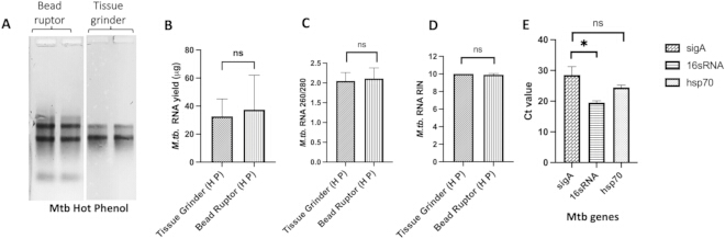
Figure 3: RNA isolation from Mtb using the hot phenol method. (A) Agarose gel electrophoresis showing total RNA extracted using bead ruptor and hand-held tissue grinder with optimized Hot Phenol method from Mtb H37Rv strain. (B–D) Comparison of isolated RNA in terms of yield, 260/280 ratio, and RNA integrity (RIN) estimation. (E) Ct values from qRT-PCR targeting sigA, 16s rRNA, and hsp70 genes. Error bars represent standard error from three biological replicates (n = 3). P-value calculated using Student’s t-test. Please click here to view a larger version of this figure.
| Hot Phenol | Trizol | RNeasy kit | |
| Yield | 60 -70 μg | 35 -50 μg | 30 - 45 μg |
| Time | 2.5-3 hrs | 2-2.5 hrs | 30-45min |
| Cost/reaction | $0.10 – $0.20 | $1.500 – $2.00 | $7.00 – $10.00 |
| Limitations | Time-consuming, strong denaturants (phenol/SDS) | Time-consuming, Highly toxic reagents like Trizol, requirement of cold centrifuge, 15X more expensive than Hot Phenol method | Expensive (72X more expensive than Hot Phenol), lower yield, column clogging |
| Advantages | Higher yield from lower volume, low cost, no need of beat beater for cell lysis, no requirement of cold centrifugation | Moderate yield and moderate cost, requirement of cold centrifugation | Moderate yield, very expensive |
| Major cost driver | Common lab reagents (TES buffer components, acidic phenol, chloroform, isopropanol, ethanol) | Commercial TRI Reagent®, chloroform or chloroform: isoamyl alcohol, isopropanol, ethanol | Proprietary Qiagen spin columns and buffers |
| RIN | 8-10 RIN | 7.5-10 RIN | 9-10 RIN |
Table 1: Comparison of cost, time, and limitations across 3 different RNA extraction procedures (Hot Phenol, TRIzol, and RNeasy methods).
Supplementary Figure 1: Optimization of RNA extraction. (A) RNA extraction using Hot Phenol and lysis using a hand-held tissue grinder. (B) RNA extraction using TRIzol. (C) RNA extraction using the RNeasy method. (D) Densitometry analysis of gel images of RNA extraction from Msmeg using different methods. Error bars represent standard error calculated using GraphPad Prism (n = 8). (E) RNA extraction using Hot Phenol from the Mtb strain. All the gel pictures are independent biological replicates. Please click here to download this file.
Supplementary Figure 2: (Upper panel) Melting curve analysis of mysA, sigE, and sigH from M. smeg, n = 3; (Lower panel) RNA integrity analysis. Please click here to download this file.
Discussion
To develop a simple and robust RNA extraction method for Mycobacterium, the Hot Phenol method was optimized. Subsequently, the extracted RNA was compared with two widely used approaches: TRIzol and a column-based kit method12,13. These techniques were compared based on RNA yield, purity, integrity, cost, and processing time to assess their practicality for downstream molecular applications. Both the Hot Phenol and TRIzol methods, though relatively time-intensive compared to the kit-based method, produced intact, high-quality RNA. All three methods yielded RNA of comparable purity and integrity; however, the Hot Phenol method consistently provided higher RNA yield. Additionally, RNA extracted using Hot Phenol and TRIzol yielded similar Ct values. However, a statistically significant difference in Ct values was observed between the Hot Phenol and RNeasy methods. Since equal amounts of RNA were used in the experiment, similar Ct values were expected across all methods. The differences observed in the case of the RNeasy method may be attributed to variations in RNA extraction chemistry. Hot Phenol and TRIzol are organic-based methods and are often more effective at solubilizing challenging transcripts, such as those encoding intrinsic membrane proteins or transcripts with high GC content. In contrast, RNeasy is a silica column–based method and may result in lower recovery of certain transcripts that do not bind efficiently to the membrane or are lost during wash steps, compared to phenol-based methods (Figure 2). Importantly, comparable RNA yields were achieved using either a hand-held tissue grinder or a bead ruptor for cell lysis, thereby allowing operational flexibility in performing cell lysis. Additionally, the extraction method was optimized to work with low culture volumes and without requiring cold centrifugation. This simplification enhances the overall ease and efficiency of sample processing.
An important factor in selecting an RNA extraction method is balancing cost, time, and output quality (Table 1). The Hot Phenol method, despite being slightly time consuming, remains highly cost-effective due to the use of common laboratory chemicals and its ability to produce high RNA yields (Msmeg RNA 60–70 µg and Mtb RNA 35–45 µg per 3 mL mid logarithmic phase culture volume) even from low culture volume of 3 mL and without the use of high-end bead ruptor equipment as used by most of the methods published until now. This makes it particularly advantageous for laboratories operating under low-resource settings. Previously published hot phenol–based RNA extraction methods differ in several key aspects. For instance, Mangan et al. employed M. bovis for RNA extraction using the hot phenol method, reporting a yield of approximately 20 µg of RNA per 109 cells. However, critical parameters such as culture volume and extraction temperature were not clearly specified. Additionally, their method includes a detergent wash using Divolab-1 prior to mechanical cell disruption, a reagent that is not readily available, followed by extraction with chloroform and isoamyl alcohol11. In another Hot Phenol method, RNA was extracted from Mtb cells, yielding approximately 39 µg of total RNA, which is comparable to the optimized method described here. However, this method required a relatively high culture volume (25 mL), increasing processing time and necessitating the use of larger centrifuges. Furthermore, the extraction involves a chloroform–isoamyl alcohol mixture, and the extraction temperature is not reported12,14. TRIzol is also widely used to obtain good RNA yield and quality at a moderate cost15; however, the yield was comparatively low. The RNeasy kit offers superior convenience and speed, making it suitable for time-sensitive workflows or less experienced users, but at a substantially higher cost than the other two methods, and the yield was significantly low. In summary, the method described here provides operational flexibility in mycobacterial cell lysis and subsequent RNA extraction. It enables users to choose the most suitable method based on their priorities, whether cost and yield or processing time, while maintaining comparable RNA quality and integrity across all three methods.
Disclosures
There were no conflicts of interest among the authors. All the authors have approved the submission.
Acknowledgements
I would like to acknowledge the support from the Department of Biotechnology (DBT), Indian Council of Medical Research (ICMR), and Science and Engineering Research Board (SERB).
Materials
| Name | Company | Catalog Number | Comments |
|---|---|---|---|
| 0.1 mm silica beads | MP Biomedicals | MP116911050 | |
| 2-propanol | Sigma | 278475 | |
| Bead Ruptor 4 | revvity | ||
| Chloroform | Sigma | 288306 | |
| Cold centrifuge | ThermoScientific | 42541217 | |
| DNase I | qiagen | 79254 | |
| EDTA | sigma | E02SS | |
| HCl | SRL | 62889 | |
| Heat block | MIULAB | MU-E02-1049 | |
| High-Capacity cDNA Reverse Transcription kit | Thermo Fischer Scientific | 4368814 | |
| Motor driven tissue grinder G10 | Coron | G22616008 | |
| NaCl | sigma | S5886 | |
| Nanophotometer N50 | IMPLEM | T51601 | |
| phenol equilibrated with 0.1 M citrate buffer, pH 4.5 | Sigma | P4682 | |
| PowerUP SYBR | Applied Biosystems | A25742 | |
| Qubit 4 fluorometer | Invitrogen | 2.32262E+12 | |
| Real-Time PCR Thermal cycler | Analytik Jena AG | 844-00503-2 | |
| RNA easy Kit | Qiagen | 74104 | |
| SDS | Sigma | S9888 | |
| Sodium acetate | Sigma | S2889 | |
| thermal cycler | Biorad | 621BR29739 | |
| TRI Reagent | Sigma | T9424 | |
| Tris base | Himedia | MB029 | |
| β-mercaptoethanol | Sigma | 516732 |
References
- Global tuberculosis report, 2024. , Available from: https://www.who.int/publications/b/74877 (2024).
- Lange, C., et al. Drug-resistant tuberculosis: An update on disease burden, diagnosis and treatment. Respirology. 23 (7), 656-673 (2018).
- Tailleux, L., et al. Probing host-pathogen cross-talk by transcriptional profiling of both Mycobacterium tuberculosis and infected human dendritic cells and macrophages. PLoS One. 3 (1), e1403(2008).
- Voskuil, M. I., et al. Inhibition of respiration by nitric oxide induces a Mycobacterium tuberculosis dormancy program. J Exp Med. 198 (5), 705-713 (2003).
- Zhang, Y., Yew, W. W. Mechanisms of drug resistance in Mycobacterium tuberculosis. Int J Tuberc Lung Dis. 13 (11), 1320-1330 (2009).
- Arnvig, K. B., Young, D. B. Identification of small RNAs in Mycobacterium tuberculosis. Mol Microbiol. 73 (3), 397-408 (2009).
- Brennan, P. J., Nikaido, H. The envelope of mycobacteria. Annu Rev Biochem. 64, 29-63 (1995).
- Belisle, J. T., Sonnenberg, M. G. Isolation of genomic DNA from mycobacteria. Methods Mol Biol. 101, 31-44 (1998).
- Manganelli, R., et al. Role of the extracytoplasmic-function sigma factor σH in Mycobacterium tuberculosis global gene expression. Mol Microbiol. 45 (2), 365-374 (2002).
- Rustad, T. R., et al. Global analysis of mRNA stability in Mycobacterium tuberculosis. Nucleic Acids Res. 41 (1), 509-517 (2013).
- Mangan, J. A., Sole, K. M., Mitchson, D. A., Butcher, P. D. An effective method of RNA extraction from bacteria refractory to disruption, including mycobacteria. Nucleic Acids Res. 25 (3), 675-676 (1997).
- Venkataraman, B., Gupta, N., Gupta, A. A robust and efficient method for the isolation of DNA-free, pure and intact RNA from Mycobacterium tuberculosis. J Microbiol Meth. 93 (3), 198-202 (2013).
- Hiebert, R. M., et al. RNA extraction and RNA-sequencing method for transcriptomic analysis of Mycobacterium tuberculosis. Biotechniques. 77 (1), 23-34 (2025).
- Kumar, K., Tharad, M., Ganapathy, S., et al. Phenylalanine-rich peptides potently bind ESAT6, a virulence determinant of Mycobacterium tuberculosis, and concurrently affect the pathogen's growth. PLoS One. 4, e7615(2009).
- Alli, A. O., et al. Optimization of RNA extraction in Mycobacterium tuberculosis for studying intracellular gene expression. Afr J Clin Exp Microbiol. 10 (2), 64-79 (2009).
Reprints and Permissions
