Here, we present a protocol to reduce DNA modifications in bacteriophages using the NgTET enzyme, enabling efficient and scarless CRISPR-Cas mutagenesis. This method facilitates the genetic engineering of phages for applications in biotechnology and phage therapy.
A subscription to JoVE is required to view this content. Sign in or start your free trial.
Method Article
Precise Phage Mutagenesis with NgTET-Assisted CRISPR-Cas Systems
In This Article
Summary
Abstract
Bacteriophages, viruses that specifically target their bacterial hosts, hold significant potential for biotechnology and medicine, especially in combating multidrug-resistant infections. However, the molecular mechanisms underlying phage infection remain largely underexplored. Precise, site-specific mutagenesis of phages is a powerful tool to elucidate gene functions and phage-host interactions.
However, a major challenge in phage genome mutagenesis is the presence of phage DNA modifications that interfere with conventional genome editing tools like CRISPR-Cas.
While CRISPR-Cas systems have been used successfully for targeted mutagenesis in various organisms, their effectiveness in phage mutagenesis is often limited by DNA modifications such as cytosine glycosylation. To overcome this barrier, we developed an efficient method that temporarily reduces the abundance of phage DNA modifications, enabling efficient CRISPR-Cas targeting and precise mutation introduction into phage genomes. Specifically, we use the Ten Eleven Translocation (TET) methylcytosine dioxygenase from Naegleria gruberi (NgTET), which iteratively demodifies methylated and hydroxymethylated cytosines in DNA. By oxidizing hydroxymethylated cytosines within phage DNA, NgTET prevents subsequent cytosine modification like glycosylation and significantly enhances the efficiency of Cas-mediated DNA cleavage.
In conclusion, the scarless and precise genome-editing approach presented here enables the efficient introduction of point mutations while maintaining the native gene architecture in phage genomes. By preserving intact transcriptional and translational frameworks, this method minimizes unintended disruptions to complex regulatory networks. This is particularly important for investigating essential or multifunctional phage proteins. The ability to generate targeted genetic modifications without introducing extraneous sequences significantly expands the experimental toolkit for phage biology. This strategy not only facilitates detailed functional studies but also enhances the potential for rational engineering of phages for therapeutic and biotechnological applications.
Introduction
Bacteriophages are viruses that specifically target bacteria. They have attracted significant research interest due to their immense potential in biotechnology and medicine, particularly in the fight against multidrug-resistant bacteria1,2,3. Despite this, phage biology remains relatively understudied. A deeper understanding of their molecular mechanisms of hijacking their bacterial hosts is essential to fully harness their therapeutic and biotechnological potential4. To investigate the molecular mechanisms underlying the efficient phage infection and further to engineer the phages for specific applications, the ability to genetically modify phage genomes is essential. Yet it remains one of the most significant challenges in the field of phage biology3,5,6.
Phage DNA is often modified by specific chemical alterations of the purine and pyrimidine bases, e.g., cytosine glycosylation and methylation. These modifications protect phage genetic material from recognition and degradation by host nucleases6. While these modifications are crucial for the phage fitness, they are a significant obstacle for phage genome engineering approaches that rely on DNA targeting systems2,3,7.
Existing mutagenesis strategies, including restriction-modification systems and Clustered Regularly Interspaced Short Palindromic Repeats (CRISPR)-CRISPR-associated (Cas) protein-based technologies, face significant challenges when modifying phage DNA due to protective epigenetic modifications like cytosine glycosylation8. CRISPR-Cas-mediated genome editing, which is highly effective in bacterial and eukaryotic cells, relies on two key steps: the precise cleavage of DNA at targeted genomic loci by the Cas nuclease, followed by DNA repair through homologous recombination with donor DNA carrying the desired mutation1. However, in phages, DNA modifications often prevent Cas nucleases from binding or cleaving the genome efficiently. Additionally, the rapid and transient nature of phage replication can further limit the efficiency of homologous recombination. These barriers make it particularly difficult to apply DNA-targeting CRISPR-Cas technology for phage mutagenesis. Phage DNA modifications have been shown to impair phage DNA targeting with CRISPR-Cas both in vitro and in vivo6,7.
To address the challenges posed by protective DNA modifications in phages, we introduce a genome mutagenesis strategy that harnesses the activity of the ten-eleven translocation (TET) methylcytosine dioxygenase, specifically NgTET. In eukaryotes, TET enzymes catalyze the stepwise oxidation of methylated cytosine through iterative processes: methylcytosine (5mdC) is first converted to hydroxymethylcytosine (5hmdC), then to formylcytosine (5fdC), and finally to carboxycytosine (5cadC) (Figure 1).
In our phage mutagenesis approach, NgTET is used to temporarily reduce the abundance of protective DNA modifications, thereby enhancing the accessibility of phage DNA to genome editing tools such as Cas enzymes9. NgTET, was selected for modulating the bacteriophage genome due to previous reports of its successful expression in active soluble form in a heterologous bacterial host, particularly E. coli. This property is essential, as NgTET must remain active during phage infection10.
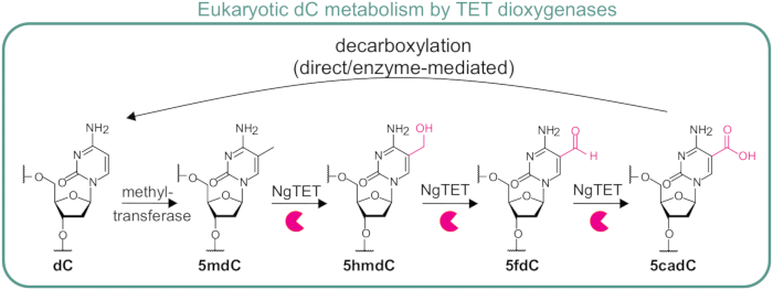
Figure 1: Stepwise Oxidation of 5-Methylcytosine by TET Dioxygenase1. The TET dioxygenase catalyzes the successive oxidation of 5-methyl-2´-deoxycytidine (5mdC) to 5hmdC, 5-formyl-2´-deoxycytidine (5fdC), and ultimately to 5-carboxyl-2´-deoxycytidine (5cadC). The final product, 5cadC, either spontaneously or enzymatically reverts to unmodified cytosine (dC), thereby contributing to dynamic epigenetic regulation. This multistep process is central to active DNA methylation in eukaryotes. Please click here to view a larger version of this figure.
Since methylcytosine and hydroxymethylcytosine are common precursors to phage DNA hypermodifications, including bulky protective structures like glycosylations, the oxidative activity of TET dioxygenase can be exploited to prevent the formation of these DNA hypermodifications9. In this TET-based genome mutagenesis approach, we heterologously express NgTET recombinantly in E. coli to oxidize hydroxymethylated cytosines in the phage genome upon infection, thereby preventing the formation of bulky glycosylation. By decreasing the abundance of these bulky DNA modifications, our approach can increase phage DNA accessibility to CRISPR-Cas nucleases. This enhanced accessibility facilitates efficient target recognition, precise DNA cleavage, and the introduction of scarless point mutations into the phage genome, distinguishing it from traditional phage mutagenesis methods that rely on gene deletions, reporter gene insertions, or the integration of artificial junctions for PCR-based mutant selection1,11. These conventional strategies have so far been the only available tools for targeted mutagenesis in phage genomes and therefore represent an important foundation for the method presented here. However, single phage proteins can possess multiple functions. Deletion of the entire gene encoding a protein of interest disrupts all associated functions. In contrast, the targeted introduction of point mutations permits the selective inactivation of specific functions, enabling detailed analysis of their roles. To date, the functions of many phage proteins remain largely uncharacterized; thus, we may overlook additional activities linked to a given gene. Therefore, a minimalistic approach that aims to remove only a single function while causing minimal changes to the overall protein is preferred12,13. The method described here enables precise genetic modifications without disrupting the overall genomic organization or function. Another major challenge of previous phage mutagenesis methods has been their low efficiency, which typically yielded mutation rates of around 0.1%1,11. With this technique, we present the first mutagenesis strategy capable of introducing single-codon changes, achieving a notable sevenfold increase in targeting efficiency. By combining it with an ONT-based high-throughput screening method, the detection of point mutations is simplified, eliminating the need to screen large phage populations to isolate one mutant. The TET-based mutagenesis strategy described here addresses a longstanding limitation in the field and enables more reliable genome editing14.
Overall, this method cannot only be applied to advance our understanding of bacteriophage infection mechanisms but also holds significant promise for synthetic biology. By enabling precise and efficient phage genome engineering, it paves the way for tailoring the phages to specific applications, thereby enhancing their potential for biotechnological and medical applications.
Access restricted. Please log in or start a trial to view this content.
Protocol
A schematic demonstrating the generation of NgTET-treated T4 phage is shown in Figure 2.
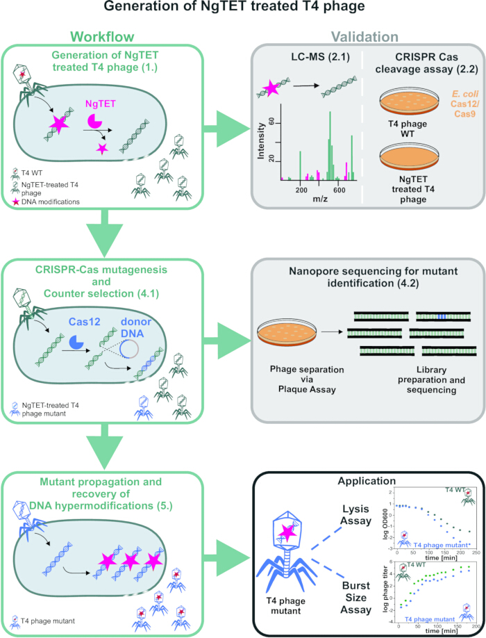
Figure 2: Schematic overview of the established workflow for T4 phage mutagenesis and mutant screening. The workflow includes the generation of the hypomodified T4 phage using NgTET, which can be validated either by LC-MS analysis or alternatively via CRISPR-Cas cleavage assay. Subsequently, the mutation of interest is introduced into the phage genome through CRISPR-Cas-mediated site-directed mutagenesis and identified by counterselection. The presence of the desired mutation can then be confirmed using Nanopore sequencing. Following validation, the mutant phage can be propagated to restore DNA hypermodifications. This enables the use of the engineered phage in downstream applications, including the analysis of infection phenotypes through lysis assays and burst size measurements. Each step in the figure is annotated with the corresponding protocol number in brackets to facilitate linking the protocol steps with the respective parts of the figure. Please click here to view a larger version of this figure.
1. Reducing phage DNA modifications using NgTET pre-treatment (~2 days)
NOTE: This step is critical for enabling efficient Cas nuclease targeting during mutagenesis by reducing the abundance of phage DNA modifications.
- Inoculate the E. coli BL21 (DE3) strain transformed with pET28a_NgTET by transferring material from a glycerol stock into 10 mL of LB medium supplemented with 30 µg/mL kanamycin. Incubate overnight at 37 °C with shaking at 160 rpm.
- The following day, use the overnight culture to inoculate a main culture to an initial OD600 of 0.1 in 50 mL of LB medium containing 30 µg/mL kanamycin. Induce the culture with 50 µM IPTG when an OD600 of 0.4 is reached.
- Grow the culture at 37 °C with shaking at 160 rpm until it reaches an OD600 of 0.8.
- Infect the culture with the phage of interest at a multiplicity of infection (MOI) of 0.1 and incubate for 4 h at room temperature with shaking at 120 rpm.
- Harvest the cells by centrifugation at 4000 × g for 20 min at 4 °C.
- Pass the supernatant through a 0.45 µm filter to obtain NgTET-treated phages with hypomodified DNA. Store the filtered phages at 4 °C for future use.
NOTE: (PAUSE POINT) Filtered phages are stable for several months at 4 °C, stored in LB medium in a glass bottle.
2. Validation
NOTE: The aim of this step is to experimentally verify the reduced abundance of phage DNA modifications.
- Treatment validation via LC-MS (~ 2 days)
- Phage purification via sucrose gradient
- To remove the residual nucleic acid from the lysate, treat 500 µL of the phage suspension (>1010 PFU/mL) with 20 U of DNase I and 2 µL of RNase A/T1 Mix (4 µg of RNase A, 10 U of RNase T1).
- Incubate the mixture at 37 °C for 30 min.
- Prepare a 0-45% sucrose gradient in TM buffer (50 mM Tris-HCl, 10 mM MgCl2, pH 7.5). Prepare the gradient using a gradient mixer or manually.
NOTE: For manual preparation, start by pipetting 5 mL of TM buffer into a gradient tube. Then, using a blunt-end cannula, slowly pipette 5 mL of the 45% sucrose solution from the bottom of the tube, allowing the 0% solution to rise gradually. - Load 500 µL of the treated phage solution on top of the gradient.
- Centrifuge at 70,000 × g for 20 min at 4 °C.
- Light the centrifugation tube from the bottom. This will allow for the visualisation of the phage, which appears as a turbid band in the gradient. Remove the phage-containing fraction carefully using a blunt cannula.
- Transfer the extracted phage fraction into a new ultracentrifugation tube.
- Add 30 mL of ice-cold TM buffer and centrifuge at 100,000 × g for 1 h at 4 °C.
- Discard the supernatant and resuspend the pellet in 500 µL of TM buffer.
- Store the resuspended phages in a glass vial at 4 °C overnight.
- Phage DNA isolation
- Add 1 µg of Proteinase K to the resuspended phages.
- Incubate for 30 min at 37 °C.
- Add 1 volume of phenol/chloroform/isoamyl alcohol (P/C/I) mixture. Mix by inversion. Separate the phases by centrifugation at 15,000 x g for 1 min at 4 °C. Transfer the aqueous phase into a new reaction tube. Repeat the extraction three times.
CAUTION: Phenol/chloroform/isoamyl alcohol (P/C/I) is highly toxic, corrosive, and volatile. Always handle it inside a fume hood while wearing appropriate protective equipment, including a lab coat, safety goggles, and double nitrile gloves. Avoid skin and eye contact, and store in a tightly sealed container in a designated chemical cabinet. Dispose of waste through approved hazardous waste channels. In case of contact, rinse immediately with water and seek medical attention. - To remove residual phenol, perform three times chloroform back-extractions with 1 volume of chloroform.
- Precipitate DNA overnight with 0.1 volume of 3 M NaOAc (pH 5.5) and 2.5 volume of ethanol at -20 °C.
- Next day, pellet the DNA by centrifugation at 15,000 × g for 1 h at 4 °C.
- Wash the DNA pellet twice with 200 µL of 70% ethanol, gently shaking, centrifuging at 15,000 × g for 15 min at 4 °C, and carefully removing the supernatant.
- Resuspend the purified DNA in ultrapure water.
- Store the DNA at -20 °C until further use.
NOTE: (PAUSE POINT) Precipitated DNA can be stored at -20 °C for several months.
- Preparation of phage DNA for LC-MS analysis
- Digest the purified DNA into single nucleosides using a Nucleoside Digestion Mix.
- Analysis of DNA composition via LC-MS.
NOTE: This step allows for validation of the effectiveness of the NgTET treatment. The protocol is adapted to the specific equipment and column available in the lab, but can be adjusted based on available resources and conditions. - Use digested DNA isolated from purified phage (step 2.1.10).
- Include the following commercially available standards for the measurements: dA, dT, dG, dC, 5hmdC, 5fdC, 5cadC. If available, include the standard of the cytosine hypermodification present on the phage DNA.
- Use a HPLC system equipped with a C18 column (150 × 2.1 mm, 100 Å, 3 µm) and a 20 × 2.1 mm guard column.
- Set the column temperature to 40 °C and the eluent flow rate to 0.2 mL/min.
- Prepare eluents as follows:
Eluent A: 10 mM Ammonium Acetate in water (pH 4.5).
Eluent B: 0.1% formic acid in methanol.
CAUTION: 0.1% formic acid in methanol is flammable and can irritate skin, eyes, and the respiratory tract. Always use in a well-ventilated area or fume hood. Wear appropriate protective equipment, including a lab coat, safety goggles, and nitrile gloves. Keep away from heat, sparks, and open flames. Store in a flammable storage cabinet. Dispose of via approved hazardous waste protocols. - Apply the following mobile phase profile: 0-1 min: Constant at 5% B; 1-5 min: Gradient from 5% to 90% B; 5-7 min: Constant at 90% B; 7-7.1 min: Gradient from 90% to 5% B; 7.1-12 min: Constant at 5% B.
- Use a mass spectrometer in negative and positive ionization modes (separate injections) with a high-temperature electrospray ionization (H-ESI) source under the following conditions:
H-ESI spray voltage: 3400 V (+), 2400 V (-), Sheath gas: 35 arbitrary units, Auxiliary gas: 7 arbitrary units, Sweep gas: 0 arbitrary units, Ion transfer tube temperature: 300 °C, Vaporizer temperature: 275 °C, Detection mode: Full scan using the Orbitrap mass analyzer, Mass resolution: 120,000, Mass range: 200-450 (m/z). - Extract ion chromatograms of the [M-H]- (dA, dT, dC, 5hmdC, 5fdC, 5cadC) and [M+H]+ (5ghmdC) forms using Tracefinder software.
- Calculate the relative abundance of each modification by normalizing the peak area of each signal to the dG peak area within the sample, using dG as a sample-specific internal standard.
- Phage purification via sucrose gradient
- Validation with CRISPR-Cas cleavage assay (~2.5 days)
NOTE: This step enables the phenotypic assessment of the success of the NgTET treatment and its impact on CRISPR-Cas cleavage. If NgTET is active, no plaques should form from the treated phage DNA, or the phage titer should be reduced, as the removal of protective DNA modifications renders the genome susceptible to CRISPR-Cas12 or 9 cleavage. In contrast, if the phage DNA is untreated, the number of the formed plaques will correspond to the titer of the phages, indicating that the hypermodified T4 phage genome is resistant to CRISPR-Cas12 or 9 cleavage.- Inoculate E. coli BL21 (DE3) from an overnight culture, transformed with both the pET28a_NgTET plasmid and a Cas12 or Cas9 expression plasmid (e.g., DS-spCas or pCpf1 without spacer). All plasmids with additional information can be found in Table 1.
- Grow the cells in LB medium supplemented with the appropriate antibiotics at 3 °C with shaking, starting from an OD600 of 0.1. Once the culture reaches an OD600 of approximately 0.4, induce NgTET expression by adding 0.05 mM IPTG and incubate for an additional 2 h at 3 °C.
- As a control, compare E. coli BL21 (DE3) strains with and without NgTET overexpression. In the absence of NgTET activity, a higher number of plaque-forming units is expected, indicating reduced CRISPR-Cas efficiency due to the presence of glycosylated 5ghmdC in the phage DNA.
- Transfer 300 µL of the E. coli cultures into a sterile tube.
- Infect the culture with T4 WT or NgTET-treated phage with MOI 0.01. For this experiment, apply a low MOI to ensure that bacteria are infected by only one phage at a time. This maximizes the efficiency of the CRISPR-Cas cleavage screening and simplifies interpretation of the results. Mix gently to ensure even distribution of the phages.
- Incubate the bacteria-phage suspension at 37 °C for 7 min.
- Add the bacteria-phage mixture to 4 mL of LB soft agar (0.75%) supplemented with antibiotics. Mix thoroughly but gently to avoid introducing bubbles.
- Pour the soft agar mixture onto a pre-warmed LB agar plate.
- Allow the plates to solidify briefly at room temperature. Incubate the plates at 37 °C overnight.
- The next day, count the resulting plaques to determine plaque-forming units (PFU).
- Inoculate E. coli BL21 (DE3) from an overnight culture, transformed with both the pET28a_NgTET plasmid and a Cas12 or Cas9 expression plasmid (e.g., DS-spCas or pCpf1 without spacer). All plasmids with additional information can be found in Table 1.
3. CRISPR-Cas mutagenesis (~ 1 week)
NOTE: CRISPR-Cas mutagenesis is based on the principle that a specific site within the phage genome is targeted by a Cas nuclease during phage infection, resulting in a double-strand break. This break is repaired through homologous recombination using donor DNA, which is provided on a second plasmid present in the infected host strain during mutagenesis. The plasmid that contains the donor DNA also encodes NgTET, which is essential to maintain accessibility of the phage DNA for the Cas nuclease by reducing the abundance of modified cytosines, as described previously. In this step, target mutations are introduced into the genome of NgTET-treated T4 phages upon infection of E. coli15. To ensure consistently low levels of DNA modifications in the T4 genome, NgTET dioxygenase is expressed by the addition of 50 µM IPTG (analogously to step 1.2) throughout the mutagenesis process.
- Generation of pET28a_NgTET_donor DNA plasmids via golden gate cloning15
- For the generation of the donor DNA, order the desired sequence as an entire gene or amplify from a genome, for example, from the phage genome, and introduce the desired mutation.
NOTE: For donor DNA design, include homologous regions flanking the target site (≈200 bp on each side for point mutations; longer regions for larger inserts). The linear donor fragment should carry BsaI recognition sites that generate compatible overhangs with the plasmid upon cleavage. In silico cloning is recommended to verify the construct design. In the representative results, all critical components for the donor DNA are illustrated in an example. - Introduce the donor DNA fragment downstream of the NgTET coding sequence and terminator in the pET28a_NgTET backbone using Golden Gate assembly.
- Prepare the assembly reactions by combining 70 fmol of pET28a_NgTET plasmid, 140 fmol of donor DNA insert, 1 µL of BsaI (20 U/µL), 5 U of T4 DNA ligase, and 2 µL of 10× ligase buffer, with nuclease-free water added to a final volume of 20 µL.
- Run reactions in 25 cycles in a PCR-cycler with the following settings: 1) Restriction, 37 °C for 1.5 min, 2) Ligation, 16 °C for 3 min, 3) Final restriction, 37 °C for 5 min, and 4) Denaturation, 60 °C for 10 min.
- Add 6 µL of the reaction mixture (from step 3.1.2) to 100 µL of chemically competent E. coli DH5α, incubate on ice for 10 min, and heat shock at 42 °C for 45 s. Immediately place the cells on ice for 1 min, then add 750 µL LB medium. Recover at 37 °C with shaking (500 rpm) for 1 h. Plate on LB agar containing kanamycin (30 µg/mL) and incubate overnight at 37 °C.
- Pick a single colony and inoculate 20 mL of LB medium supplemented with kanamycin (30 µg/mL). Incubate overnight at 37 °C with shaking at 160 rpm.
- From 5 mL of the overnight culture, pellet the cells and isolate plasmid DNA using a standard plasmid extraction method. Elute the plasmid in 40 µL of nuclease-free water.
- Confirm correct cloning by sequencing the insertion site of the plasmid using Sanger sequencing.
- For the generation of the donor DNA, order the desired sequence as an entire gene or amplify from a genome, for example, from the phage genome, and introduce the desired mutation.
- Construction of spacer-containing CRISPR-Cas plasmids (pCpf1-sp as a Model)15
NOTE: The plasmid described in this chapter enables the first step of mutagenesis by introducing a site-specific double-strand break in the phage genome. While the procedure is demonstrated here with the CRISPR-Cas12 system (pCpfl-sp plasmid), alternative nucleases such as CRISPR-Cas9 (DS-spCas plasmid) can also be employed15 .- Design a 20-nt spacer targeting the mutagenesis site (PAM: TTTV). Identify and design suitable protospacers and corresponding spacers using bioinformatic tools.
- Synthesize two 5′-phosphorylated oligonucleotides. Ensure each carries 10 nt of the desired spacer sequence (together forming the full 20 nt spacer) and an additional 10 nt region complementary to the insertion site within the pCpf1-sp plasmid.
- Insert the designed CRISPR-Cas spacer into the pCpf1-sp plasmid by PCR amplification using the primers from step 2. Set up the reaction as follows (final volume 50 µL): 5 µL 10× GC buffer, 1 µL 10 mM dNTPs, 1 µL forward primer (10 µM), 1 µL reverse primer (10 µM), 2.5 µL DMSO, plasmid template corresponding to 2 fmol, 1 µL Phusion DNA polymerase (2 U/µL), and nuclease-free water to 50 µL.
- Place the reaction tubes in a thermocycler and run the following program: initial denaturation at 98 °C for 30 s; 25 cycles of 98 °C for 10 s, annealing at the appropriate temperature (X °C) for 30 s, and extension at 72 °C for 6 min; followed by a final extension at 72 °C for 10 min. Hold the samples at 4 °C until further processing.
- Mix 5 µL of the PCR reaction with DNA loading buffer (10×) and load onto a 1% agarose gel. Run the gel at 130 V for 20 min and visualize the DNA bands using UV trans illumination. If amplification is successful, purify the PCR product using a suitable cleanup kit and elute in 20 µL of nuclease-free water.
- Circularize the plasmid by ligation using 100 ng of the amplified product mixed with 1× T4 DNA ligase buffer and 60 U T4 DNA ligase in a total reaction volume of 15 µL. Incubate for 1 h at room temperature. Transform 100 µL of chemically competent E. coli with the ligation product as described in step 3.1.3. Plate the cells on LB agar containing 50 µg/mL streptomycin and incubate overnight at 37 °C.
- Pick a single colony from the plate and inoculate it into 20 mL of LB medium supplemented with 50 µg/mL streptomycin. Incubate overnight at 37 °C with shaking at 160 rpm. From 5 mL of the overnight culture, harvest the cells by centrifugation and isolate plasmid DNA using a plasmid extraction method of choice. Elute the plasmid in 40 µL of nuclease-free water.
- Confirm successful cloning by sequencing the plasmid at the insertion site using Sanger sequencing.
- Phage mutagenesis
- Transform E. coli BL21 (DE3) cells with two plasmids: pET28a_NgTET_donor-DNA and pCpf1-sp (Cas12)/DS-SPcas (Cas9) spacer (generated in steps 3.1 and 3.2). Mix 1 µL of each plasmid with 100 µL of chemically competent E. coli BL21 (DE3). Follow the protocol described in step 3.1.3. Plate the transformed cells on agar containing 50 µg/mL streptomycin and 30 µg/mL kanamycin, and incubate overnight at 37 °C.
- Inoculate the transformed E. coli strain (E. coli BL21 (DE3) pET28a_NgTET_donor-DNA and pCpf1-sp(Cas12)/DS-SPcas(Cas9)_spacer) into 10 mL of LB medium supplemented with 30 µg/mL kanamycin and 50 µg/mL streptomycin. Incubate overnight at 37 °C with shaking at 160 rpm.
- The next day, dilute the overnight culture to an initial OD600 of 0.1 in 50 mL of LB medium containing 30 µg/mL kanamycin and 50 µg/mL streptomycin.
- Grow the culture at 37 °C with shaking at 160 rpm until an OD600 of 0.8 is reached. Lower the temperature to room temperature (RT) and reduce the agitation speed to 130 rpm.
- To enhance phage adsorption and DNA injection, add MgCl2 and CaCl2 to final concentrations of 1 mM each, as these divalent cations facilitate efficient phage-host interactions.
- Infect the culture with NgTET-pretreated T4 phages, and as a negative control, with NgTET-untreated T4 phages at an MOI of 0.1. Continue incubation at RT for 4 h.
- After incubation, transfer the cultures to a 50 mL conical tube and centrifuge at 4000 × g at 4 °C for 20 min to pellet the cells. Filter the supernatants through a 0.45 µm filter to remove any residual bacterial cells. Use the filtered supernatants for plaque assays (described in step 2.2) to determine phage concentration or perform counterselection.
4. Mutant identification
NOTE: The purpose of counterselection is to decrease the proportion of wild-type phages remaining in the population after mutagenesis. For this, phages obtained from the mutagenesis step (step 3.3) are used to infect E. coli carrying the same CRISPR-Cas13 system, which is encoded by another plasmid pBA560 (additional information in Table 1) and the same spacer sequence applied during mutagenesis. In this setup, Cas nucleases selectively recognize and cleave wild-type phage DNA, while phages carrying the desired mutations remain unaffected. This selective pressure suppresses wild-type replication and enriches the population for mutant phages14.
- Counterselection with Cas13
- Infect E. coli Cas13a_spacer (plasmid pBA560) with the phages for counter-selection. This step facilitates counterselection by degrading phage RNA that has not undergone the intended mutation.
- Perform the counter-selection under the same conditions used during mutagenesis (see Steps 3.3.1-3.3.5). Use E. coli expressing a non-targeting Cas13a spacer as a negative control to confirm that plaque reduction is spacer-specific.
- After incubation, filter the supernatant containing counter-selected phages through a 0.45 µm filter to remove bacterial debris.
NOTE: (PAUSE POINT) Filtered phages are stable for several months at 4 °C, stored in LB medium in a glass bottle. - Use the counterselected and filtered phages for a plaque assay on an E. coli B strain to isolate individual plaques for downstream validation.
- Next generation sequencing (~ 1 week)
NOTE: The method for the highly multiplexed sequencing approach involves a two-step PCR process to generate multiplexed amplicon DNA suitable for long-read next-generation sequencing16.- Separate the phages from the mutagenesis by plating them out as described in step 2.2. Include proper controls by using phages obtained from mutagenesis performed both in the presence and absence of NgTET expression. As a control, CRISPR-Cas9 and Cas12 targeting without NgTET should not result in the introduction of point mutations. In contrast, co-expression of NgTET is expected to enable successful editing, leading to higher mutagenesis efficiencies.
- To isolate the single phages, pick individual plaques from the plate and transfer them into a tube containing 100 µL of Pi-Mg buffer (26 mM Na2PHO4, 68 mM NaCl, 22 mM KH2PO4, 1 mM MgSO4, pH 7.5) and 1 µL of CHCl3.
CAUTION: Chloroform is toxic, volatile, and a suspected carcinogen, even in small volumes. Handle only in a fume hood and wear full protective equipment, including a lab coat, safety goggles, and nitrile gloves. Avoid inhalation and skin contact. Store in a tightly sealed container in a ventilated, flammable chemical cabinet. Dispose of chloroform waste via hazardous waste collection. - Use 1 µL of isolated phage T4 in Pi-Mg buffer as the template for the screening.
- Prepare the reaction mix with 0.125 µM of each primer (around the insertion position) and 1x high-accuracy polymerase master mix, adjusting the total volume of the amplification reaction to 10 µL. Run a PCR reaction with 30 amplification cycles.
- Perform a second PCR to attach the barcodes. Therefore, use 1 µL of a 1:10 dilution of the product from the first PCR as a template.
- Prepare the reaction mix with 0.3 µM barcoding primers and a high-fidelity hot-start polymerase mix in a total volume of 7 µL. Perform PCR with 20 amplification cycles.
NOTE: (PAUSE POINT) PCR-Products can be stored at -20 °C for several weeks. - Pool all barcoded PCR products and purify them using magnetic bead-based cleanup according to standard protocols for next-generation sequencing library preparation.
- Capture DNA using magnetic beads, wash twice with 80% ethanol, and elute in 100 µL of elution buffer (5 mM Tris-HCl, pH 8.5).
- Measure the DNA concentration using spectrophotometric and fluorometric methods, applying either broad-range or high-sensitivity assay as appropriate.
NOTE: (PAUSE POINT) DNA can be stored at -20 °C for several weeks. - Generate sequencing libraries using a ligation-based protocol according to the manufacturer's instructions, starting with 1 µg of input DNA.
5. Mutant propagation recovery of DNA modifications and proof
NOTE: Once the desired phage mutant is identified, it is propagated in wild-type E. coli to restore DNA modification levels similar to those of the WT phage. This ensures that the mutant phage can be utilized in subsequent biological experiments without any compromise, apart from the introduced mutation.
- Prepare a 10 mL culture of E. coli B strain in LB medium and incubate overnight at 37 °C with shaking at 160 rpm.
- The following day, inoculate a fresh 50 mL LB medium with the overnight culture to achieve an initial OD600 of 0.1.
- Allow the culture to grow at 37 °C with shaking at 160 rpm until it reaches an OD600 of 0.8.
- Add 50 µL of the mutant phage stock (4.2.2) to the culture. Remove the phase carefully to avoid transferring CHCl3.
- Incubate the infected culture overnight at room temperature with gentle shaking at 120 rpm.
- After incubation, centrifuge the culture at 4000 × g to remove bacterial cells. Filter the supernatant through a 0.45 µm filter to collect the phage.
- Transfer the filtered mutant phage to a glass container and store at 4 °C until further use.
NOTE: (PAUSE POINT) Filtered phages are stable for several months at 4 °C, stored in LB medium in a glass bottle. - As an optional validation of the regeneration of DNA modifications, specifically glycosylated cytosines, perform an LC-MS analysis as described in step 2.1.
- For this purpose, purify the recovered phage DNA from the phage progeny as outlined in step 2.1.1, isolate as in step 2.1.2, and subsequently prepare for LC-MS analysis as described in step 2.1.3.
- Analyze ion chromatograms and calculate the relative abundance of each modification by normalizing the peak area of each signal to the dG peak area within the sample, using dG as a sample-specific internal standard as described in steps 2.1.3.
NOTE: Including T4 wild-type phage, T4 phage treated with NgTET, and T4 phage treated with the inactive NgTET D234A variant as controls is recommended, in order to directly validate and compare the abundance of 5ghmdC10.
Access restricted. Please log in or start a trial to view this content.
Results
Identification of the phage band in the gradient.
The visibility of the phage band in the gradient depends on the phage concentration in the initial sample loaded on the gradient. When the phage is sufficiently concentrated (~>1 × 10¹¹ PFU/mL), the band can be clearly seen by illuminating the gradient from the bottom with a light source (see Figure 3).
However, at low phage concentrations, the band may not be easily visible. In t...
Access restricted. Please log in or start a trial to view this content.
Discussion
This study presents a robust and adaptable strategy for introducing precise point mutations into bacteriophage genomes, including single-nucleotide changes. A central element of the workflow is the integration of stringent validation steps at each stage, which is essential for ensuring both the accuracy and efficiency of genome editing. This is particularly critical given the multistep nature of the workflow and the dynamic interactions between phages and their bacterial hosts, which introduce numerous variables that can...
Access restricted. Please log in or start a trial to view this content.
Disclosures
KH and NP filed a European Patent Application for "Engineering of Phages", European Patent Application No. 23 175 257.7. The other authors declare no competing interests.
Acknowledgements
This project has received funding from the German Research Council (DFG; SPP 2330 project number 464500427, RTG 2355 project 11, and RTG 2937 (project number 505997786) from the Max Planck Society (Max Planck Research Group Leader funding to K.H.).
Access restricted. Please log in or start a trial to view this content.
Materials
| Name | Company | Catalog Number | Comments |
|---|---|---|---|
| 1.5 mL reaction tubes | Sarstedt | 72.690.001 | |
| 15 mL Canocial Tubes | Sarstedt | 62.554.502 | |
| 15 mL Open-Top Thinwall Polypropylene Tubes | Beckman Coulter Life Sciences | 361706 | 16 mm x 95 mm (for ultracentrifugation) |
| 50 mL canonical tubes | Sarstedt | 62.547.254 | |
| 5x GC buffer | Thermo Fisher Scientific | F519L | |
| 6x TriTrack DNA Loading Dye | Thermo Fisher Scientific | R1161 | |
| Agarose | Meridian BIOSCIENCE | BIO-41025 | Molecular Grade |
| Avanti J-30I Centrifuge | Beckman Coulter Life Sciences | no catalog number | Serien-Nr. JKY14M01, https://www.beckman.de/centrifuges/ultracentrifuges/optima-xe |
| Boric acid | Merck | 10043-35-3 | |
| BsaI-HF v2 | NEB | R3733S | |
| Calciumchlorid Hexahydrat | ACROS ORGANICS | 7774-34-7 | 97 %, extra pure |
| Cell density meter | Harvard Biochrom | 80211630 | |
| ChemiDoc MP | Biorad | 12003154 | |
| Chloroform | VWR | 650498 | |
| Cuvettes | Sarstedt | 67.742 | |
| Dimethyl sulfoxide (DMSO) | Sigma Aldrich | D4540 | |
| Di-Sodium hydrogen phosphate | neoFroxx GmbH | 7558-79-4 | pharma grade |
| Disposable Glass Pasteur Pipettes | VWR | 612-1702 | 230 mm |
| DNase I | Roche | 4716728001 | 10 U/µL |
| dNTP mix (10 mM each dNTP) | Sigma-Aldrich | 72004 | |
| DS_SPcas_ModA | In-house product. | https://github.com/MaikTungsten/CRISPRT4/tree/main/Plasmid_maps | Plasmid construct that contains CRISPR-Cas12/ModA sgRNA |
| DS-spCas (CRISPR-Cas9 system) | Addgene | 48645 (addgene accessory number) | Plasmid for Construction of Spacer-Containing CRISPR-Cas Plasmids (CRISPR-Cas9 system) for phage mutagenesis (step 3.2) |
| E. coli B Strain Strain was used for T4 infection | DSMZ | no catalog number | Escherichia coli (Migula 1895) Castellani and Chalmers 1919 (DSM 613, ATCC 11303) |
| E. coli BL21 (DE3) Expression strain, competent cell | Invitrogen | C600003 | Expression strain, competent cells. |
| E. coli DH5α Cloning strain, competent cells | Invitrogen | 18265017 | Cloning strain, competent cells. |
| EDTA | Roth | 8043.2 | |
| Ethanol (absolute) | VWR | 200-578-6 | |
| Flongle Flow Cell | Oxford Nanopore Technologies | no catalog number | R10.4.1. chemistry, https://nanoporetech.com/document/flongle |
| Gene JET Plasmid Miniprep Kit | Thermo Fisher Scientific | 10242490 | |
| GeneRuler 1 kb Plus DNA ladder | Thermo Fisher Scientific | SM1331 | |
| GeneRuler ULR DNA ladder | Thermo Fisher Scientific | SM1213 | |
| Gradient Master 108 | BIOCOMP | no catalog number | https://biocompinstruments.com/our-approach/gradient-forming |
| Heraeus Pico 17 Centrifuge | Thermo Scientific | 75002410 | |
| Infors HT Incubator Minitron | Infors AG | no catalog number | https://infors-ht.com/de/produkte/inkubationsschuettler/minitron#productspecs |
| Isopropyl-β-D-1-thiogalactopyranoside (IPTG) | Sigma-Aldrich | 367-93-1 | |
| Kanamycin | Carl Roth | T832.1 | |
| LB-Agar | Carl-Roth | X969.1 | |
| LB-medium (Luria/Miller) | Carl-Roth | X968.1 | |
| Magnesium chloride hexahydrate | Sigma-Aldrich | 7791-18-6 | |
| Magnesium sulfate | Sigma-Aldrich | 7487-88-9 | |
| MinION device | Oxfrod Nanopore Technologies | no catalog number | https://store.nanoporetech.com/eu/minion.html |
| ModA E165A_fw | IDT | GAATTAGTTTCAGATGA ACAAGCGGTAATGATAC CAGCT | Forward primer for introduction of point mutation for ModA E165A and to introduce sticky ends for Golden Gate Cloning (Step 3.1) |
| ModA E165A_rev | IDT | CATATTACGATAACGA TGACTATCCGGAAAC | Reverse primer for introduction of point mutation for ModA E165A and to introduce sticky ends for Golden Gate Cloning (Step 3.1) |
| ModA_sgRNA_Cas12_fw | IDT | TCTTGTTCATCTACAAC AGTAGAAATTAATTTAAA GTTCTTAGACCCG | Forward primer for generating spacer for CRISPR-Cas12/ CRISPR-Cas9/ CRISPR-Cas13 systems for phage mutagenesis and counterselection (step 3.2 and step 4.1) |
| ModA_sgRNA_Cas12_rev | IDT | AGTAATGATAAAGAACT TTAAATAATTTCTACTGT TGTAGATGCTAC | Reverse primer for generating spacer for CRISPR-Cas12/ CRISPR-Cas9/ CRISPR-Cas13 systems for phage mutagenesis and counterselection (step 3.2 and step 4.1) |
| NanoDrop ND-1000 Spectrophotometer | Thermo Fisher Scientific | AZY2019944 | |
| NucleoMag Kit | Machery Nagel | REF 744970.50 | |
| Nucleoside digestion mix | New England Biolabs | M0649S | |
| pBA560 (CRISPR-Cas13 system) | Addgene | 186236 (addgene accessory number) | Plasmid encoding CRISPR-Cas13 system for counterselection (step 4.1) |
| pCpfl-sp plasmid (CRISPR-Cas12 system) | Addgene | 122186 | |
| pCpfl-sp plasmid (CRISPR-Cas12 system) | Addgene | 122186 (addgene accessory number) | Plasmid for Construction of Spacer-Containing CRISPR-Cas Plasmids (CRISPR-Cas12 system) for phage mutagenesis (step 3.2) |
| PCR reaction vessels | Sarstedt | 72.991.002 | |
| PeqGREEN | VWR | 732-3196 | |
| pET28a_NgTET | In-house product. | no catalog number | plasmid is available upon request, the plasmid map can be found at the following https://zenodo.org/records/10615143 |
| pET28a_NgTET | In-house product. | no catalog number | Plasmid for construction of pET28a_NgTET_donor DNA via Golden Gate Cloning (step 3.1), plasmid is available upon request, the plasmid map can be found at the following https://zenodo.org/records/10615143 |
| Petri Plates | Sarstedt | 82.1473.001 | 92 x 16 mm with cams |
| Phusion DNA Polymerase | Thermo Fisher Scientific | F-530XL | 2 U/µL |
| Potassium Phosphate | Sigma Aldrich | 7778-77-0 | Monobasic |
| PowerPac Universal Power Supply | Bio Rad | 1645070 | |
| Proteinase K | Carl Roth | 7528.1 | 100 mg/mL |
| QIAquick PCR Purification Kit | QIAGEN | 28506 | |
| Quantus Fluormeter, QuantiFluor dsDNA system | Promega | E2670 | |
| RNase A/T1 | Thermo Fisher Scientific | EN0551 | mix 2 mg/mL of RNase A 5000 U/mL of RNase T1 |
| Roti-Aqua P/C/I for DNA extraction | Carl Roth | X985.1 | |
| Sodium acetate | Sigma-Aldrich | 127-09-3 | |
| Sodium chloride | Sigma-Aldrich | 7647-14-5 | |
| SQK-LSK109 Ligation sequencing Kit | Oxford Nanopore Technologies | no catalog number | https://nanoporetech.com/document/gDNA-q-sqk-lsk109 |
| Steritop filters, 0.22 µm pore size | Millipore | S2GPT05RE | 250 mL and 500 mL |
| Streptomycin 50 mg/L | Carl Roth | HP66.1 | |
| Succrose | NeoFroxx GmbH | 57-50-1 | for molecular biology |
| Swinging-Bucket Aluminum Rotor | Beckman Coulter Life Sciences | JS[1]24.15 | |
| T4 DNA Ligase 5 U/µL | Thermo Fisher Scientific | EL0011 | |
| T4 DNA Ligase Buffer | Thermo Fisher Scientific | B69 | |
| Test Tubes, heavy wall, rimless | Karl Hecht GmbH | 42775054 | 100 mm x 18 mm |
| Thermal cycler, T100 | Biorad | 1861096 | |
| Thermomixer comfort | Eppendorf | 5382000015 | |
| Trizma Base | Sigma Aldrich | 77-86-1 | |
| Wather bath, VWB2 | VWR | 462-0557 | 12 L volume |
References
- Pozhydaieva, N., et al. Temporal epigenome modulation enables efficient bacteriophage engineering and functional analysis of phage DNA modifications. PLoS Genet. 20 (9), e1011384(2024).
- Schroven, K., Aertsen, A., Lavigne, R. Bacteriophages as drivers of bacterial virulence and their potential for biotechnological exploitation. FEMS Microbiol Rev. 45 (1), fuaa041(2021).
- Moye, Z. D., Woolston, J., Sulakvelidze, A. Bacteriophage applications for food production and processing. Viruses. 10 (4), 205(2018).
- Wolfram-Schauerte, M., Pozhydaieva, N., Viering, M., Glatter, T., Hofer, K. Integrated omics reveal time-resolved insights into T4 phage infection of E. coli on proteome and transcriptome levels. Viruses. 14 (11), 2502(2022).
- Usman, S. S., Uba, A. I., Christina, E. Bacteriophage genome engineering for phage therapy to combat bacterial antimicrobial resistance as an alternative to antibiotics. Mol Biol Rep. 50 (8), 7055-7067 (2023).
- Vlot, M., et al. Bacteriophage DNA glucosylation impairs target DNA binding by type I and II but not by type V CRISPR-Cas effector complexes. Nucleic Acids Res. 46 (2), 873-885 (2018).
- Tao, P., Wu, X., Tang, W. C., Zhu, J., Rao, V. Engineering of bacteriophage T4 genome using CRISPR-Cas9. ACS Synth Biol. 6 (10), 1952-1961 (2017).
- Mohanraju, P., et al. Diverse evolutionary roots and mechanistic variations of the CRISPR-Cas systems. Science. 353 (6299), aad5147(2016).
- Zemach, A., McDaniel, I. E., Silva, P., Zilberman, D. Genome-wide evolutionary analysis of eukaryotic DNA methylation. Science. 328 (5980), 916-919 (2010).
- Hashimoto, H., et al. Structure of a Naegleria Tet-like dioxygenase in complex with 5-methylcytosine DNA. Nature. 506 (7488), 391-395 (2014).
- Shitrit, D., et al. Genetic engineering of marine cyanophages reveals integration but not lysogeny in T7-like cyanophages. ISME J. 16 (2), 488-499 (2022).
- Dedrick, R. M., et al. Engineered bacteriophages for treatment of a patient with a disseminated drug-resistant Mycobacterium abscessus. Nat Med. 25 (5), 730-733 (2019).
- Marinelli, L. J., et al. BRED: a simple and powerful tool for constructing mutant and recombinant bacteriophage genomes. PLoS One. 3 (12), e3957(2008).
- Adler, B. A., et al. Broad-spectrum CRISPR-Cas13a enables efficient phage genome editing. Nat Microbiol. 7 (12), 1967-1979 (2022).
- Pozhydaieva, N., Höfer, K. Utilizing Golden Gate assembly to streamline CRISPR-Cas/NgTET-based phage mutagenesis. Methods Mol Biol. 2850, 329-343 (2025).
- Ramírez Rojas, A. A., et al. DuBA.Flow─a low-cost, long-read amplicon sequencing workflow for the validation of synthetic DNA constructs. ACS Synth Biol. 13 (2), 457-465 (2024).
- Lagerbäck, P., et al. Bacteriophage T4 endonuclease II, a promiscuous GIY-YIG nuclease, binds as a tetramer to two DNA substrates. Nucleic Acids Res. 37 (18), 6174-6183 (2009).
- Lagerbäck, P., Carlson, K. Amino acid residues in the GIY-YIG endonuclease II of phage T4 affecting sequence recognition and binding as well as catalysis. J Bacteriol. 190 (16), 5533-5544 (2008).
- Shembrey, C., et al. Principles of CRISPR-Cas13 mismatch intolerance enable selective silencing of point-mutated oncogenic RNA with single-base precision. Sci Adv. 10 (51), eadl0731(2024).
- Molina Vargas, A. M., et al. New design strategies for ultra-specific CRISPR-Cas13a-based RNA detection with single-nucleotide mismatch sensitivity. Nucleic Acids Res. 52 (2), 921-939 (2024).
- Shi, P., Wu, X. Programmable RNA targeting with CRISPR-Cas13. RNA Biol. 21 (1), 1-9 (2024).
- Levin, B. R., et al. The population and evolutionary dynamics of phage and bacteria with CRISPR-mediated immunity. PLoS Genet. 9 (3), e1003312(2013).
- Hershey, A. D., Chase, M. Independent functions of viral protein and nucleic acid in growth of bacteriophage. J Gen Physiol. 36, 39-56 (1952).
- Bair, C. L., Rifat, D., Black, L. W. Exclusion of glucosyl-hydroxymethylcytosine DNA containing bacteriophages is overcome by the injected protein inhibitor IPI. J Mol Biol. 366 (3), 779-789 (2007).
Access restricted. Please log in or start a trial to view this content.
Reprints and Permissions
