Here, we introduce a novel approach-Laparoscopic Extracorporeal Knot-Tying for Uterine Vessel Occlusion during Hysterectomy with Cervical Cerclage in Large Uteri. A laparoscopic extracorporeal knot-pusher is used to perform cervical cerclage, achieving complete occlusion of uterine arteries. Thereafter, the corpus uteri are excised to enhance visibility and facilitate the operative field.
Method Article
Laparoscopic Extracorporeal Knot-Tying for Uterine Vessel Occlusion during Hysterectomy with Cervical Cerclage in Large Uteri
* These authors contributed equally
In This Article
Summary
Abstract
Large benign uterine pathologies affect millions of women globally and impose substantial socioeconomic costs. Although hysterectomy remains the definitive treatment, minimally invasive surgery (MIS) methodologies, including vaginal procedures and laparoscopy, have largely supplanted open laparotomy. However, significant uterine enlargement compromises pelvic workspace, obscures surrounding anatomical structures, which may impair the surgeon's ability to operate effectively, and increase the risks of hemorrhage and adjacent-organ injury. Consequently, many gynecological surgeons regard uteri ≥ 17-week equivalent as a contraindication to total laparoscopic hysterectomy (TLH). We devised a novel approach known as Laparoscopic Extracorporeal Knot-Tying for Uterine Vessel Occlusion during Hysterectomy with Cervical Cerclage (LEKTUVOHCC) in Large Uteri, comprising a cervical cerclage using an extracorporeal laparoscopic knot-pusher for en bloc occlusion of uterine arteries and corpus uteri excised to debulk the uterus and optimize the operative field. We successfully performed this procedure in 31 cases at our hospital. We recorded operative time (OT), intraoperative blood loss (IBL), and intra- and postoperative complications, and patient satisfaction over a median follow-up of 12 months. The median operative time was 127 (100, 213) min; the mean IBL was 80 (50, 200) mL. Complete LEKTUVOHCC was achieved in all cases (31/31, 100%), with patient-reported satisfaction in 30/31 (96.77%). No significant complications were observed during or after surgery. This approach effectively overcomes the hemostatic and visualization challenges in laparoscopic hysterectomy for large uteri, such as the dissection of uterine blood arteries and the reduction of IBL, thereby eliminating the risk of blood transfusion and lowering healthcare costs. This technique is reproducible, facilitates rapid adoption, and represents a significant advance in MIS.
Introduction
Hysterectomy stands as the second most prevalent gynecological procedure available for females within their reproductive age to address benign disorders. Annually, almost 600,000 females in the United States receive hysterectomies1,2. The principal surgical methods for hysterectomies are vaginal, abdominal, or laparoscopic techniques3. However, abdominal hysterectomy (AH) remains the default for cases involving uteri ≥ 17 weeks' size despite its association with prolonged hospital stays, increased postoperative pain, higher wound-related morbidity, and suboptimal aesthetic outcomes. In contrast, minimally invasive surgery (MIS), encompassing vaginal and laparoscopic approaches, affords significant benefits, including reduced analgesic requirements, expedited mobilization, accelerated recuperation, decreased hospital stays, and advantageous aesthetic outcomes, all of which substantially enhance patient satisfaction by alleviating long-term mental and physical stress4,5.
Large uteri present unique challenges to MIS. Although AH is generally reserved for uteri measuring ≥ 17 weeks' gestational size6, it should be employed only after MIS approaches fail to yield satisfactory outcomes. Large pelvic uteri compromise trocar triangulation and obstruct clear visualization of critical anatomy, thereby heightening the risks of hemorrhage and adjacent-organ injury7. To mitigate these constraints, several adjunctive strategies have been proposed: transvaginal uterine bivalving, myomectomy preceding the hysterectomy, and direct morcellation following uterine artery ligation8. Nevertheless, these techniques extend prolonged operative time (OT), require advanced dissection skills, and do not consistently achieve reliable hemostasis.
We have devised an innovative surgical technique termed Laparoscopic Extracorporeal Knot-Tying for Uterine Vessel Occlusion during Hysterectomy with Cervical Cerclage (LEKTUVOHCC, hereafter referred to as "the technique"), a novel protocol optimized for large uteri. This innovative procedure employs a knot-pusher for cervical cerclage, achieving en bloc occlusion of both uterine arteries at the cervical isthmus. Thereafter, the corpus uteri are excised to restore pelvic exposure and facilitate safe parametrial dissection. By combining definitive vascular control with targeted specimen reduction, this technique overcomes the dual challenges of restricted deep-pelvic exposure and difficult hemostasis, markedly reducing intraoperative blood loss (IBL) and obviating the need for blood transfusion. This technique requires minimal additional instrumentation, is simple to understand, and is readily reproducible in diverse clinical settings.
The primary objective of this study was to assess the technical feasibility and safety of LEKTUVOHCC in patients with uteri of gestational size ≥ 17 weeks, and to delineate procedural nuances that facilitate its safe and effective implementation in routine clinical practice.
Protocol
The study protocol received approval from the Institutional Review Board of The University of Hong Kong-Shenzhen Hospital (IRB No. [2025]069), and all participants provided written informed consent for publication. Detailed specifications of the surgical instruments, sutures, and other materials are listed in the Table of Materials.
1. Planning for LEKTUVOHCC
- Subject all candidates to a standardized preoperative workup, which includes pelvic examination in the lithotomy position; transvaginal ultrasonography to determine uterine volume.
- Set the following inclusion criteria: 1) Uterine size ≥ 17 weeks' gestational and ≤ 28 weeks' gestational equivalent on ultrasound; 2) clinical indication for the technique; 3) proficiency in the written and spoken local language (Chinese in this study).
- Set the following exclusion criteria: 1) uterine size < 17 weeks' gestational equivalent; 2) contraindications to laparoscopy (e.g., severe cardiopulmonary compromise); 3) known or suspected gynecologic malignancy; 4) concurrent cesarean hysterectomy or pelvic organ prolapse repair; 5) predominant cervical leiomyomas or broad-ligament leiomyomas; 6) inability to comply with follow-up or communicate in the local language (Chinese in this study).
- Enroll the patients meeting all inclusion and none of the exclusion criteria, and proceed to perform the technique.
- Discuss the anticipated benefits and specific risks, including uncontrolled bleeding that may require blood transfusion, conversion to open laparotomy, adjacent-organ injury, and surgical-site infection.
- After obtaining informed consent, administer enoxaparin sodium injection (100 AXaIU/kg, see Table of Materials) for perioperative thrombosis prophylaxis.
- Have an anesthesiologist administer endotracheal general anesthesia and monitor vital signs.
NOTE: Continual assessment of the patient's circulation, oxygenation, ventilation, and temperature is essential throughout the anesthesia period. - Administer perioperative prophylactic antibiotics intravenously 30 min before surgery; use cefuroxime (1.5 g, see Table of Materials) following hospital protocols unless there is an allergic history.
2. Procedure
- After sterilizing the skin, confirm the optimal site for trocar insertion, particularly for the initial trocar (Figure 1). Insert the first 10 mm trocar at a point midway between the umbilicus and the xiphoid process to avoid dense adhesions. Establish pneumoperitoneum with CO2 at 12 mmHg and a flow rate of 20 L/min. Under laparoscopic guidance, place one 5 mm port in the right lower quadrant and two 5 mm ports in the left lower quadrant.
- Examine the abdominal cavity via laparoscopy to determine the feasibility of the technique.
- Perform coagulation and resection of the infundibulopelvic ligament if adnexectomy is indicated.
- Sew a stitch and tie knots at the uterine lateral wall with a suture, for pulling the uterus to expose the pelvic anatomical structures (Figure 2A). Pull the suture to the left to expose the right anatomical structures, such as the right round ligament and broad ligament, and vice versa to expose the left anatomical structures.
- Conduct coagulation and transection of the utero-ovarian ligament if salpingectomy is indicated (Figure 2B). Electrocoagulate and sever the round ligament (Figure 2C), then separate the anterior and posterior layers of the broad ligament subsequently (Figure 2D).
- Utilize the Knot-Pusher (see Table of Materials) to form a Roeder's knot with an absorbable synthetic polyglactin braided suture (size 1, see Table of Materials) around the cervical isthmus (Figure 3A,B). Inject diluted vasopressin into the space between the uterine fibroid and myometrium (Figure 3C), pull the Roeder's knot farther forward to occlude the uterine vessels completely as the uterine body blanched and firmed, with its color turning to pale, even, dark purple (Figure 3D).
- Transect the uterine corpus 1.0-1.5 cm above the cerclage line using scissors (Figure 4A). Execute Loop ligation around the residual cervical stump to reinforce hemostasis with the extracorporeal Roeder's knot. (Figure 4B).
- Incise the vesico-uterine peritoneum with a unipolar hook to expose the uterine arteries (Figure 5A). Coagulate and divide the uterine vessels (Figure 5B).
- Perform a circumferential colpotomy at the vaginal fornix with a monopolar hook (Figure 6A), morcellate the uterus completely with scissors (Figure 6B), and extract the specimen transvaginally under direct laparoscopic visualization (Figure 6C).
- Perform peritoneal and vaginal stump closure with an absorbable synthetic polyglactin braided suture (size 2-0, see Table of Materials).
NOTE: The technique is shown in a schematic diagram (Figure 7A), and the photo of the extracorporeal knot typing device is displayed in Figure 7B.
3. Postoperative management
- Record intraoperative and postoperative parameters, including the operation time , intraoperative blood loss (IBL) based on vacuum drainage, and associated perioperative complications.
- Instruct patients to avoid vaginal intercourse for 3 months following surgery.
- Perform follow-up of patients 2 weeks, 6 months, and 1 year after surgery.
- Calculate the objective success rate and subjective success rate.
- Ask the patients to complete the Patient Global Impression of Improvement (PGI) questionnaire to evaluate the postoperative condition relative to the condition before the surgical intervention. Use a single question to rate the condition on a scale from "1. Very much better" to "7. Very much worse".
NOTE: We considered an objective success rate of our medical care if completion of the technique was achieved without conversion to an abdominal hysterectomy, and a subjective success rate of our medical care if the PGI was ≤ 2.
- Ask the patients to complete the Patient Global Impression of Improvement (PGI) questionnaire to evaluate the postoperative condition relative to the condition before the surgical intervention. Use a single question to rate the condition on a scale from "1. Very much better" to "7. Very much worse".
Results
We successfully performed Laparoscopic Extracorporeal Knot-Tying for Uterine Vessel Occlusion during Hysterectomy with Cervical Cerclage (LEKTUVOHCC) in 31 patients over the past 2 years. Their median age was 48 (44, 49) years, and the median BMI was 21.25 (21.35, 25.42) kg/m². Six patients (19.35%) were nulliparous, 12 of 31 (38.71%) had a vaginal parity of 0, and 9 of 31 (29.03%) had a history of cesarean delivery. The uterus measured a median of 20 (18, 22) weeks' gestational equivalent. Ten patients (32.26%) had a history of prior abdominal surgery, and 12 (38.71%) presented with preoperative anemia. Surgery was indicated for menometrorrhagia in 18/31 (58.06%) and compressive symptoms in 11/31 (35.48%). The median intraoperative blood loss (IBL) was 80 (50, 200) mL, and only one patient (3.23%) required an intraoperative transfusion. The median perioperative hemoglobin (HGB) decline was 5 (1, 15) g/dL. The median operative time (OT) was 127 (100, 213) min. The results indicated an objective success rate of 100% and a subjective success rate of 96.77% (see Table 1, Table 2, and Table 3). The learning curve effect is demonstrated by significant reductions in median OT (116 min vs 214 min) and median IBL (60 mL vs 150 mL) after the first 15 cases (p < .05; Table 4). Postoperatively, patients remained hospitalized for a median of 3 (2,5) days. Over a median follow-up of 12 (7, 19) months, no perioperative complications were observed (Table 1).
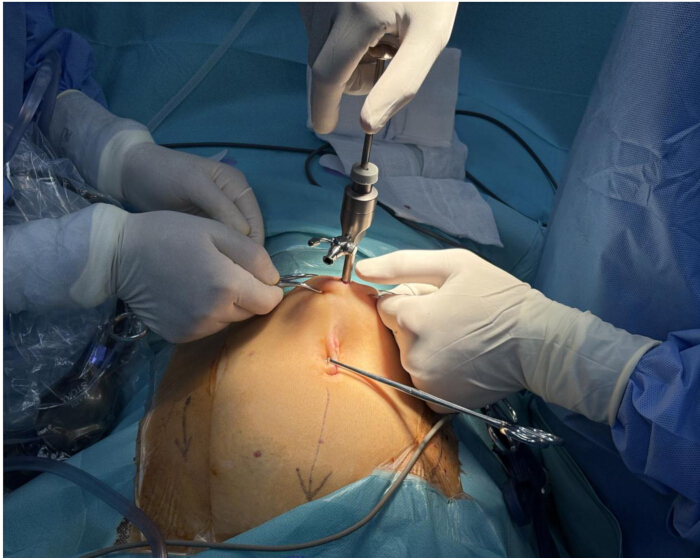
Figure 1: Laparoscopic placement of the primary 10 mm trocar midway between the umbilicus and the xiphoid process. Please click here to view a larger version of this figure.
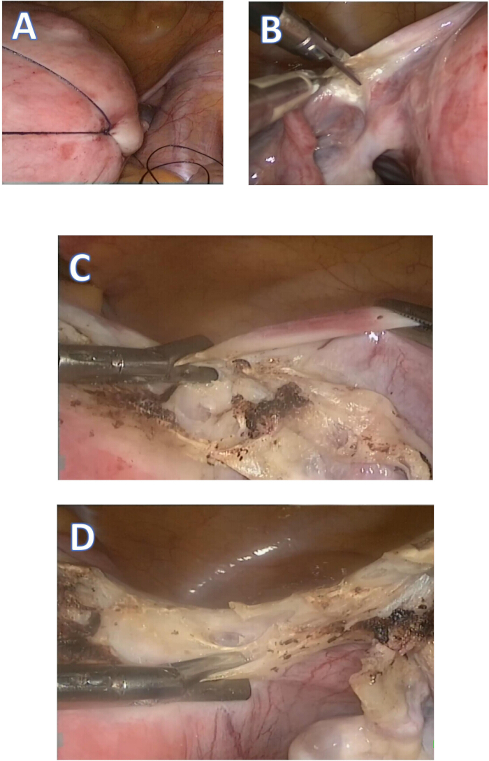
Figure 2: Coagulation and transection of the utero-ovarian, round, and broad ligaments. (A) Pull the suture to the left to expose the right anatomical structures. (B) Coagulation and transection of the left utero-ovarian ligament. (C) Transection of the right round ligament. (D) Transection of the right broad ligament Please click here to view a larger version of this figure.
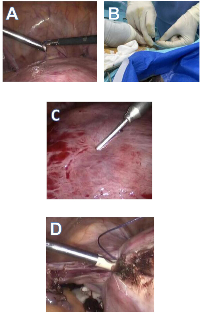
Figure 3: Surgical steps of laparoscopic extracorporeal knot tying method for uterine vessel occlusion. (A) Place the suture around the cervical isthmus. (B) Form an extracorporeal Roeder's knot. (C) Inject diluted vasopressin into the space between the uterine fibroid and myometrium. (D) Pull the Roeder's knot farther forward to occlude the uterine vessels completely. Please click here to view a larger version of this figure.
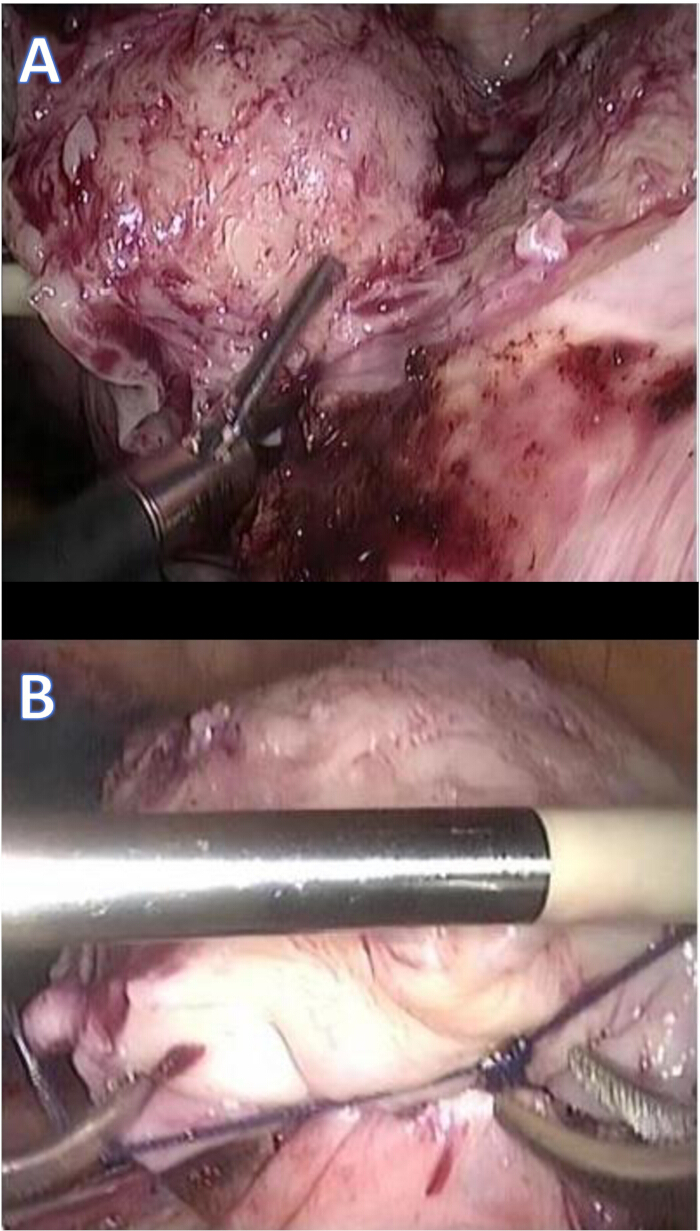
Figure 4: Sequential uterine debulking and reinforcement of the cervical cerclage. (A) Transect the uterine corpus 1.0-1.5 cm above the cerclage line using scissors. (B) Execute Loop ligation around the residual cervical stump to reinforce hemostasis with the extracorporeal Roeder's knot. Please click here to view a larger version of this figure.
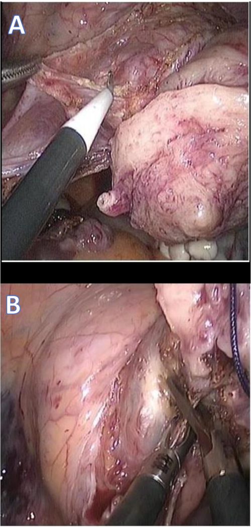
Figure 5: Surgical steps for revealing the uterine arteries and severing the uterine vessels. (A) Incise the vesico-uterine peritoneum with a unipolar hook to expose the uterine arteries. (B) Coagulate and divide the uterine vessels. Please click here to view a larger version of this figure.
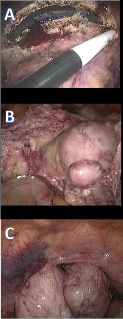
Figure 6: Colpotomy and specimen retrieval. (A) Perform a circumferential colpotomy at the vaginal fornix with a monopolar hook. (B) Morcellate the uterus completely with scissors. (C) Extract the specimen transvaginally under direct laparoscopic visualization. Please click here to view a larger version of this figure.
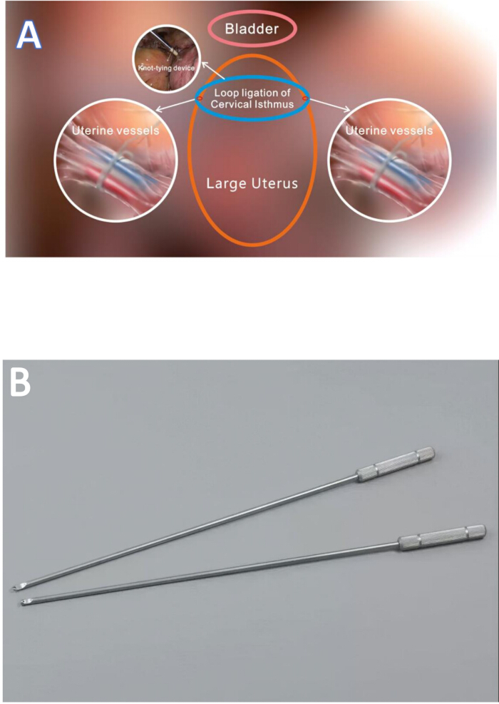
Figure 7: Schematic diagram of the approach and the photo of extracorporeal knot tying device. (A) A schematic diagram of LEKTUVOHCC. (B) The photo of the extracorporeal knot-tying device. Abbreviation: LEKTUVOHCC = Laparoscopic Extracorporeal Knot-Tying for Uterine Vessel Occlusion during Hysterectomy with Cervical Cerclage. Please click here to view a larger version of this figure.
| Characteristic | Basic parameters and surgical outcomes (n=31) |
| Age | 48 (44, 49) years |
| BMI | 21.25 (21.35, 25.42) |
| Total parity n(%) | |
| 0 | 6 (19.35%) |
| 1 | 11 (35,48%) |
| ≥2 | 14 (45.16%) |
| Vaginal parity n(%) | |
| 0 | 12 (38.71%) |
| 1 | 5 (16.13%) |
| ≥2 | 11 (35.48%) |
| Cesarean parity n(%) | |
| 0 | 22 (70.97%) |
| 1 | 6 (19,35%) |
| ≥2 | 3 (9.68%) |
| The median uterine size | 20 (18, 22) weeks of gestation |
| History of previous abdominal surgery n(%) | 10 (32.26%) |
| Anemia n(%) | 12 (38.71%) |
| Menometrorrhagia n(%) | 18/31 (58.06%) |
| Compressive symptoms n(%) | 11/31 (35.48%) |
| Intraoperative blood loss | 80 (50, 200) mL |
| Intraoperative transfusion rates n(%) | 1 (3.23%) |
| Decline in hemoglobin level | 5 (1, 15) g/dL |
| Operation time | 127 (100, 213) min |
| Postoperative hospital duration | 3 (2,5) days |
| Duration of follow-up | 12 (7, 19) months |
| Complications | None |
| Objective success rate | 100% |
| Subjective success rate | 96.77% |
Table 1: Baseline demographics and surgical outcomes in 31 patients undergoing LEKTUVOHCC. Variables include age, BMI, parity, uterine size (weeks' gestational equivalent), history of prior abdominal surgery, preoperative anemia, OT, IBL, perioperative hemoglobin decline, length of hospital stay, and objective and subjective success rates. Abbreviations: LEKTUVOHCC = Laparoscopic Extracorporeal Knot-Tying for Uterine Vessel Occlusion during Hysterectomy with Cervical; BMI = body mass index; OT = operative time; IBL = intraoperative blood loss.
| Surgical Approach | Number of Pateints |
| No conversion to laparotomy | 31 |
| Conversion to laparotomy | 0 |
| Objective Success Rate | 100% |
Table 2: Calculation of the objective success rate. Objective success is defined as completion of laparoscopic hysterectomy without conversion to open laparotomy; all 31 procedures were completed successfully, yielding a 100% success rate.
| PGI Scale | Number of Patients |
| PGI ≤ 2 | 30 |
| PGI >2 | 1 |
| Subjective Success Rate (PGI≤2) | 96.77% |
Table 3: Evaluation of subjective success rate. Subjective success rate was calculated by dividing the number of patients with the Patient global impression of improvement ≤ 2 by the total number of patients. Patient case 7 who had an IBL of 750 mL gave the answer "3. slightly better" on the PGI questionnaire survey; the remaining 30 patients were satisfied with the procedure and gave the answer "1. Very much better" or "2. Much better" on the PGI questionnaire survey. Abbreviation: PGI, Patient Global Impression.
| Surgical Outcomes | Group A (n = 15) | Group B (n = 16) | P Value |
| Operative time, min | 214 (163, 245) | 116 (102, 141) | .037 |
| Intraoperative blood loss, mL | 150 (100, 250) | 60 (30, 80) | .002 |
Table 4: Surgical outcomes regarding the learning curve in the procedure. Comparative analysis of early (cases 1-15, Group A) versus later (cases 16-31, Group B) LEKTUVOHCC cohorts. The learning curve effect is demonstrated by significant reductions in median OT (116 min vs 214 min) and median IBL (60 mL vs 150 mL) after the first 15 cases (p < .05). Abbreviation: LEKTUVOHCC, Laparoscopic Extracorporeal Knot-Tying for Uterine Vessel Occlusion during Hysterectomy with Cervical; OT = operative time; IBL = intraoperative blood loss.
Discussion
Many gynecologic surgeons regard uteri of ≥17-week size as a relative contraindication to total laparoscopic hysterectomy (TLH) due to the markedly distended, tortuous uterine vasculature, which complicates secure vascular control and elevates conversion rates9. The core technical challenge in these cases is achieving safe, reliable occlusion of the uterine arteries. Currently, bipolar electrocoagulation and titanium vascular clamps are the primary modalities for vessel control; however, both demand extensive dissection to skeletonize the uterine vessels and ureters, thereby prolonging operative time and increasing procedural complexity, especially when the vessel caliber is large10. Additionally, bipolar energy risks collateral thermal injury to the ureter, while mechanical clamping may result in vessel wall slippage and secondary bleeding upon clamp release. These drawbacks have limited the applicability of TLH in patients with substantially enlarged uteri.
We introduce Laparoscopic Extracorporeal Knot-Tying for Uterine Vessel Occlusion during Hysterectomy with Cervical Cerclage (LEKTUVOHCC), a novel minimally invasive technique designed for markedly enlarged uteri. Our findings confirmed that this technique is both feasible and effective even in uteri approximating a 28-week gestational size. The procedure began with the placement of an extracorporeal laparoscopic knot-pusher to fashion a Roeder's knot around the cervical isthmus, thereby achieving en bloc occlusion of the uterine arteries. Once vascular control is secured, the uterine corpus is transected supracervically to rapidly debulk the specimen. This staged approach restores deep pelvic exposure, optimizes visualization of critical structures, and enables precise dissection throughout the operation.
Since the novel surgery we designed involves placing the cervical ligature around the cervical isthmus, if there were cervical fibroids or uterine broad ligament fibroids, they may prevent the cervical ligature from being positioned accurately and tightened properly, increasing the risk of cervical ligature slipping off. To ensure the effectiveness of the ligature and guarantee the patient's safety, as well as avoid the risk of massive bleeding due to improper tightening or ligature slippage, we decided to exclude patients with these two types of special location fibroids.
Our data indicated that this technique achieved a median intraoperative blood loss (IBL) of just 80 mL) markedly lower than the averages reported by Purohit et al. and Tchartchian et al.11,12. The blood transfusion rate was 3.23% in our study, which was much lower than reported by Mamik et al. with a median IBL of 275 mL and a transfusion rate of 8.4%13. By substantially reducing IBL, this technique minimizes reliance on allogeneic transfusion, which carries well-documented risks of infectious and immunologic complications14 and helps to conserve scarce erythrocyte supplies, addressing broader public health concerns related to blood product shortage15.
The analysis of our institutional learning curve further demonstrated that both IBL and operative time (OT) declined significantly after approximately 15 cases. In the latter half of our cohort (cases 16-31), the median IBL and OT were significantly reduced compared to the first 15 cases (116 min vs 214 min, 60 mL vs 150 mL), indicating that proficiency in this technique for large uteri can be attained after a relatively short series of procedures. This contrasts with the 50-60 cases typically required to master conventional laparoscopic hysterectomy16 and the roughly 20 cases needed to achieve proficiency with robotic hysterectomy in large uteri17. Such a shortened learning curve not only expedites the safe dissemination of the technique but also enhances surgical efficiency, further mitigating the risks associated with prolonged OT and IBL.
The key features of this technique include the following:
The best site for trocar insertion
The procedure began by establishing a 10 mm optical trocar midway between the umbilicus and xiphoid process, which optimized cranial visualization and instrument triangulation. Three additional 5 mm ports provided direct access to the utero-ovarian and broad ligaments while preserving ergonomic hand positioning.
Division of the utero-ovarian, round, and broad ligaments
With the pneumoperitoneum in place, the utero-ovarian, round, and broad ligaments were sequentially coagulated and transected. This ligamentous division fully mobilized the adnexa, cleared the anterior and lateral uterine surfaces, and created a working space for subsequent cervical cerclage.
Intracervical Roeder's knot cerclage for en bloc uterine artery occlusion
An extracorporeal knot-pusher was used to position a size-1 braided polyglactin suture around the cervical isthmus, where a Roeder's knot is tied and tensioned at the 10 o'clock position. Once advanced fully, uniform blanching of the uterus body confirmed complete arterial occlusion. To ensure sustained hemostasis, two additional Roeder's knots were sequentially applied to the residual cervical stump. In our series, only one patient case 7, whose uterus measured approximately 28 weeks' gestational equivalent, had an IBL of 750 mL and required a blood transfusion. Investigation revealed that the initial knot had been under-tensioned, resulting in incomplete vessel closure. This experience led us to standardize knot-tension monitoring and refine our tying technique, after which no further transfusions were necessary.
Injection of diluted vasopressin into the uterine myometrium before loop ligation
Prior to cervical cerclage, diluted vasopressin was injected into the myometrium to induce vigorous myometrial contraction. This vasopressin-mediated constriction "milks" intramural blood into the systemic circulation, rendering the uterine wall pale and turgid. At the point of maximal blanching, a Roeder's knot was advanced around the cervical isthmus to effect en bloc occlusion of the uterine arteries, thereby preventing retrograde bleeding into the impending specimen. Conceptually analogous to an in situ autologous blood transfusion, this maneuver facilitated the return of retained uterine blood to the systemic circulation of the patient prior to the transection of the uterine corpus. Although a minor perioperative hemoglobin drop is anticipated, two patients in our series exhibited postoperative HGB gains, most likely reflecting the reinfused intramural volume. Compared with conventional TLH, this vasopressin-augmented protocol substantially reduced net IBL.
Supracervical debulking and bladder mobilization prior to uterine vessel division
To develop the surgical spaces, the uterine corpus was first removed, and bladder reflection was then performed by incising the vesico-uterine peritoneum, which fully exposes the uterine vessels for en bloc coagulation and division.
Conclusion
Laparoscopic Extracorporeal Knot-Tying for Uterine Vessel Occlusion during Hysterectomy with Cervical Cerclage (LEKTUVOHCC) offers a robust, minimally invasive solution for uteri equivalent to ≥17 weeks' gestation. By integrating extracorporeal Roeder's knot ligation at the cervical isthmus, achieving en bloc uterine artery occlusion, with staged supracervical debulking, this technique substantially minimizes IBL, obviates allogeneic blood transfusion, and streamlines operative workflow. This two-stage protocol preserves standard laparoscopic ergonomics, shortens postoperative recovery, and reduces healthcare resource utilization. Our series demonstrated that procedural proficiency and concomitant reductions in operative time (OT) and intraoperative blood loss (IBL) occurred after approximately 15 cases, reflecting a comparatively brief learning curve versus conventional laparoscopic or robotic hysterectomy for large uteri.
The novel procedure does not require any special equipment. All that is needed is a simple knot-tying device, which is very cheap and readily available, so it is worth promoting in remote areas or in grassroots hospitals.
Limitations of this single-center study include its relatively small cohort and nonrandomized design. To corroborate these promising results, we plan a multicenter, randomized controlled trial with an expanded sample size to rigorously evaluate the technique's safety profile, efficacy, and cost-effectiveness in the management of large benign uterine pathology.
Disclosures
The authors have no relevant financial or non-financial conflicts of interest to declare.
Acknowledgements
The authors wish to thank the surgical nursing team and research coordinators at The University of Hong Kong-Shenzhen Hospital for their invaluable assistance with patient management and data acquisition. This work received no external financial support.
Materials
| Name | Company | Catalog Number | Comments |
|---|---|---|---|
| Cefuroxime sodium for injection | YOUCARE , China | H20063758 | Prophylactic antibiotics |
| Extracorporeal knot typing device | MEDTRAINING , China | MT005 | Diameter x Length 5 x 330 mm |
| Enoxaparin sodium for injection | TECHDOW , China | H20056847 | Perioperative thrombosis prophylaxis |
| Trocars 5 mm | KARL STORZ, Germany | 30160X | Endoscopic equipment |
| Trocars 10 mm | KARL STORZ, Germany | 30160 H2 | Endoscopic equipment |
| VICRYL 1 | ETHICON, USA | VCP752D | Absorbable braided suture |
| VICRYL 2-0 | ETHICON, USA | VCP317H | Absorbable braided suture |
References
- Gendy, R., Walsh, C. A., Walsh, S. R., Karantanis, E. Vaginal hysterectomy versus total laparoscopic hysterectomy for benign disease: a meta-analysis of randomized controlled trials. Am J Obstet Gynecol. 204 (5), e388.e1-8(2011).
- Harvey, S. V., Pfeiffer, R. M., Landy, R., Wentzensen, N., Clarke, M. A. Trends and predictors of hysterectomy prevalence among women in the United States. Am J Obstet Gynecol. 227 (4), e611.e1-e611.e12 (2022).
- American College of Obstetricians and Gynecologists. Choosing the route of Obstetricians and Gynecologists. Choosing the route of hysterectomy for benign disease ACOG Committee Opinion No. 701. Obstet Gynecol. 129 (6), e155-e159 (2017).
- Lambat-Emery, S., Boulvain, M., Petignat, P., Dubuisson, J. Operative complications and outcomes comparing small and large uterine weight in case of laparoscopic hysterectomy for a benign indication. Front Surg. 8, 755781(2021).
- Wang, P., Uzianbaeva, L., Hughes, N., Mehdizadeh, A. Hysterectomy for large uterus by minimally invasive surgery (MIS). JSLS. 28 (3), e2024.00017(2024).
- Schmitt, J. J., et al. Prospective implementation and evaluation of a decision-tree algorithm for route of hysterectomy. Obstet Gynecol. 135 (4), 761-769 (2020).
- Uccella, S., et al. Laparoscopic versus open hysterectomy for benign disease in uteri weighing >1 kg: a retrospective analysis on 258 patients. J Minim Invasive Gynecol. 25 (1), 62-69 (2018).
- Sinha, R., et al. Total laparoscopic hysterectomy for large uterus. J Gynecol Endosc Surg. 1 (1), 34-39 (2009).
- Pfeuti, C. K., Zhi, L., Hoffman, M. K. Uterine weight and perioperative morbidity in robotic-assisted versus conventional laparoscopic hysterectomy. JSLS. 27 (4), e2023.00042(2023).
- Sezgin, B., Camuzcuoğlu, A., Küçük, M., Camuzcuoğlu, H. Bilateral uterine artery coagulation in laparoscopic hysterectomy for benign disease in uteri more than 1000 g. J Obstet Gynaecol Res. 46 (1), 133-139 (2020).
- Purohit, R., Sharma, J. G., Meher, D. A vagino-laparoscopic strategy for hysterectomy of large uteri to ease surgical challenges. Int J Womens Health. 15, 369-379 (2023).
- Tchartchian, G., Bojahr, B., Krentel, H., De Wilde, R. L. Evaluation of complications, conversion rate, malignancy rate, and surgeon's experience in laparoscopic assisted supracervical hysterectomy (LASH) of 1274 large uteri: a retrospective study. Acta Obstet Gynecol Scand. 101 (12), 1450-1457 (2022).
- Mamik, M. M., et al. Hysterectomy techniques and outcomes for benign large uteri: a systematic review. Obstet Gynecol. 144 (1), 40-52 (2024).
- Mladinov, D., Frank, S. M. Massive transfusion and severe blood shortages: establishing and implementing predictors of futility. Br J Anaesth. 128 (2), e71-e74 (2022).
- Pickett, C. M., et al. Surgical approach to hysterectomy for benign gynaecological disease. Cochrane Database Syst Rev. 8 (8), CD003677(2023).
- Sinha, R., Swarnasree, G., Rupa, B., Madhumathi, S. Laparoscopic hysterectomy for large uteri: outcomes and techniques. J Minim Access Surg. 15 (1), 8-13 (2019).
- Lee, J., Kim, S. Charting proficiency: the learning curve in robotic hysterectomy for large uteri exceeding 1000 g. J Clin Med. 13 (15), 4347(2024).
Reprints and Permissions
