The preparation of stable isotope-labeled vitamin A, necessary for the retinol isotope dilution technique, needs to conform to food safety standards. The protocol shows dosage preparation in vegetable oil at concentrations that will be suitable for the chosen delivery system and the applied analytical platform.
Method Article
Formulation of Vitamin A Tracer Doses for Administration to Human Subjects using Retinol Isotope Dilution Technique
In This Article
Summary
Abstract
The retinol isotope dilution (RID) technique relies on the administration of isotopically labeled vitamin A tracers to human subjects. Here, we present a method to guide research teams on how to prepare vitamin A stable isotope tracers for both research studies and population surveys. The article provides an overview of the preparation, sourcing of materials, equipment required, and procedures to ensure that the tracers are safe for human consumption. Vitamin A stable isotope tracers can be obtained commercially and are usually labeled with either deuterium [2H] or carbon-13 [13C] with >95% all-trans isomeric purity and >99% isotopic enrichment. The preparation of the vitamin A stable isotope tracers involves dissolving the tracer in edible vegetable oil through sonication and heat. Tracers need to be prepared at a concentration that is well within the volumetric limits of the delivery system. Finally, the tracer is aliquoted into sterile amber glass vials, and tracer concentrations are confirmed spectrophotometrically.
Introduction
To enable effective monitoring of intervention programs that aim to reduce vitamin A deficiency, the retinol isotope dilution (RID) method has been successfully applied to provide information on total body vitamin A stores (TBS) in various population groups1,2,3,4. While serum retinol5, serum retinol binding protein (RBP)6,7,8 and modified relative dose response (MRDR) tests9 are often employed to confirm vitamin A deficiency, RID is the only method that accurately quantifies total body vitamin A stores across the full spectrum -- from subclinical deficiency to toxicity3,10. By administering a known dose of labeled vitamin A and measuring its dilution in plasma after equilibration, the RID method provides precise estimates of TBS and liver vitamin A reserves11,12. Importantly, the RID method remains reliable even in populations with inflammation, making it indispensable for evaluating intervention efficacy and assessing toxicity risks in overlapping supplementation and fortification programs13,14.
To carry out the RID method, isotopically labeled vitamin A needs to be administered as an oral dose of retinyl ester, which in most cases is retinyl acetate dissolved in edible vegetable oil. Most laboratories will obtain isotope-labeled vitamin A from commercial suppliers1,3,7,15,16,17,18,19,20,21,22. Nearly all commercially available vitamin A isotope tracers are labeled using stable isotopes of carbon-13 [13C] or deuterium [2H] (Table 1). The choice of the applied isotope dose depends on the analytical instrument used to determine the serum or plasma concentration of mass-labeled retinol. While gas chromatography-mass spectrometry (GC-MS) and liquid chromatography-tandem mass spectrometry (LC-MS/MS) can analyze both [13C] and [2H]22,23,24,25,26,27, gas chromatography-combustion-isotope ratio mass spectrometry (GC-C-IRMS) can only analyze [13C] labeled retinol tracers4,25,28. The limits of detection of the chosen analytical instrument, the target group (children or adults), and the length of the investigation will determine the size of the dose that needs to be applied3,25,29. To date, applied isotope doses have ranged from 0.3 - 4.3 mg retinol equivalent (RE; equivalent to 1 - 15 µmol RE) in children3,4,30 and from 0.6 - 10.0 mg RE (equivalent to 2 - 35 µmol RE) in adults2,3,31, with more recent studies applying lower doses ranging between 0.3 -0.6 mg RE (1 - 3 µmol RE) for children and 0.6 - 2.0 mg RE (2 - 7 µmol RE) in adults. Although fears were raised that a high mass of vitamin A in the dose may perturb the underlying kinetics of vitamin A, a recent analysis using compartmental modeling confirmed that isotope doses as high as 7 µmol RE for children or 14 µmol RE for adults should not affect the accuracy of TBS predictions29. Here, we will demonstrate how to prepare an isotope-labeled tracer for oral application for the RID method, with consideration of food safety concerns and obtaining accurate dose concentrations. The isotope dose preparation is vital for the RID method.
Protocol
Study 1 was approved by the National Research Ethics Service, North-East Sunderland Committee (REC 09/H0904/20) before registration with the UK Clinical Research Network (UKCRN: 7413). Study 2 was approved by the Research Ethics Board of the University of the Philippines, Manila (IRB ID: UPMREB 2016-282-01), the Institutional Review Board at UC, Davis (IRB ID: 903681-2), and registered at ClinicalTrials.gov (ID: NCT03030339). Study 2 was part of the GloVitAS project funded by the Bill and Melinda Gates Foundation (OPP1115464: Assessing the risk of vitamin A toxicity due to large-scale food fortification and other interventions). Georg Lietz was the lead investigator for the GloVitAS project and oversaw the implementation of the field study. An overview of the study protocol is given in Figure 1.
1. Inclusion and exclusion criteria
- For study 1, the given criteria were: Inclusion criteria: Include healthy male and female volunteers with an age range of 18-45 years. Exclusion criteria: Exclude volunteers with pregnancy, smoking, high blood pressure, diabetes, BMI >30, liver/kidney/gastrointestinal disease, lipid metabolic disorders, and consumption of multivitamins (containing vitamins A, C, E) or β-carotene supplements 3 months prior to the study start.
- For study 2, the given criteria were: Inclusion criteria: Include 12-18-month-old children who remained in the study area for the duration of the study. Exclusion criteria: Exclude if children did not receive high-dose vitamin A supplementation and lived in areas that were not exposed to vitamin A-fortified foods in local markets.
2. Determining the total amount of stable isotope tracer for RID
- Decide the amount of stable isotope tracer each subject should receive taking into account isomeric purity. It is recommended to use vitamin A stable isotopes with >95% all-trans purity since biological activity and metabolic pathways differ significantly between geometric isomers. Calculate the dose level based on retinol equivalents (RE). An example of how this is calculated is given in Table 2.
- Multiply the dose level by the total number of subjects. Base total number of subjects on power calculations for required sample size but increase based on expected attrition (see Table 2 for example). Increase total amount of tracer needed by taking pipetting losses of up to 20% into account.
NOTE: As a rule, the isotope preparation needs to include an excess, as it is not possible to recover all the material from the vial. Losses will further occur due to the viscosity of the oil that remains on the outside of the pipette tip. To obtain accurate volumes of the dose during delivery, excess oil on the outside of the pipette tip needs to be wiped off. Operators can minimize pipetting losses if the pipette tip is not dipped too deep into the oil.
3. Determining the amount of vegetable oil needed to dissolve the stable isotope tracer
- Decide which vegetable oil to use and the volume of the dose per subject (Table 2). Determine the amount of added oil by the delivery system used during dosing of subjects.
NOTE: We use sunflower oil due to the high vitamin E content, which aids the stability of the vitamin A preparation32,33. However, other vegetable oils have been employed by other groups to satisfy local dietary norms. Since the RID relies on accurate delivery of stable isotope-labeled tracers, it is recommended to use a positive displacement pipette for dose application prepared in edible oil. Most recent studies have used volumes up to 250 µL, particularly if the dose is administered to children1,7,23. - Calculate the dose concentration of the stable isotope tracer in [mg/mL] vegetable oil. Determine the total volume of vegetable oil needed. Calculate the weight of oil required, based on the calculated volume and the relative density of the vegetable oil (see Table 2 for an example).
NOTE: For accuracy, it is recommended to weigh the oil for stable isotope tracer preparation. Once the total volume of required oil has been calculated, the stable isotope-labeled tracer can be ordered. It is important to request the isotope tracer to be shipped in a bottle of sufficient volume that will enable the research team to weigh out the vegetable oil directly into the bottle containing the isotope-labeled tracer.
4. Preparation of stable isotope-labeled tracer in dedicated areas for food preparation
- Carefully open the lid of the amber bottle containing the pre-determined total amount of the stable isotope tracer and place it on an analytical balance, which is then tared to zero.
- Add the oil dropwise until the desired weight is achieved (with an accuracy of ± 0.1 g) using a sterile 10 mL syringe.
NOTE: Since some retinyl-acetate crystals may be trapped inside the cap of the amber bottle, the bottle needs to be opened very carefully. After adding the vegetable oil to the amber bottle, the cap is carefully placed back on the bottle, which is then shaken carefully by hand to resuspend any retinyl acetate crystals that remain inside the cap. - Place the bottle in a sonicating water bath with ultrasonic power: 14 W/L and operating frequency: 44 kHz. Fill with water to above the level of oil in the bottle, but below the lid level, to prevent water from inadvertently entering the preparation.
- Sonicate the bottle for 15 min before careful mixing by hand, followed by sonicating for a further 15 min. Care must be taken that the temperature of the sonicating water bath does not exceed 50 °C.
NOTE: Using sunflower oil for preparation, we have found that isomerization to cis retinoids does not occur when the dose is exposed to 50 °C for 30 min (see Figure 2). However, preparations should be analyzed by HPLC to assess the extent of thermal isomerization. - Place the bottle in a heated water bath at 50 °C for 30 min to completely dissolve the microcrystals.
- Take a 20 µL sample of oil to check for the presence of microcrystals under a light microscope at 200x magnification in a laboratory. If microcrystals are present, repeat the sonicating and heating procedure.
- Once complete dissolution is achieved, aliquot the total oil preparation into 2 - 4 mL sterile glass amber vials. Use positive displacement pipettes for this procedure to avoid cross-contamination.
NOTE: The volume of stable isotope tracer added to the glass amber vials can be guided by the daily recruitment rate, e.g., if 5 subjects are recruited per day and the dose volume is 150 µL, the vial needs to contain enough volume to account for 5 doses plus pipetting losses: 5 x 150 µL x 1.2 = 900 µL. - Determine stable isotope concentration in triplicate. Reserve 200 µL of pure vegetable oil (used for preparation of a spectrophotometric blank) and 200 - 350 µL of the stable isotope dose in separate 2 mL amber vials during the preparation of the stable isotope labeled tracer.
- If the dose concentration is ≤ 0.5 mg RE, reserve 350 µL of the stable isotope in a separate 2 mL amber vial. For dose concentrations > 0.5 mg RE, reserve 200 µL of the stable isotope dose in a separate 2 mL amber vial.
- Blanket the vials with an inert gas (Ar) to delay potential oxidation of the isotope-labeled tracer, close tightly, and store in a cryobox labeled with isotope concentration, dose volume (determined afterwards), preparation date, and project name. Store the cryobox in a -20 °C food freezer until shipment. Under these conditions, the tracer dose is stable for at least 2 years.
5. Determination of stable isotope tracer concentration in the laboratory
- Dilute the stable isotope tracer in 3 volumetric flasks with n-hexane. Fill volumetric flasks with n-hexane to 1 cm below the calibration line before adding the tracer dose.
- Determine the dilution of the stable isotope tracer by the tracer dose concentration. If the dose concentration of the stable isotope tracer is ≤ 0.5 mg RE, dilute 100 µL of the stable isotope dose in a 50 mL volumetric flask with n-hexane. If the dose concentration is between 0.5 - 2 mg RE, then 50 µL of the stable isotope dose in a 50 mL volumetric flask with n-hexane. If the dose concentration is >2 mg RE, then 50 µL of the stable isotope dose in a 100 mL volumetric flask with n-hexane.
- Directly dispense 50 or 100 µL of oil into the hexane and the flask made to 50 or 100 mL volume with additional hexane using a glass Pasteur pipette. Invert the flask 10x to mix the oil with the hexane.
CAUTION: Hexane should be handled in a fume cabinet, wearing appropriate PPE. Waste hexane is collected in a dedicated container for specialized disposal by an approved contractor.
- Prepare the spectrophotometric blank flask by pipetting the same volume of vegetable oil as the stable isotope tracer (50 or 100 µL) and dispensing it into hexane. Ensure that the applied dilution (50 or 100 mL) is the same as for the stable isotope tracer. Invert the flask 10x to mix the oil with the hexane.
NOTE: For pipetting either stable isotope tracer or pure oil, a 100 µL positive displacement pipette is used. To ensure accurate volumes are pipetted, it is critical that the tip is wiped downwards using lint-free paper to remove residual tracer/oil from the outside of the tip. Care must be taken when wiping the direct replacement pipette tip so that the paper does not come into contact with the tip opening, as oil will be drawn from within the tip. - Measure the stable isotope tracer concentrations in a quartz cuvette using a spectrophotometer set to a wavelength of 325 nm, using the vegetable oil in hexane as the spectrophotometric blank (see Table 3 for an example).
- Start measurements using the vegetable oil in hexane as the spectrophotometric blank. Clean the quartz UV cuvette between each measurement by rinsing it 3x with n-hexane. After all samples have been measured, carefully clean the cuvette using pure n-hexane.
- Determine the concentration of the stable isotope tracer using the average AU readings and apply the isotope-specific molar extinction coefficient using the Beer-Lambert law. From this, calculate the volume of the prepared stable isotope tracer needed per dose (see example in representative results below and in Table 3).
Results
Total isotope tracer dose size calculation
For a study of a sample size of 100 subjects, the total amount of [13C10]retinyl acetate was calculated to identify the quantity of [13C10]retinyl acetate needed to be synthesized (Table 2). The amount is based on power calculations for required samples size (n = 100), expected attrition (n=15; 15% attrition), pipetting losses (20%) and [13C10]retinyl acetate concentration per dose (0.4 mg RE; based on the sensitivity of the applied mass spectrometer, the length of the experiment and the age group of the subjects). The obtained total amount of RE needed (55.2 mg RE) is then converted into the required weight for [13C10]retinyl acetate (63 mg).
To calculate the volume of sunflower oil needed to dissolve [13C10]retinyl acetate at the desired concentration, we first set the dose delivery volume to 150 µL/dose (0.15 mL/dose). Following this, the concentration of [13C10]retinyl acetate in 1 mL of sunflower oil is calculated (Table 2). Dividing the required weight for [13C10]retinyl acetate (63 mg) by the required dose concentration gives the total volume of sunflower oil needed. Finally, the weight of sunflower oil is determined by using the relative density of sunflower oil.
The accurate target dose concentration of [13C10]retinyl acetate in oil was confirmed by measuring the stable isotope tracer concentrations using a spectrophotometer based on the Beer-Lambert law (Table 3). Taking the dilution of the stable isotope tracer during the measurement into consideration (dilution factor of 500) gives a concentration of 2.58 µmol/µL [13C10]retinyl acetate in oil. The final volume of the prepared stable isotope tracer needed for a 0.4 mg RE dose was therefore determined to be 155 µL (Table 3).
Checking for potential isomerization of the tracer dose
The isomeric purity of the obtained tracer dose was determined by checking for cis isomerization using High Performance Liquid Chromatography (HPLC; Figure 2). To perform this, a sample from the diluted tracer solution from step 5.1.2 was dried down under nitrogen gas and the residue redissolved into 100% ethanol for injection. HPLC analysis of 3 repeat samples was carried out using the method of Liu et al.34 to check for any cis-isomerization (see Figure 2). Purity of the tracer dose should be monitored throughout the study to make sure that the dose is not degrading.
Effect of tracer dose concentrations on fraction of dose over time
Application of different dose levels will lead to varying fraction of the ingested dose over time, which could affect the ability of the analytical platform to detect [13C10]retinol in all subjects at required time points. Applying a dose of either 1 mg or 0.4 mg of [13C10]retinyl acetate in oil in adults (Figure 3A) or children (Figure 3B) indicated higher fraction of dose after administering 1mg compared to 0.4mg of [13C10]retinyl acetate and confirmed the ability of the applied LC-MS/MS to detect [13C10]retinol in subjects up to 28 days post dosing. This corroborates that the information on the fraction of dose at varying time points is critical to decide if the applied dose level is high enough to obtain a signal above the level of detection for the applied analytical platform, and that this information should ideally be obtained during a pilot study with representative samples.
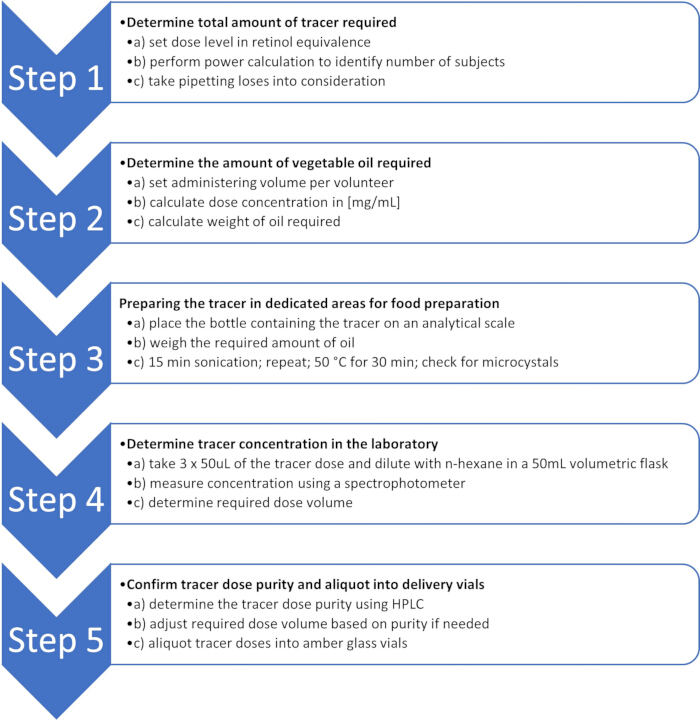
Figure 1: Flow chart for preparation of vitamin A tracer doses. Key steps to carry out stable isotope dose preparation including determination of required amounts, concentration of obtained tracer dose and confirmation of dose purity. Please click here to view a larger version of this figure.
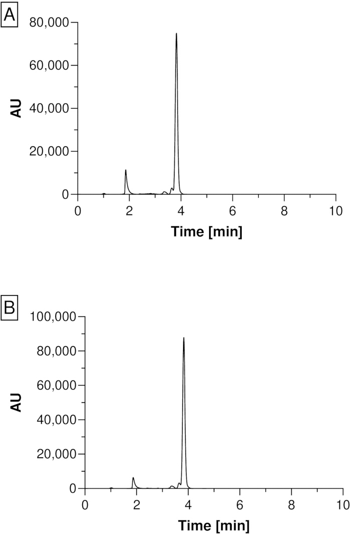
Figure 2: HPLC chromatogram of the tracer dose to investigate the isomerization of retinyl acetate. Potential isomerization of [13C10]retinyl acetate after (A) sonication and (B) heating to 50 °C for 30 min each was assessed using HPLC analysis of the isotope dose after each step. Analysis was carried out on an HPLC with diode array detection using the method of Liu et al.34. Please click here to view a larger version of this figure.
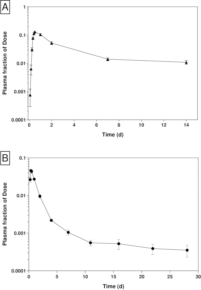
Figure 3: Observed means of the plasma retinol fraction of dose over time after ingestion of [13C10]retinyl acetate. Fraction of dose from (A) 45 UK adults16 and (B) 112 Filipino children1 who were administered either a 1 mg or 0.4 mg [13C10]retinyl acetate dose, respectively. Fraction of dose = {plasma [13C]retinol concentration (µmol/L) X estimated plasma volume (L) / [13C]dose (µmol). Data displayed as geometric mean ± SEM. Please click here to view a larger version of this figure.
| Stable isotope | Supplier/Manufacturer |
| [D4]-retinyl acetate | Buchem BV/ReseaChem GmbH/ Cambridge Isotope Laboratory |
| [D6]-retinyl acetate | Buchem BV/ReseaChem GmbH/ Cambridge Isotope Laboratory |
| [D8]-retinyl acetate | Buchem BV/ReseaChem GmbH/ Cambridge Isotope Laboratory |
| [13C3]-retinyl acetate | Buchem BV/ReseaChem GmbH |
| [13C4]-retinyl acetate | Buchem BV/ReseaChem GmbH/ Cambridge Isotope Laboratory |
| [13C8]-retinyl acetate | Buchem BV/ReseaChem GmbH/ Cambridge Isotope Laboratory |
| [13C10]-retinyl acetate | Buchem BV/ReseaChem GmbH/ Cambridge Isotope Laboratory |
Table 1: Commercially available stable vitamin A isotope tracers. List of 13C and 2H labelled retinyl acetate tracers from different suppliers.
| Parameters | ||
| Sample size [n]: | 100 | |
| Attrition [n] (%): | 15 (15%) | |
| Pipetting losses: | 20% | |
| MW [13C10]retinyl acetate: | 338.5 µg/µmol | |
| MW [13C10]retinol: | 296.46 µg/µmol | |
| Dose size: | 0.4 mg RE of [13C10]retinyl acetate per volunteer | |
| Calculation | ||
| Total amount of RE required: | = dose size in mg RE x sample size x attrition x pipetting losses | |
| = 0.4 mg RE x 100 x 1.15 x 1.2 | ||
| = 55.2 mg RE | ||
| Total amount of [13C10]retinyl acetate required: | = mg RE x MW [13C10]retinyl acetate / MW [13C10]retinol | |
| = 55.2 mg RE x 338.5 µg/µmol / 296.46 µg/µmol | ||
| = 63.0 mg [13C10]retinyl acetate | ||
| Dose concentration of [13C10]retinyl acetate in oil: | [13C10]retinyl acetate [mg/ml] = | |
| (mg RE x MW [13C10]retinyl acetate / MW [13C10]retinol) / 0.15 ml | ||
| = (0.4 mg RE x 338.5 µg/µmol / 296.46 µg/µmol) / 0.15 ml | ||
| = 3.04 mg/ml | ||
| Total volume of sunflower oil required: | = Total [13C10]retinyl acetate weight / dose concentration in oil | |
| = 63.0 mg / 3.04 mg/ml | ||
| = 20.7 ml oil | ||
| Weight of sunflower oil needed: | = Total volume of sunflower oil x relative density sunflower oil | |
| = 20.7 ml x 0.92 g/ml | ||
| = 19.0 g sunflower oil | ||
Table 2: Example of stable isotope tracer dose size calculations. Indication of parameters and calculations to determine tracer dose and vegetable oil amounts.
| Parameters | |
| Molar extinction coefficient (E1%1cm) of [13C10]retinyl acetate in hexane: | 52718 L mol-1 cm-1 x 1 cm |
| Dilution factor: | 500 (100µL of oil preparation in 50mL of hexane) |
| Measured average absorbance: | 0.917 |
| MW [13C10]retinol: | 296.46 µg/µmol |
| Calculation | |
| Concentration (mol/L) of [13C10]retinyl acetate in hexane: | Concentration (mol/L) = Absorption / E1%1cm x Length of cuvette (1cm) |
| = 0.917 / 52718 L mol-1 cm-1 x 1 cm = 1.739 x 10⁻⁵ mol/L | |
| Converting concentration into µmol/µL: | Concentration (mol/L) = Concentration (µmol/µL) |
| 1.739 x 10⁻⁵ mol/L is equivalent to 1.739 x 10⁻⁵ µmol/µL | |
| Correcting for dilution: | 1.739 x 10⁻⁵ µmol/µL x 500 = 8.697 x 10⁻³ µmol/µL in oil |
| Weight per µL (µg/µL): | = [13C10]retinyl acetate µmol/µL in oil x MW [13C10]retinol |
| = 8.697 x 10⁻³ µmol/µL x 296.45 µg/µmol = 2.58 µg/µL | |
| Volume per dose: | = Volume (µL/ 400 µg dose) = 400µg / 2.58 µg/µL = 155 µL |
Table 3: Determination of stable isotope tracer dose concentration. Required parameters and calculations to determine tracer dose concentrations and required volumes for oral dosing.
Discussion
Stable isotope tracers used in human studies are generally considered to be safe, with no reported adverse biological or physiological effects at the applied concentrations25,35. However, since the stable isotope tracer needs to be orally administered, it is essential that the tracer dose conforms to food safety standards. This can be achieved by demanding that suppliers and manufacturers of stable isotope-labeled vitamin A carry out food safety tests on the supplied isotopes7,20,24. Likewise, it is imperative that isotope preparation and storage are only carried out in dedicated areas for food preparation. Furthermore, all equipment used for preparing stable isotopes must be dedicated to isotope preparation only and kept in areas for food preparation.
Retinyl acetate labelled isotopes have previously been dissolved using ethanol, then dried under nitrogen gas before being dissolved into vegetable oil36. Here, we present a method that allows the dissolution of the isotope-labeled tracer into vegetable oil using a combination of sonication and heat. Although this method is easier to perform in a food preparative area, care must be taken when applying heat to dissolve the tracer in vegetable oil. All-trans-retinol is isomerized to 13-cis-retinol during heat treatment, which accelerates with higher temperatures37. Isomerization of retinol further increases through exposure to light32. Moreover, the stability of retinol is influenced by the lipid oxidation property of the vegetable oil32,38. Since α-tocopherol content in vegetable oil has a positive impact on vitamin A retention32, and sunflower oil has one of the highest contents of α-tocopherol33, we opted to use sunflower oil in preparing the tracer doses. The decision to apply up to 50°C heat to dissolve the tracer was based on the application of heat to dissolve retinol crystals in cosmetic products without significant loss39. Although we have not seen significant degradation of retinyl acetate under the described protocol, it is vital to check for cis isomerization of the tracer dose right after the heat treatment (Figure 2). We further recommend that the stability of the tracer dose should be monitored throughout the study to confirm the applied tracer dose concentration.
The tracer must be prepared at a concentration well within the volumetric limits of the delivery system used to administer the required dose. Since the RID relies on accurate delivery of stable isotope-labeled tracers, it is recommended to use a positive displacement pipette for dose application. Although dose sizes of up to 1 mL have been applied in the past to adults16,20,40, it is easier to apply volumes between 100 - 250 µL, as these volumes have been shown to be well tolerated by infants and children1. Alternatively, doses can also be administered in capsule form17, but it is essential to select capsules specifically designed to prevent oil leakage and to ensure acceptability within the study population. Finally, applied dose levels depend on the age group of the target population29, the ability of the applied analytical platform to detect low levels3 and the total body stores of the target population, with a lower plasma fraction of dose at higher total body stores1.
As indicated in the example above, [13C10]retinyl acetate at a concentration of 2.58 µg/µL in sunflower oil will provide a dose of 0.4 mg RE tracer in 155 µL of sunflower oil, which can be delivered accurately using a 250 µL positive displacement pipette. To obtain concentrations that are within the volumetric limits of the delivery system, it is critical that the vegetable oil chosen to solubilize the tracer is accurately weighed using an analytical balance (accurate to 0.1 g) into the bottle containing the stable isotope tracer. For example, if a total of 63 mg [13C10]retinyl acetate is to be prepared at a concentration of 3.04 mg/mL in sunflower oil, and the specific gravity of sunflower oil is 0.92 g/mL at 25 ◦C, then 19.0 g (20.7 mL x 0.92 g/mL) of oil needs to be weighed into the bottle containing the tracer. Although the final concentration of the stable isotope tracer will be confirmed using a spectrophotometer reading, accurate weighing of the isotope tracer and the applied oil will guarantee that the obtained concentrations are close to the chosen delivery volume for each dose.
It is advisable to aliquot oil preparations based on the anticipated daily requirement to ensure that all doses in a vial are used within 2 weeks after opening the vial. It is best to keep the oil preparations on ice packs in the field during use and refreeze them as soon as possible after the dosing is completed. Although the oil preparations are not affected by repeated freeze and thaw cycles, they need to always be protected from light and frozen when not in use.
In summary, this protocol details the precise preparation and quality control of tracer doses for the RID method, ensuring accurate dosing, stability, and safety. The method calculates tracer requirements, optimizes solubilization in vegetable oil, and verifies purity and concentration using spectrophotometry and HPLC. Rigorous handling and storage guidelines guarantee reliable tracer integrity for accurate isotope-based assessments.
Disclosures
The authors have nothing to declare.
Acknowledgements
Support for this work was provided by the Bill & Melinda Gates Foundation (Project Number: OPP1115464) and the International Atomic Energy Agency (IAEA) in Vienna (Project Number E4.30.30).
Materials
| Name | Company | Catalog Number | Comments |
|---|---|---|---|
| 2ml amber glass vials for aliquoting of doses | Fisher | 11573690 | Depending on the sample size of the study and the amount of volunteers that are being dosed on a daily basis, it may be more efficient to use larger, e.g. 4ml, amber vials |
| 100ml volumetric flasks | Fisher | 11323644 | Pyrex Borosilicate Glass Class A Certified Volumetric Flask |
| 50ml volumetric flasks | Fisher | 11313644 | Pyrex Borosilicate Glass Class A Certified Volumetric Flask |
| Analytical weighing scales | Fisher | 13592970 | |
| Capillary pistons (pipette tips) CP100 10x96 TIPACK | Gilson | FD148314 | |
| Capillary pistons CP1000ST 2x91 | Gilson | F148180 | |
| Capillary pistons CP250ST 6x96 | Gilson | F148714 | |
| Cardboard cryobox | Starlab | A9623-8181 | |
| Hellma cuvette UV quartz | SLS | CEL1600 | |
| Hexane 95% n-Hexane for HPLC CertiFied HPLC | Fisher | H/0406/17 | |
| Lids for 2ml amber glass vials | Fisher | 11573542 | |
| Microman E M1000E Pipette (positive displacement) | Gilson | FD10006 | |
| Microman E M100E Pipette (positive displacement) | Gilson | FD10004 | |
| Microman E M250E Pipette (positive displacement) | Gilson | FD10005 | |
| Microscope | Nikon | Eclipse E400 | |
| Microscope glass slides | Sigma | 41122601 | |
| Sonicating water bath | VWR | USC100T | |
| Spectrophotometer | Thermo Fisher Scientific | A51119600C | |
| Stable isotope labelled retinyl acetate tracer | Buchem BV | N/A | A range of stable isotopes can be used for the RID method. A list of different commercially available isotopes are given in Table 1. |
| Unstirred water bath | SLS | SLS1176 | |
| Vegetable oil | Any grocery store | N/A | It is important to use fresh vegetable oil with sufficient shelf life for preparing isotope doses. |
| Volac unplugged glass pasteur pipette 150mm | SLS | PIP4150 |
References
- Ford, J. L., et al. Use of Model-Based Compartmental Analysis and a Super-Child Design to Study Whole-Body Retinol Kinetics and Vitamin A Total Body Stores in Children from 3 Lower-Income Countries. J Nutr. 150 (2), 411-418 (2020).
- Haskell, M. J., Jamil, K. M., Peerson, J. M., Wahed, M. A., Brown, K. H. The paired deuterated retinol dilution technique can be used to estimate the daily vitamin A intake required to maintain a targeted whole body vitamin A pool size in men. J Nutr. 141 (3), 428-432 (2011).
- Lietz, G., et al. Current Capabilities and Limitations of Stable Isotope Techniques and Applied Mathematical Equations in Determining Whole-Body Vitamin A Status. Food Nutr Bull. 37, S87-S103 (2016).
- Pinkaew, S., et al. Triple-Fortified Rice Containing Vitamin A Reduced Marginal Vitamin A Deficiency and Increased Vitamin A Liver Stores in School-Aged Thai Children. J Nutr. 144 (4), 519-524 (2014).
- VMNIS: Serum retinol concentrations for determining the prevalence of vitamin A deficiency in populations. , World Health Organisation. https://www.who.int/publications/i/item/WHO-NMH-NHD-MNM-11.3 (2011).
- Engle-Stone, R., et al. Plasma retinol-binding protein predicts plasma retinol concentration in both infected and uninfected Cameroonian women and children. J Nutr. 141 (12), 2233-2241 (2011).
- Engle-Stone, R., et al. Filipino Children with High Usual Vitamin A Intakes and Exposure to Multiple Sources of Vitamin A Have Elevated Total Body Stores of Vitamin A But Do Not Show Clear Evidence of Vitamin A Toxicity. Curr Dev Nutr. 6 (8), nzac115(2022).
- Larson, L. M., et al. Approaches to Assess Vitamin A Status in Settings of Inflammation: Biomarkers Reflecting Inflammation and Nutritional Determinants of Anemia (BRINDA) Project. Nutrients. 10 (8), (2018).
- Sheftel, J., Tanumihardjo, S. A. Systematic Review and Meta-Analysis of the Relative Dose-Response Tests to Assess Vitamin A Status. Adv Nutr. 12 (3), 904-941 (2021).
- Tanumihardjo, S. A., et al. Biomarkers of Nutrition for Development (BOND)-Vitamin A Review. J Nutr. 146 (9), 1816-1848 (2016).
- Furr, H. C., et al. Vitamin A concentrations in liver determined by isotope dilution assay with tetradeuterated vitamin A and by biopsy in generally healthy adult humans. Am J Clin Nutr. 49 (4), 713-716 (1989).
- Haskell, M. J., et al. Assessment of vitamin A status by the deuterated-retinol-dilution technique and comparison with hepatic vitamin A concentration in Bangladeshi surgical patients. Am J Clin Nutr. 66 (1), 67-74 (1997).
- Green, M. H., Ford, J. L., Green, J. B. Development of a Compartmental Model to Investigate the Influence of Inflammation on Predictions of Vitamin A Total Body Stores by Retinol Isotope Dilution in Theoretical Humans. J Nutr. 151 (3), 731-741 (2021).
- Suri, D. J., et al. Association between Biomarkers of Inflammation and Total Liver Vitamin A Reserves Estimated by (13)C-Retinol Isotope Dilution among Preschool Children in 5 African Countries. J Nutr. 153 (13), 622-635 (2023).
- Furr, H. C., et al. Stable isotope dilution techniques for assessing vitamin A status and bioefficacy of provitamin A carotenoids in humans. Public Health Nutr. 8 (6), 596-607 (2005).
- Green, M. H., et al. A Retinol Isotope Dilution Equation Predicts Both Group and Individual Total Body Vitamin A Stores in Adults Based on Data from an Early Postdosing Blood Sample. J Nutr. 146 (10), 2137-2142 (2016).
- Green, M. H., et al. Use of Population-Based Compartmental Modeling and Retinol Isotope Dilution to Study Vitamin A Kinetics and Total Body Stores among Ghanaian Women of Reproductive Age. Curr Dev Nutr. 8 (11), 104484(2024).
- Haskell, M. J., et al. Population-based plasma kinetics of an oral dose of [2H4]retinyl acetate among preschool-aged, Peruvian children. Am J Clin Nutr. 77 (3), 681-686 (2003).
- Lopez-Teros, V., et al. Use of a "Super-child'' Approach to Assess the Vitamin A Equivalence of Moringa oleifera Leaves, Develop a Compartmental Model for Vitamin A Kinetics, and Estimate Vitamin A Total Body Stores in Young Mexican Children. J Nutr. 147 (12), 2356-2363 (2017).
- Oxley, A., et al. An LC/MS/MS method for stable isotope dilution studies of beta-carotene bioavailability, bioconversion, and vitamin A status in humans. J Lipid Res. 55 (2), 319-328 (2014).
- Ribaya-Mercado, J. D., et al. Quantitative assessment of total body stores of vitamin A in adults with the use of a 3-d deuterated-retinol-dilution procedure. Am J Clin Nutr. 77 (3), 694-699 (2003).
- Tang, G. W., Qin, J., Hao, L. Y., Yin, S. A., Russell, R. M. Use of a short-term isotope-dilution method for determining the vitamin A status of children. Am J Clin Nutr. 76 (2), 413-418 (2002).
- Oxley, A., et al. Determination of Vitamin A Total Body Stores in Children from Dried Serum Spots: Application in a Low- and Middle-Income Country Community Setting. J Nutr. 151 (5), 1341-1346 (2021).
- Oxley, A., Lietz, G. Use of stable isotopes to study bioconversion and bioefficacy of provitamin A carotenoids. Methods Enzymol. 670, 399-422 (2022).
- Preston, T. Existing and emerging technologies for measuring stable isotope labelled retinol in biological samples: isotope dilution analysis of body retinol stores. Int J Vitam Nutr Res. 84 (Suppl 1), 30-39 (2014).
- Tang, G. W., Qin, J., Dolnikowski, G. Deuterium enrichment of retinol in humans determined by gas chromatography electron capture negative chemical ionization mass spectrometry. J Nutr Biochem. 9 (7), 408-414 (1998).
- Wang, J., et al. Vitamin A equivalence of spirulina beta-carotene in Chinese adults as assessed by using a stable-isotope reference method. Am J Clin Nutr. 87 (6), 1730-1737 (2008).
- Gannon, B., et al. Biofortified orange maize is as efficacious as a vitamin A supplement in Zambian children even in the presence of high liver reserves of vitamin A: a community-based, randomized placebo-controlled trial. Am J Clin Nutr. 100 (6), 1541-1550 (2014).
- Green, M. H., Lopez-Teros, V., Green, J. B. Does the Amount of Stable Isotope Dose Influence Retinol Kinetic Responses and Predictions of Vitamin A Total Body Stores by the Retinol Isotope Dilution Method in Theoretical Children and Adults. J Nutr. 152 (1), 86-93 (2022).
- Ribaya-Mercado, J. D., et al. Bioconversion of plant carotenoids to vitamin A in Filipino school-age children varies inversely with vitamin A status. Faseb J. 14 (4), A486-A486 (2000).
- Valentine, A. R., Davis, C. R., Tanumihardjo, S. A. Vitamin A isotope dilution predicts liver stores in line with long-term vitamin A intake above the current Recommended Dietary Allowance for young adult women. Am J Clin Nutr. 98 (5), 1192-1199 (2013).
- Hemery, Y. M., et al. Influence of light exposure and oxidative status on the stability of vitamins A and D3 during the storage of fortified soybean oil. Food Chem. 184, 90-98 (2015).
- Shahidi, F., de Camargo, A. C. Tocopherols and Tocotrienols in Common and Emerging Dietary Sources: Occurrence, Applications, and Health Benefits. Int J Mol Sci. 17 (10), (2016).
- Liu, Z., Lee, H. J., Garofalo, F., Jenkins, D. J. A., El-Sohemy, A. Simultaneous Measurement of Three Tocopherols, All-trans-retinol, and Eight Carotenoids in Human Plasma by Isocratic Liquid Chromatography. J Chromatograph Sci. 49 (3), 221-227 (2011).
- Davies, P. S. W. Stable isotopes: their use and safety in human nutrition studies. Eur J Clin Nutr. 74 (3), 362-365 (2020).
- Haskell, M. J., Ribaya-Mercado, J. D. Handbook on Vitamin A Tracer Dilution Methods to Assess Status and Evaluate Intervention Programs. , Harvest Plus Technical Monographs. (2005).
- Han, S. H., et al. Quantitative characterization of degradation behaviors of antioxidants stabilized in lipid particles. Talanta. 71 (5), 2129-2133 (2007).
- Park, H., Mun, S., Kim, Y. R. UV and storage stability of retinol contained in oil-in-water nanoemulsions. Food Chem. 272, 404-410 (2019).
- Retinol 50C, 15D and 10S. , BASF. https://www.skinident.world/fileadmin/img/spanish-pictures/pdf/BASF_Vitamin_A.pdf (2005).
- Green, M. H., et al. Plasma Retinol Kinetics and beta-Carotene Bioefficacy Are Quantified by Model-Based Compartmental Analysis in Healthy Young Adults with Low Vitamin A Stores. J Nutr. 146 (10), 2129-2136 (2016).
Reprints and Permissions
