This protocol demonstrates the use of an implantable cardioverter-defibrillator (ICD) to reliably induce ventricular arrhythmias, perform internal cardioversion, and obtain intracardiac ECG recordings in a porcine model.
Method Article
Porcine Cardiac Arrest Model Using an Implantable Defibrillator
In This Article
Summary
Abstract
This study presents a reproducible porcine cardiac arrest model utilizing an implantable cardioverter-defibrillator (ICD) for reliable induction and cardioversion of ventricular fibrillation (VF), as well as intracardiac ECG monitoring during resuscitation. Large animal models are vital for translational research in cardiac arrest; however, conventional VF induction and cardioversion techniques -- primarily using external electrodes and defibrillation -- are often hampered by motion artifacts, inconsistent conversion rates, and high animal resource use. This protocol describes repeated cycles of VF induction and cardioversion within the same animal, providing precise control while minimizing physiological stress and animal numbers, in accordance with the 3R principles (Replacement, Reduction, and Refinement). The ICD-based method allows for accurate rhythm monitoring by intracardiac ECG throughout resuscitation, with improved artifact resistance compared to surface ECG. Eleven pigs underwent repeated VF induction and cardioversion, with high success rates and recovery of spontaneous circulation. The model enables standardized and reliable data acquisition for cardiac arrest studies, ensuring experimental consistency and facilitating reproducible investigation of pathophysiology and resuscitation strategies while minimizing animal use.
Introduction
Pigs and humans share significant anatomical and physiological similarities, making porcine models essential for resuscitation research1. Techniques to induce ventricular fibrillation (VF) and cardiac arrest include: direct current (DC) application by spinal needles inserted into the myocardium using a 9 V battery, or sinus generator2; transcutaneous or subcutaneous alternating current (AC)3,4; intramyocardial application with a pacer electrode using either direct current (DC) (9 V battery), alternating current (AC; oscilloscope/function generator), or an external pacemaker5,6,7,8. While all these methods effectively induce VF, conversion to sinus rhythm (SR) is typically performed through external transcutaneous application of a DC shock and return of spontaneous circulation (ROSC) is often suboptimal. Moreover, external shocks impose significant physiological and tissue stress and potential injury9,10, limiting the number of repeat interventions per animal. Consequently, VF studies often require a large number of animals to detect statistically significant differences, which conflicts with ethical imperatives to reduce animal use.
Previous research in humans has shown that VF can be induced using an implantable cardioverter-defibrillator (ICD) system11. To our knowledge, repeated VF inductions and conversions using an ICD system have not been studied in a porcine model. An electrocardiogram (ECG) is used to detect the conversion from ventricular fibrillation to sinus rhythm. During cardiopulmonary resuscitation (CPR), high-quality standard surface ECG may be unreliable or challenging to obtain due to motion artifacts and artifacts related to the electrode-skin interface and the electrical properties of the electrodes12,13,14.
In line with the 3R principles of animal research (Replacement, Reduction, and Refinement)8, and our ongoing research on flow detection during cardiac arrest15,16,17, we aimed to develop a predictable and reproducible porcine cardiac arrest model for repeated VF induction and conversion to SR within a single animal. Additionally, we aimed to use the ICD device to obtain intracardiac ECG readings during CPR. By reducing variability and improving CPR success, this method addresses key limitations of existing models, minimizes animal use, and facilitates more reliable cardiac arrest research. This refined approach ensures consistency and offers significant advantages over traditional techniques, enabling researchers to achieve reliable results while adhering to ethical standards in experimental design. The model presented is technically advanced and requires access to an ICD and a programming unit, as well as fluoroscopy or ultrasound for guiding pacemaker lead insertion. Due to the size of the ICD, pacemaker leads, and vascular introducers, the model is applicable to pigs weighing at least 25 kg.
Protocol
The experiments described in this protocol were approved by the Norwegian Animal Research Authority (FOTS-ID 25415) and performed in accordance with EU Directive 2010/63/EU on the Protection of Animals Used for Scientific Purposes. Eleven Norwegian Landrace pigs (Sus scrofa domesticus), comprising 10 males and one female, with a mean age of 56 ± 1.5 days and a mean weight of 30 ± 3 kg, were used. The experimental setup is illustrated in Figure 1.
1. Animal retrieval, anesthesia, and monitoring
- Animal retrieval
- Coordinate with a local farmer to obtain pigs of appropriate size and weight for the planned experiment.
- To promote animal welfare, retrieve animals immediately prior to the procedure . Provide them ad libitum access to food and water until retrieval .
- Sedation on the farm
- Administer a mixture of the following intramuscular drugs for a 30 kg pig: Ketamine 2500 mg (25 mL of Belatamin VET 100 mg/mL), Midazolam 10 mg (2 mL of Midazolam 5 mg/mL), Atropine 1 mg (1 mL of Atropin).
- Confirm adequate sedation by verifying that the pig is asleep, recumbent, and unresponsive to stimulation before transport.
- Transport the sedated pig in a secure, ventilated cage. Ensure protection against hypothermia using heat blankets. Minimize transportation time.
- Laboratory preparation
- Weigh the pig. Insert two 20G peripheral intravenous (IV) catheters into the ears using standard aseptic technique. If needed, place a warm water-filled glove on constricted veins to promote vasodilation. Roll and place elastic or gauze bandages within the outer portion of the pig's ear and secure with tape to stabilize the IV catheters.
- Wash and shave the neck and jaw. Administer ketamine (50 mg IV) or thiopental (50-100 mg IV) as needed to maintain sedation. Avoid high doses of thiopental to ensure spontaneous breathing.
- Initial monitoring and positioning
- Position the pig prone on the operating table. Attach a neonatal pulse oximeter to the tail and electrocardiogram (ECG) electrodes to the torso. Verify clear readings.
- Begin IV infusions as follows:
Catheter 1: Thiopental (Pentocur Abcur) diluted in 5% glucose or 0.9% NaCl to a final concentration of 25 mg/mL. Infusion rate 0.16 mL/kg/h (equivalent to 4 mg/kg/h)
Catheter 2: A mixture of midazolam and morphine diluted in 5% glucose or 0.9% NaCl to a final concentration of 0.15 mg/mL and 2 mg/mL (equivalent to 0.15 mg/kg/h and 2 mg/kg/h, respectively). Infusion rate 1 mL/kg/h. On the same catheter, administer Ringer's acetate (Ringer-Acetate) 4-10 mL/kg/h.
CAUTION: Do not mix pentobarbital with other drugs to avoid crystal formation in IV catheters.
- Oxygenation and deep anesthesia induction
- Provide oxygen (6 L/min) through pig-specific-facemask connected to a self-expanding ventilation bag.
- Induce deep anesthesia with pentobarbital (200-300 mg IV) and confirm cessation of spontaneous breathing. Initiate mask ventilation.
- Intubation
- Use a mouth gag to open the pig's mouth. With the pig prone, insert a Miller straight laryngoscope blade #5 to locate the epiglottis and open the laryngeal introitus. Monitor saturation (SpO2) using the attached pulse oximeter and ventilate as needed if saturation drops below 80%.
- For endotracheal intubation, insert the bougie until a coughing reflex indicates the carina. Advance a 7.0 mm outer diameter endotracheal tube over the bougie, rotating the tube 90° counterclockwise to ease passage past the vocal cords. Remove the bougie and inflate the cuff.
- Ventilate the pig using a self-expanding bag connected to the capnograph. Confirm tube placement by observing CO2 return and auscultating lung sounds. Secure the tube with tape.
- If placement fails -- indicated by absent ventilation sounds and no expiratory end tidal CO2 (ETCO2) -- remove the tube and repeat the process.
- Connect the tube to a ventilator. Set volume-controlled ventilation as:
Tidal volume: 8-10 mL/kg
Respiratory rate: ~17/min
Inspired oxygen fraction (FiO2): 21%
Positive end expiratory pressure (PEEP): 5 cmH2O - Monitor SpO2 and end ETCO2. Adjust ventilation frequency to maintain blood pH of ~7.45. Avoid hyperventilation to prevent alkalosis. Most pigs have a physiologically higher base excess (BE) of +5 to +10, thus aiming for a higher than human ETCO2 to maintain pH stability.
2. Instrumentation
- Arterial catheter placement
- Turn the pig into a supine position and fixate limbs with a gauze bandage.
- Disinfect the neck, jaw, groin, and belly with chlorhexidine 5 mg/mL in ethanol and cover with sterile drapes.
- Stretch a hind leg to extend the iliac artery. Use ultrasound to locate the iliac artery and insert a 5 French Pulse Index Continuous Cardiac Output (PiCCO) arterial catheter using the Seldinger technique.
- Connect the catheter to a prefilled pressure transducer and flush set. Zero the transducer and confirm a good arterial pressure tracing. Release the hind leg.
- Bladder catheterization
- Identify the bladder at the lowest nipple line using ultrasound. Perform a mini laparotomy using diathermy to expose the bladder.
CAUTION: Diathermy equipment generates high temperatures, posing a risk of thermal injury. Avoid contact with the electrode to prevent personal injury and ensure precise control, and avoid contact with surrounding tissues to prevent unintended damage. Diathermy produces potentially carcinogenic surgical smoke. Minimize use and ensure effective local smoke evacuation and room ventilation. - Apply a purse-string suture (0.5 cm diameter) on the bladder. Make a 0.5 cm incision using diathermy within the suture and insert a 12 Charrière catheter.
- Inflate the catheter balloon with sterile water and tighten the suture. Attach the catheter to a collection bag.
- Reinsert the bladder into the peritoneal cavity and close the laparotomy with staples.
- Identify the bladder at the lowest nipple line using ultrasound. Perform a mini laparotomy using diathermy to expose the bladder.
- Central venous catheter (CVC) placement
- Tilt the pig in Trendelenburg position and increase ventilator PEEP to 10 cmH2O to distend neck veins.
- Use ultrasound to locate the left external jugular vein and insert a three-lumen CVC using the Seldinger technique.
- Verify venous backflow on all lumens. Transfer sedation and fluid infusions to the CVC.
- Connect a pressure transducer to the third lumen and confirm central venous pressure (CVP) tracing.
- Vascular introducer and implantable cardioverter-defibrillator (ICD) electrode placement
- Locate the right external jugular vein using ultrasound.
- Insert a 7 Fr vascular introducer using the Seldinger technique. Confirm venous backflow and flush with saline. (Introducer size may vary depending on the type of ICD lead - consult with the relevant producer).
- Decrease PEEP to 5 cmH2O and level the operating table. Confirm introducer tip placement by transesophageal echocardiography or fluoroscopy.
- Insert a single coil ICD electrode through the vascular introducer. Advance the electrode into the apex of the right ventricle.
- Observe adequate electrode position using transthoracic or transesophageal echocardiography or fluoroscopy, as available.
- Connect the ICD lead to a pacing system analyzer (PSA) using the dedicated connector lead. Confirm placement in the right ventricle by measuring intracardiac R-waves of >6 mV.
- Expose helix to fasten the tip of the ICD lead to the endocardium of the right ventricle (apex or distal septum).
- Confirm adequate position of the lead through measurements of pacing threshold (<2.5 V; 0.4 ms) and impedance (500-1500 Ω for pacing, 50-100 Ω for shock coil). Reposition the lead if necessary.
- Make a 4 cm horizontal skin incision below the left clavicle using diathermy. Using a pair of forceps and the fingers, create a small subcutaneous pocket, large enough to contain the ICD.
- Connect the lead to the ICD and insert the device into the pocket. It is important that the ICD lead has a good position in the right ventricle and that the retractable screw is firmly attached to the myocardium. If the measured R-wave drops or the pacing threshold increases considerably, reposition the lead. Furthermore, if the impedance (pacing and/or shock) is out of range, check that the connector end of the lead has been fully advanced into the header of the ICD and that the set screw has been securely fastened.
3. Pacemaker/defibrillator programming
- Switch from Pacing System Analyzer (PSA) mode to Programmer mode on the PSA. Connect the ICD to the programmer using the supplied antenna and confirm wireless telemetry.
- The ICD programming software opens automatically. Repeat measurements of intracardiac R-wave, threshold, and impedances, and reposition the ICD-lead if necessary.
- Program the pacing mode to desired settings (e.g., VVI 60 bpm, output 3.5 V; pulse width 0.4 ms; autosensing enabled).
- Program tachyarrhythmia therapy parameters to desired values, including number of zones, number of R-waves required for therapy, and type of therapy, e.g., 2 zones with ventricular tachycardia (VT) 190-240 and ventricular fibrillation (VF) > 240. In the VT zone, 32 intervals, two attempts of anti-tachycardia pacing before a 30 J cardioversion, and in the VF zone, 12 intervals before a DC shock.
NOTE: Detection time for arrhythmia depends on the programmed parameters, including heart rate (cycle length) and the number of consecutive R-waves required for diagnosis. For example, if the heart rate is 240 bpm and 12 consecutive R-waves are required to diagnose VF, the detection time will be significantly shorter than if the heart rate is 180 bpm and 32 R-waves are required. This applies to settings where therapy is delivered automatically, as in clinical practice. In contrast, when using the programmer's test mode, therapy can be delivered after a predefined time interval (up to 20 s) or manually, where no therapy is given until the operator activates it. In these latter modes, arrhythmia detection is manual rather than automatic. - On the right side of the programmer screen, select the Test icon, then open the Fibber and NIPS folder. Press the Ventricular Fibber icon to enable induction of VF using various methods, the most reliable for VF being DC Fibber11 -- a constant DC voltage of 7.5 V for 2 s. Other options for VF are shock on T-wave or 50 Hz pacing.
- When VF is induced, there are three options for delivering therapy (lower left corner of screen). Use Automatic, in which the device will automatically deliver a shock according to the programmed tachy therapy parameters in the ICD. Other options are Timed, where the ICD will deliver a shock after an interval programmable between 3 s and 20 s, or Manual, where therapy is only given when the operator presses the Deliver Therapy icon.
- Ventricular tachycardia can be induced by clicking the Ventricular NIPS icon. Select the Burst icon and desired cycle length for induction, e.g., 300 ms, which corresponds to a heart rate of 200. Press Hold to Apply Burst for up to 20 s (maximum) to induce VT. If unsuccessful, the procedure can be repeated with a shorter cycle length, e.g., 280 ms or 260 ms.
- If VT or VF is induced, therapy is delivered according to the programmed settings in the Tachy folder. To access the settings, click on the Parameters icon on the device. Ensure the restoration of SR.
4. Establish non-invasive continuous carotid Doppler monitoring
- To monitor the carotid flow, use a combination of the intra-arterial pressure tracing from the left carotid artery and, if available, supplemented by a non-invasive ultrasound-guided flow velocity measurement in the right carotid artery using a hands-free and continuous Doppler, for example, the RescueDoppler15.
- Attach the Doppler probe perpendicular to the left carotid artery. Ensure proper skin contact using generous amounts of ultrasound gel.
- Check the ultrasound monitor to ensure a good carotid trace. Fixate the probe thoroughly using adhesive tape.
5. Conduct cardiac arrest studies
- To monitor the cardiac rhythm, use the intracardiac ECG obtained from the pacemaker and the superficial ECG tracings obtained from the patient monitor.
- To monitor the ROSC, use the invasive arterial pressure in the left carotid artery obtained from the patient monitor and the non-invasive ultrasound-guided flow velocity measurement in the right carotid artery obtained from the RescueDoppler.
- Induce VF or VT according to the study protocol using one of the available methods in the programmer. For Abbott devices, the most reliable method for VF induction is the DC Fibber: a constant DC voltage of 7.5 V applied for 2 s. Alternative methods for VF induction include shock on the T-wave or 50 Hz pacing . Induce VT by rapid overdrive pacing, typically at a cycle length of 300-250 ms (corresponding to a heart rate of 200-240 bpm).
- Observe cessation of carotid flow on the carotid Doppler trace. Maintain the no-flow period as per the study protocol.
- Perform manual CPR, mechanical compressions, or other interventions as per study protocol.
- When desired, convert the pig to sinus rhythm by delivering a manual DC shock through the implantable defibrillator. Program the energy level and adjust based on the animal's size. For fully grown pigs, the maximum energy setting is typically 36-41 J, depending on the manufacturer. In smaller animals, 10-20 J may be sufficient.
NOTE: In humans, an ICD usually delivers an energy of 30-40 J when treating a VF. In 30 kg pigs, less energy may suffice, increasing the longevity of the ICD, which can then be reused in other animals. Delivered energy is programmable, but less than 15 J is not recommended, as a lack of success and the need for repeated shocks with higher energy will lead to a longer period of asystole and lower reproducibility between animals. - To ensure a high conversion rate, terminate VF within 120 s. If the animal is in VT, achieve conversion either by anti-tachycardia pacing (ATP) -- delivering low-voltage impulses at a rate faster than the tachycardia -- or by a synchronized DC shock (cardioversion).
NOTE: Commercially available ICDs cannot be programmed to indefinitely withhold therapy. For safety reasons, they will automatically deliver a committed shock once the maximum detection time has elapsed, provided the rhythm remains VF.
CAUTION: Avoid direct contact with the animal when delivering a DC shock. Although the shock is administered by implanted electrodes and the risk of electrical injury is minimal, standard safety precautions must still be followed. - Let the animal rest, ensure normalization of physiological parameters, including blood pressure, ETCO2, and pH. Normal rest time is approximately 5-10 min or until vital parameters have returned as close as possible to baseline values.
- Repeat steps 5.3 to 5.6 as many times as needed according to the study protocol.
6. Euthanasia and disposal of waste
- After finalizing the study, euthanize the pig by administering either 50 mmol potassium chloride (50 mL of potassium chloride 1 mmol/mL) or 0.1 mg/kg Pentobarbital (Exagon vet 400 mg/mL).
CAUTION: Euthanasia drugs are potent and lethal agents. Handle with strict care and follow local safety protocols. - Remove all foreign bodies, including the pacemaker, electrodes, catheters, tubes, and lines from the animal cadaver. Dispose of all sharp instruments (e.g., syringes) in an approved sharps container.
CAUTION: Prior to explanting the ICD, ensure that Disable therapy is programmed to prevent unintended shocks during handling, storage, and preparation for reuse. Treat the animal cadaver and all single-use equipment as biological hazardous material. Dispose of them in accordance with local biosafety and waste management regulations.
Results
Intentionally timed induction of VF and subsequent cardioversion to SR with ROSC was considered a positive outcome of the method. Failure to either induce VF or cardiovert was considered a negative outcome. Using the VF fibber mode consistently induced VF or VT in all animals, as desired (Table 1). Further, using the defibrillator mode consistently cardioverted all pigs. Both induction of VF and cardioversion were remotely controlled from the programming unit; thus, no external sinus generator or defibrillator was needed. All animals were repeatedly cardioverted to sinus rhythm with ROSC several times throughout each experiment (Table 1)15,17. The resting interval between each VF sequence was a minimum of 5 min.The success rate for achieving ROSC after induced VF was 92% across all sequences in all animals (Table 1). VF requiring two DC shocks for conversion to ROSC occurred twice in one animal. In two animals that had an intentionally large myocardial infarction induced by the injection of microbeads in the coronary arteries before the induction of VF15, VF induction led to PEA. In one animal, DC shock resulted in immediate transition to asystole. Standard CPR was subsequently initiated in all three animals, but none achieved ROSC. In this series of 11 porcine experiments utilizing ICD-based induction and cardioversion of ventricular fibrillation, each animal successfully underwent repeated cycles of VF and resuscitation. The ICD protocol consistently yielded high cardioversion success rates with minimal physiological stress and rapid recovery of spontaneous circulation. Intracardiac ECG monitoring provided artifact-free rhythm documentation throughout resuscitation, enabling precise detection of arrhythmia and interventions. These results demonstrate the technique's reliability, reproducibility, and potential for detailed outcome analysis, such as measuring conversion rates, recovery intervals, and ECG signal quality, thereby supporting robust experimental investigation of resuscitation strategies
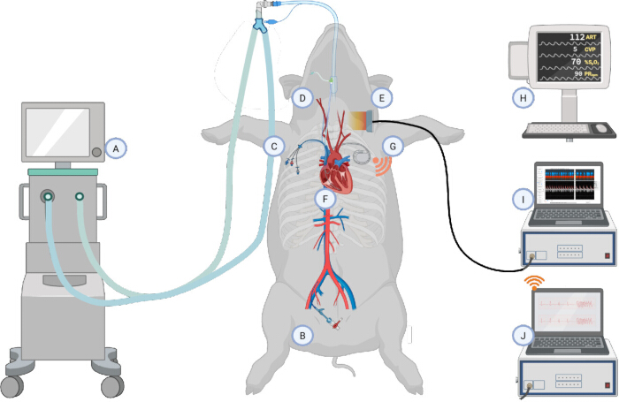
Figure 1: Experimental setup. (A) The experimental animals were intubated with an endotracheal tube in the prone position and connected to a ventilator. In the supine position, under ultrasound guidance, the following vascular access was established: (B) A PiCCO arterial line was inserted into the femoral artery, (C) a central venous catheter was placed in the right external jugular vein, and (D) a vascular sheath introducer was inserted into the right internal jugular vein. Additionally, a suprapubic catheter was inserted into the bladder (not shown here), and (E) a RescueDoppler carotid flow probe was placed over the left carotid artery. (F) Through the vascular sheath, a pacemaker electrode was advanced into the right ventricle and its position verified using echocardiography or fluoroscopy. (G) An implantable cardioverter-defibrillator unit (ICD) was inserted in a subcutaneous pocket under the left clavicle and connected to the pacemaker electrode. (H) The animals were connected to a multimodal monitor recording ECG, tail plethysmography (SpO₂), arterial blood pressure, central venous pressure, and end-tidal CO₂. (I) The RescueDoppler sensor was connected to a RescueDoppler recorder, providing continuous carotid flow tracings. (J) The ICD device was connected through telemetry to an ICD programmer. Figure created using BioRender. Please click here to view a larger version of this figure.
| Pacemaker-induced arrhythmia | CPR procedures performed | Arrhythmia conversion rate (%) | |||
| Pig* | Ventricular fibrillation (n) | Ventricular tachycardia (n) | Manual compression (n) | Mechanical compression (n) | |
| 1 | 12 | 4 | 6 | 2 | 91.6 |
| 2 | 14 | 7 | 0 | 5 | 100 |
| 3 | 9 | 7 | 0 | 9 | 100 |
| 4 | 8 | 4 | 5 | 5 | 100 |
| 5 | 18 | 0 | 12 | 5 | 94.4 |
| 6 | 8 | 0 | 8 | 0 | 87.5 |
| 7 | 15 | 8 | 9 | 6 | 100 |
| 8 | 18 | 0 | 9 | 9 | 94.4 |
| 9 | 14 | 2 | 9 | 5 | 100 |
| 10 | 27 | 0 | 13 | 11 | 100 |
| 11 | 19 | 0 | 12 | 4 | 84.2 |
Table 1: Summary of ICD-induced VF/VT and subsequent cardioversion. *Eleven pigs underwent experiments. On average, 18 arrhythmias, including ventricular fibrillation (VF) and ventricular tachycardia (VT)- were induced per animal using an implantable cardioverter-defibrillator (ICD). The mean VT/VF duration was 0.75 min. CPR was performed in selected VF episodes according to earlier study protocols15,16,17. Animals were then converted to sinus rhythm (SR) by direct current (DC) shock via the ICD. Each animal was stabilized for 5 min before the next arrhythmia was induced.
Discussion
To our knowledge, the presented model utilizing an implantable defibrillator to both induce and cardiovert ventricular fibrillation (VF) in pigs has not previously been described in the literature. VF is commonly induced using either sinusoidal waveform generators or direct current from an external battery through epicardial electrodes. Cardioversion is typically performed using external defibrillators. While these methods may reliably induce VF, failure to cardiovert animals using external defibrillation has been reported in up to 45% of cases2.
ECG monitoring during CPR is challenging due to compression-related artifacts that may obscure the underlying rhythm18. However, an implantable defibrillator with intracardiac ECG enables continuous rhythm monitoring during chest compressions, allowing for more accurate real-time rhythm assessment and reliable post-compression analysis.
In contrast, this model reliably cardioverted all animals, with multiple successful conversions per experiment. This enabled the repeated use of individual animals across experimental protocols, thereby reducing the number of animals sacrificed and improving overall resource efficiency. Additionally, the model eliminated the need for external defibrillation patches, freeing space on the thorax and improving access during instrumentation.
The model is unique in its ability to study the physiological effects of short, complete circulatory arrests in intact animals, where cardiac restart is achieved by a gentle, single internal shock. This feature enables controlled investigations of ischemic tolerance in vital organs such as the kidneys and gastrointestinal tract, redistribution of blood flow after global ischemia, and the interplay between endogenous sympathetic activation, ischemic stress responses, and the administration of adrenergic drugs. The model, therefore, provides a valuable experimental platform for exploring organ-specific and systemic responses to cardiac arrest, causing whole-body ischemia under highly standardized conditions. Beyond electrode placement, maintaining physiological stability is crucial for reproducibility. Continuous monitoring of temperature, ventilation, and anesthesia depth ensures normothermia, adequate oxygenation, and stable hemodynamics throughout the experiment.
The presented representative results demonstrate the reliability and reproducibility of the ICD-controlled VF induction and cardioversion in a controlled experimental setting. The ability to induce and terminate VF remotely enabled rapid and precise timing, with minimal external interference, which is valuable for studying cardiac arrest physiology and resuscitation dynamics. The outcome analysis focused on the consistency of VF induction, the number of successful cardioversions, and the stability of ROSC.
In this study, we adhered to the 3Rs principle by generating statistically robust data while minimizing the number of animals used. Owing to the high rate of successful resuscitations, ventricular fibrillation and cardioversion could be induced multiple times in each subject. By employing repeated-measures statistics within subjects and mixed-effects models accounting for both fixed and random effects, the study could be conducted using only eleven animals.
Limitations
The primary limitation of the model lies in the requirement for specialized equipment and operator expertise. A pacemaker generator, transvenous pacing lead, programming unit, and a trained operator are all essential. Accurate placement of the pacing lead is critical and requires imaging guidance, typically by transthoracic echocardiography or fluoroscopy.
Consequently, implementation of this model may be challenging in resource-limited settings. However, once established, it offers a highly effective and reproducible method to induce and terminate VF, minimizing the occurrence of non-convertible fibrillation and enhancing experimental control. This model is broadly relevant to research groups conducting in vivo resuscitation studies in large animal models.
This porcine model was developed to assess the feasibility of automated carotid Doppler monitoring during cardiac resuscitation, and not for testing clinical resuscitation strategies. The study focused on acute physiological responses and was conducted as a terminal protocol, which excluded long-term follow-up or histological evaluation. Additionally, we used only 11 animals in this study. Although multiple repeated experiments were performed in each animal, inter-individual variability may not have been fully captured. Furthermore, the study did not include a control group using traditional methods. Although such methods were referenced as having a high failure rate, the lack of direct comparison within the same experimental framework limits the ability to demonstrate the relative advantages of the proposed technique. Finally, the model requires access to specialized equipment such as an ICD, programmer, and Doppler ultrasound, which may limit its applicability in resource-constrained settings. Additionally, the surgical protocol is technically demanding, involving multiple vascular access points, bladder stoma creation, and ICD implantation, which may present a steep learning curve for inexperienced operators.
In this experimental setup, intracardiac ECG recordings could not be exported from the programming unit; therefore, data interpretation relied solely on visual monitoring. Future studies should incorporate a method enabling direct export of ECG traces to facilitate quantitative analysis.
Disclosures
Charlotte Björk Ingul is a consultant for Cimon Medical, the company that owns the technology associated with RescueDoppler. The remaining authors declare no conflicts of interest.
Acknowledgements
We used Perplexity and Microsoft Copilot to assist with English language proofreading. All content was reviewed and approved by the authors. This project has received funding from Nord University, Norwegian University of Science and Technology, and the Norwegian Research Council.
Materials
| Name | Company | Catalog Number | Comments |
|---|---|---|---|
| Abbott Durata 7120 ICD Single Coil Lead | Abbott, Abbot Park, IL | 7120 | |
| Abbott Gallant VR ICD | Abbott, Abbot Park, IL | CDVRA500Q | |
| Abbott Merlin Programmer | Abbott, Abbot Park, IL | 3650 | |
| Central venous catheter Certofix trio | B. Braun Medical, Melsungen, Germany | 4167408-07 | |
| Endotracheal tube Portex #7.0 | Smiths Medical, Rockland, MA | 100/150/070 | |
| Exagon VET (Pentobarbital 400 mg/mL) | VetViva Richter, Wels, Austria | N/A | |
| Heparin 5000 IU/mL | Panpharma, Luitré, France | ETI3M807-1 | |
| Ketamine 50 mg/mL | Abcur AB, Helsingborg, Sweden | LB-12355-01 | |
| Midazolam 5 mg/mL | B. Braun Medical, Melsungen, Germany | 753-12612876-0621 | |
| Morphine | Abcur AB, Helsingborg, Sweden | LB-122184-01 | |
| Normal Saline flush solution | Fresenius Kabi, Uppsala, Sweden | B202382-05 | |
| PentoCur (Thiopental) | Abcur AB, Helsingborg, Sweden | LB-122564-01 | |
| Periphal IV catheter 20G Vasofix | B. Braun Medical, Melsungen, Germany | 4268113B | |
| PiCCO Monitoring kit (pressure transducer, flush system and thermistor) | Pulsion, Feldkirchen, Germany | PV8215 | |
| PiCCO catheter 5 Fr 20 cm | Pulsion, Feldkirchen, Germany | PV2015L20-A | |
| Pressure transducer and flush kit TruWave | Edwards Lifescience, Irvine, CA | PX272 | |
| RescueDoppler | Cimon Medical, Trondheim, Norway | N/A | Proprietary, not commercially available |
| Ringer acetate | Fresenius Kabi, Uppsala, Sweden | B203150-02 | |
| Urinary cather 12 Ch | Various | N/A | |
| Vascular sheath introducer Prelude 7.0 Fr | Merit Medical, South Jordan, UT | PSI-7F-11-035-18G | |
| Vue Ultrasound Gel | Optimum medical, Leeds, United Kingdim | 1157 |
References
- Cherry, B. H. Modeling cardiac arrest and resuscitation in the domestic pig. World J Crit Care Med. 4 (1), 1(2015).
- Burgert, J. M., Johnson, A. D., Garcia-Blanco, J. C., Craig, W. J., O'Sullivan, J. C. An Effective and Reproducible Model of Ventricular Fibrillation in Crossbred Yorkshire Swine (Sus scrofa) for Use in Physiologic Research. Comp Med. 65 (5), 444-447 (2015).
- Schwarz, B., et al. Neither vasopressin nor amiodarone improve CPR outcome in an animal model of hypothermic cardiac arrest. Acta Anaesthesiol Scand. 47 (9), 1114-1118 (2003).
- White, N. J., et al. Coagulopathy during cardiac arrest and resuscitation in a swine model of electrically induced ventricular fibrillation. Resuscitation. 82 (7), 925-931 (2011).
- Euler, D. E., Whitman, T. A., Roberts, P. R., Kallok, M. J. Low Voltage Direct Current Delivered Through Unipolar Transvenous Leads: An Alternate Method for the Induction of Ventricular Fibrillation. Pacing Clin Electrophysiol. 22 (6), 908-914 (1999).
- Ruemmler, R., Ziebart, A., Garcia-Bardon, A., Kamuf, J., Hartmann, E. K. Standardized Model of Ventricular Fibrillation and Advanced Cardiac Life Support in Swine. J Vis Exp. (155), e60707(2020).
- Xanthos, T., et al. Cardiopulmonary arrest and resuscitation in Landrace/Large White swine: a research model. Lab Anim. 41 (3), 353-362 (2007).
- Greenwood, J. C., et al. Carbon monoxide as a cellular protective agent in a swine model of cardiac arrest protocol. PLoS One. Wang, M. 19 (5), e0302653(2024).
- Vogel, U., Wanner, T., Bültmann, B. Extensive pectoral muscle necrosis after defibrillation: nonthermal skeletal muscle damage caused by electroporation. Intens Care Med. 24 (7), 743-745 (1998).
- Guensch, D. P., et al. Evidence for Acute Myocardial and Skeletal Muscle Injury after Serial Transthoracic Shocks in Healthy Swine. PLoS One. Akar, F. G. 11 (9), e0162245(2016).
- Rubenstein, J. C., Gupta, M. S., Kim, M. H. Effectiveness of VF induction with DC fibber versus conventional induction methods in patients on chronic amiodarone therapy. J Interv Card Electrophysiol. 38 (2), 137-141 (2013).
- Fitzgibbon, E., Berger, R., Tsitlik, J., Halperin, H. R. Determination of the noise source in the electrocardiogram during cardiopulmonary resuscitation. Crit Care Med. 30 (4), S148-S153 (2002).
- Babaeizadeh, S., Firoozabadi, R., Han, C., Helfenbein, E. D. Analyzing cardiac rhythm in the presence of chest compression artifact for automated shock advisory. J Electrocardiol. 47 (6), 798-803 (2014).
- Ruiz de Gauna, S., et al. Rhythm analysis during cardiopulmonary resuscitation: past, present, and future. BioMed Res Int. 2014, 386010(2014).
- Faldaas, B. O., et al. Hands-free continuous carotid Doppler ultrasound for detection of the pulse during cardiac arrest in a porcine model. Ressuc Plus. 15 (100412), 1-10 (2023).
- Faldaas, B. O., et al. Real-time feedback on chest compression efficacy by hands-free carotid Doppler in a porcine model. Ressusc Plus. 18, 100583(2024).
- Faldaas, B. O., et al. A hands-free carotid Doppler can identify spontaneous circulation without interrupting cardiopulmonary resuscitation: an animal study. Intensive Care Med Exp. 12 (1), 121(2024).
- Ruiz De Gauna, S., et al. Rhythm Analysis during Cardiopulmonary Resuscitation Past, Present, and Future. BioMed Res Int. 2014, 1-13 (2014).
Reprints and Permissions
