This protocol describes a self-made multiring thread method to provide controlled internal traction during gastric endoscopic submucosal dissection. The technique facilitates effective mucosal flap traction in desired directions and forces, enabling easy, safe, and cost-effective dissection.
A subscription to JoVE is required to view this content. Sign in or start your free trial.
Method Article
Self-Made Internal Traction Method For Endoscopic Submucosal Dissection of Early Gastric Cancer
In This Article
Summary
Abstract
This protocol describes a self-made multiring thread method to provide controlled internal traction during endoscopic submucosal dissection (ESD) for early gastric cancer. ESD is a minimally invasive treatment that enables en bloc resection; however, the procedure can be technically challenging when adequate visualization and access to the submucosal layer are limited. Conventional traction methods, such as the dental floss clip technique, may be hindered by interference with the endoscope and instability of clip attachment. To address these limitations, a multiring thread device was developed to enable stable and adjustable traction. The device allows precise control of traction direction and force without interfering with endoscope maneuverability. This protocol outlines the preparation and deployment of the multiring thread during the procedure. The method facilitates improved exposure of the submucosal layer, enhances procedural efficiency, and supports safe dissection. This approach provides a simple, cost-effective, and reproducible technique for improving outcomes in gastric ESD.
Introduction
Gastric endoscopic submucosal dissection (ESD) has been widely used as a minimally invasive treatment for gastric neoplasms, including early gastric cancer and adenoma1. Indications are determined based on endoscopic and pathological findings, such as lesion size, depth of invasion, ulceration (UL), and histological type2. However, complete resection of some lesions remains technically challenging using conventional ESD techniques, resulting in prolonged procedure time, piecemeal resection, incomplete resection, and perforation3. Adequate visualization of the submucosal layer is essential for safe and effective dissection, and insufficient traction is a major limiting factor.
Various traction methods have been developed to improve visualization and facilitate dissection within the broader field of endoscopic resection techniques. The dental floss clip (DFC) method is commonly used because of its simplicity and low cost4; however, it may interfere with endoscope maneuverability and can become unstable during traction. Internal traction methods have been introduced to overcome these limitations5,6, but commercially available devices are often expensive and require additional preparation, limiting their widespread use. The goal of this protocol is to describe a self-made multiring thread method that enables controlled internal traction during gastric ESD. This technique is easy to prepare and cost-effective. This method is particularly applicable for lesions located in the middle or lower stomach, on the lesser curvature, or near the pylorus, as well as for lesions associated with severe fibrosis. The method allows rapid establishment of stable traction of the mucosal flap, which may facilitate safe and highly efficient submucosal dissection.
Access restricted. Please log in or start a trial to view this content.
Protocol
This retrospective cohort study was approved by the Ethics Committee of Kitano Hospital (258005, August 19, 2025) and was conducted in accordance with the Declaration of Helsinki. Informed consent was obtained through opt-out forms on our website (https://kitano.bvits.com/rinri/publish.aspx).
1. Prepare the multiring thread
- Prepare a 2.5 mL plastic disposable syringe with an outer diameter of 10–12 mm.
- Cut approximately 40 cm of 4-0 nylon thread. Adjust the length based on the number of rings required and lesion characteristics.
- Tie three tight overhand knots around the syringe to form the first ring (Figure 1).
CAUTION: Avoid excessive tension when tying the nylon thread to prevent breakage or deformation of the ring structure. - Rotate the first ring by 180° along its longitudinal axis (Figure 2).
- Repeat the knotting method described in Step 1.3 to form an additional ring connected to the first ring (Figure 3).
- Form three rings for typical gastric lesions. Increase the number of rings for larger lesions or when a longer traction distance is required (Figure 4).
NOTE: The number of rings and the thread length depend on lesion location and procedural requirements. Three rings are generally sufficient to grasp the mucosal flap and the opposing mucosa and maintain stable traction. - Prepare additional multiring threads in advance when needed. Store prepared threads in a sterile container at room temperature. Use within an appropriate time frame based on institutional practice (e.g., up to approximately 30 days). Cut the loops to the desired length and number immediately before or during the procedure.
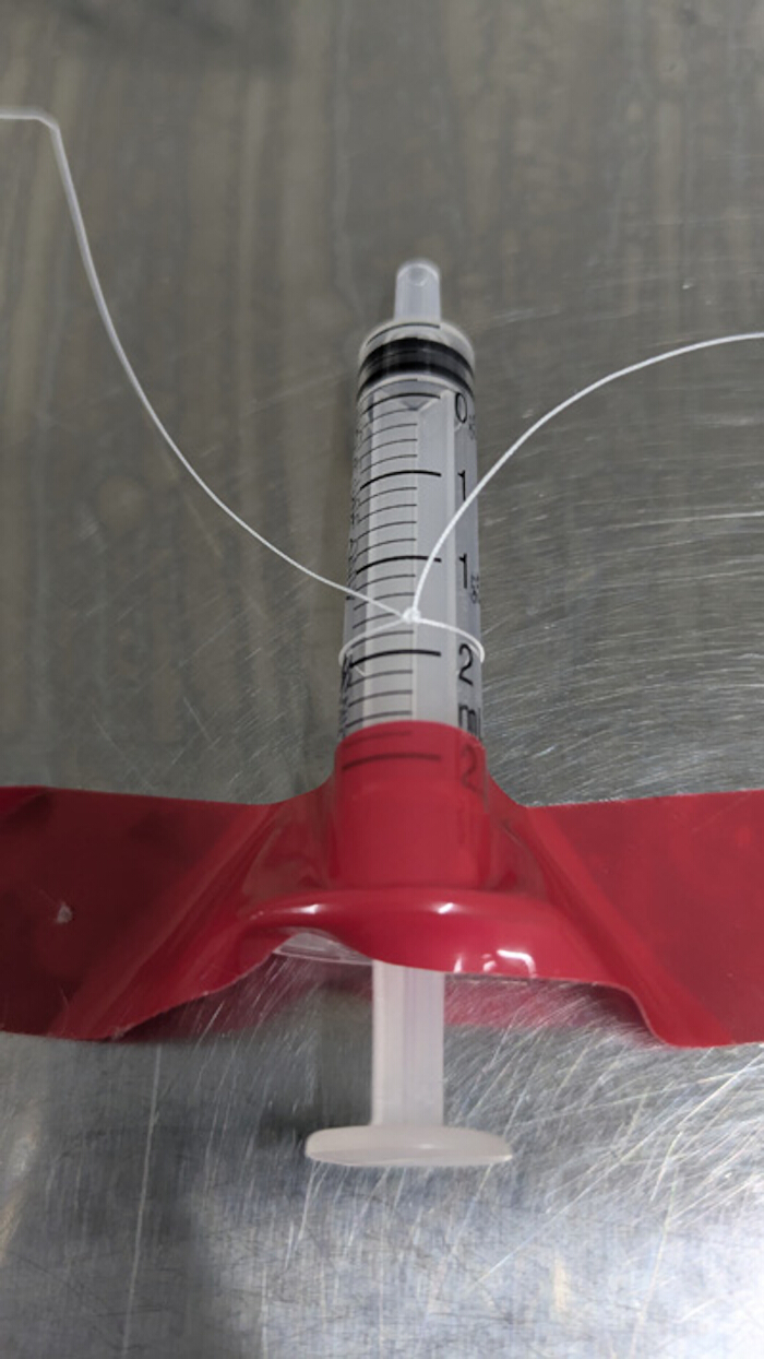
Figure 1. Formation of the first ring of the multiring thread. The 4-0 nylon thread is tied around a 2.5 mL syringe three times to form the initial ring used for traction. Please click here to view a larger version of this figure.
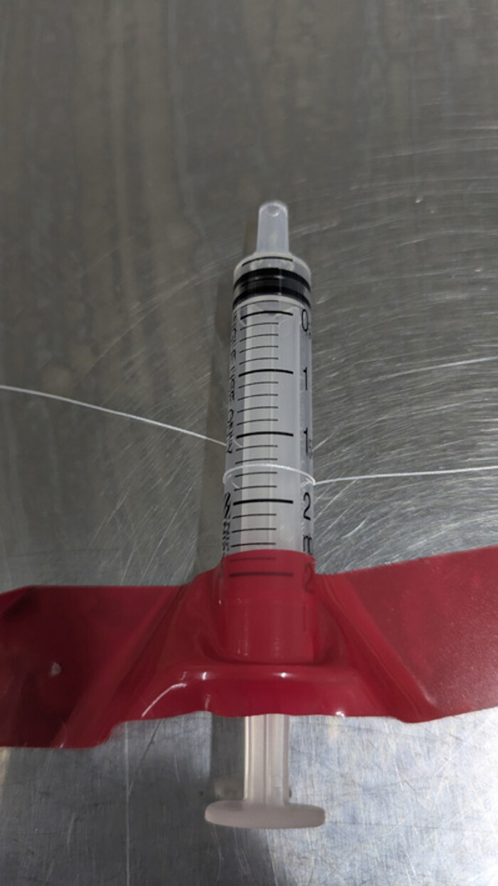
Figure 2. Rotation of the first ring. The first ring is rotated 180° after formation to enable proper alignment of subsequent rings. Please click here to view a larger version of this figure.
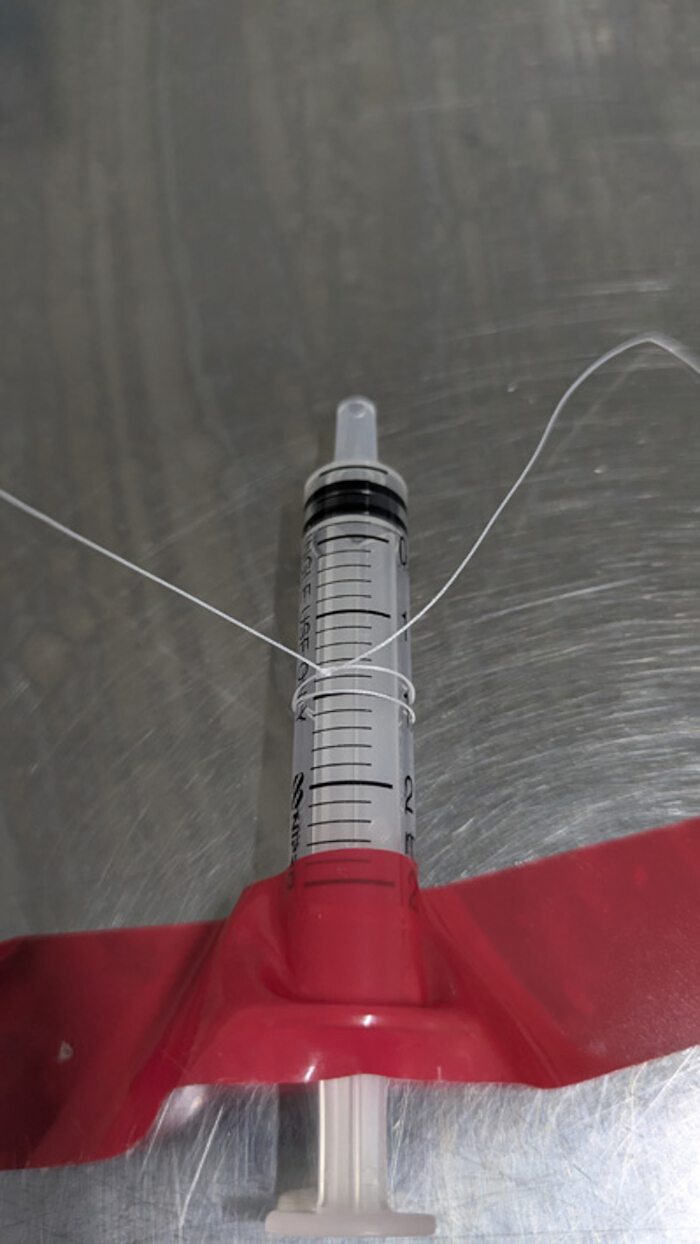
Figure 3. Formation of an additional ring. The nylon thread is tied again around the syringe to create a second ring connected to the first ring. Please click here to view a larger version of this figure.
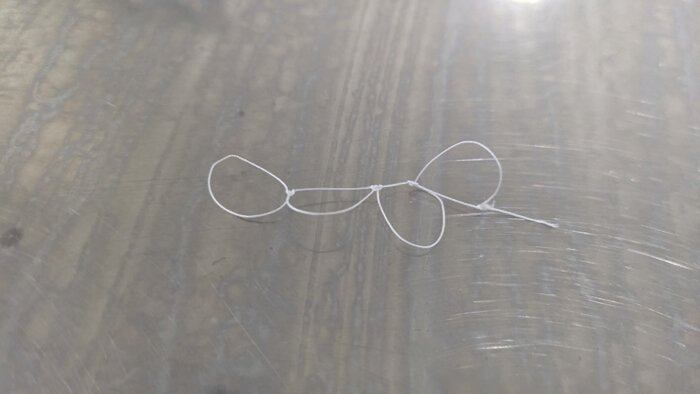
Figure 4. Completed multiring thread structure. Three connected rings are formed in series to create the multiring thread used for internal traction. Please click here to view a larger version of this figure.
2. Gastric ESD procedure
- Perform the procedure under conscious sedation or general anesthesia according to institutional protocols using a standard therapeutic endoscope with a working channel of ≥2.8 mm. Continuously monitor vital signs, including heart rate, blood pressure, oxygen saturation, and respiratory status, throughout the procedure. Ensure that appropriate resuscitation equipment and trained personnel are available.
- Perform circumferential marking using an ESD knife. Place markings approximately 3 mm from the lesion border. Increase the marking distance to 5–10 mm for lesions suspected of lateral spread.
- Perform a circumferential mucosal incision and dissect to the deep submucosal layer while injecting a submucosal fluid cushion. Continue dissection until the deep submucosal layer is clearly exposed, as indicated by visualization of the white muscular layer and blue-stained submucosal fibers.
NOTE: After applying the traction device, visualization and access to the rear side of the lesion may be limited. Ensure adequate submucosal trimming before device deployment. - Form a mucosal flap by dissecting the front side of the submucosa. Create a flap large enough to securely accommodate clip attachment (Figure 5).
- Place two or three marking points on the mucosa to indicate the intended traction site when traction direction is difficult to determine (Figure 6).
NOTE: The ideal traction point is typically located opposite and anterior to the mucosal flap.
- Place two or three marking points on the mucosa to indicate the intended traction site when traction direction is difficult to determine (Figure 6).
- Inject 2–3 mL (or as needed to achieve adequate lifting) of a submucosal lifting solution (e.g., saline, glycerol solution, or hyaluronic acid) beneath the mucosal flap immediately before applying the traction device.
NOTE: Adequate lifting facilitates clip attachment and reduces the risk of grasping the muscular layer.
CAUTION: Avoid excessive injection to prevent overdistension or mucosal tearing. - Use a reopenable endoscopic clip compatible with the working channel (≥2.8 mm) and capable of repeated opening and closing. Then grasp the end ring of the multiring thread prepared in Step 1.
- Advance the clip holding the multiring thread through the endoscope working channel (Figure 7).
CAUTION: Advance the clip carefully through the endoscope channel to avoid damage to the device or channel. - Attach the clip to the mucosal flap (Figure 8).
CAUTION: Avoid grasping the muscular layer to prevent perforation.
NOTE: If the muscular layer is inadvertently grasped, use grasping forceps to gently disengage and reposition the clip. - Advance a second reopenable clip through the endoscope working channel.
- Grasp the ring opposite the attached end of the multiring thread using the second clip.
- Attach the second clip to the normal mucosa opposite the lesion at a position that provides optimal exposure of the submucosal layer (Figure 9).
NOTE: Use previously placed markings as guidance. Adjust the traction direction based on lesion location to optimize visualization of the submucosal layer. - Continue submucosal dissection while maintaining traction. Approach from the front side and dissect the exposed submucosal layer (Figure 10).
NOTE: If the submucosal layer becomes obscured or the mucosal flap collapses, apply an additional clip to the normal mucosa closer to the mucosal flap.
CAUTION: Avoid excessive traction force to prevent tissue tearing or clip dislodgement. - Continue dissection until the lesion is fully separated from the surrounding tissue.
NOTE: Confirm complete resection by ensuring no residual lesion remains at the margins.
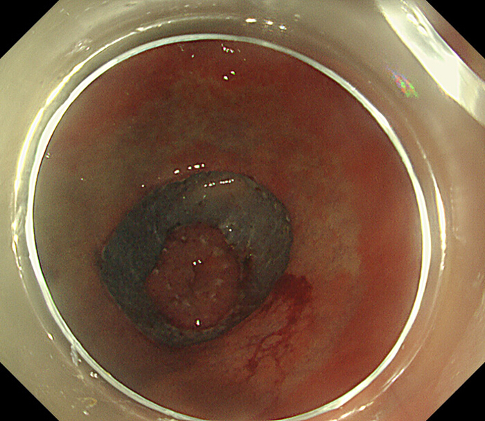
Figure 5. Folded mucosal flap before traction. The mucosal flap remains folded, limiting visualization and access to the submucosal layer. Please click here to view a larger version of this figure.
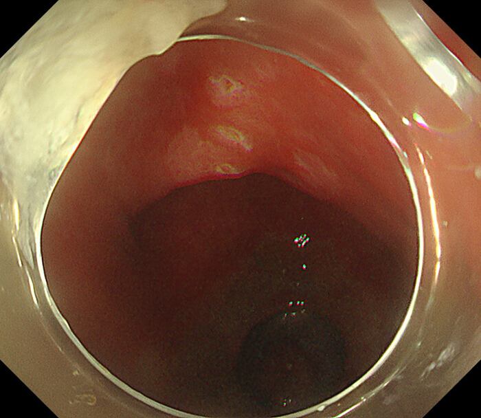
Figure 6. Identification of the traction point. Marking points are placed on the mucosa to determine the optimal location for traction. Please click here to view a larger version of this figure.
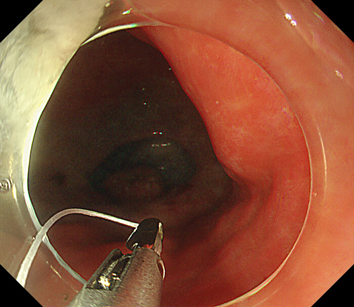
Figure 7. Delivery of the multiring thread. A reopenable clip grasps the end ring of the multiring thread and is advanced through the endoscope channel. Please click here to view a larger version of this figure.
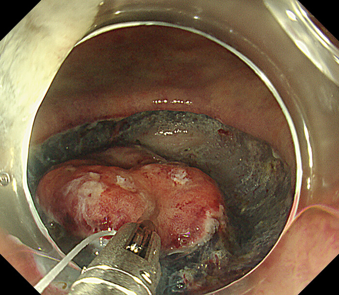
Figure 8. Attachment of the traction device to the mucosal flap. The clip holding the multiring thread is applied to the mucosal flap to initiate traction. Please click here to view a larger version of this figure.
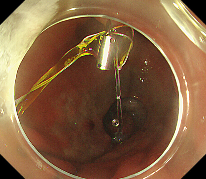
Figure 9. Fixation of the traction device to the opposing mucosa. A second clip secures the free end of the multiring thread to the target mucosal site, establishing traction. Please click here to view a larger version of this figure.
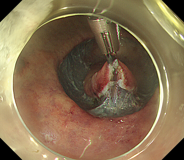
Figure 10. Expanded mucosal flap after traction. Application of the traction device extends the mucosal flap, improving visualization and facilitating submucosal dissection. Please click here to view a larger version of this figure.
3. Post-complete resection
- Advance grasping forceps through the endoscope working channel and remove the second clip from the mucosa.
CAUTION: Remove the clip carefully to avoid mucosal injury or dislodging the resected specimen. - Retrieve the resected lesion together with the traction device.
Access restricted. Please log in or start a trial to view this content.
Results
We retrospectively collected data on internal traction methods at a single institution. Patients who underwent gastric ESD using the internal traction method between January 2025 and September 2025 were consecutively enrolled. Use of the method was confirmed in recorded videos. Data were collected from medical records, endoscopic reports, and videos. Patients aged <20 years were excluded.
The study included patients taking antithrombotic agents. Management of these agents was conducted in ...
Access restricted. Please log in or start a trial to view this content.
Discussion
We designed and applied this self-made device for traction of mucosal flaps during gastric ESD. The internal traction method is mainly used for colonic ESD11,12. This self-made internal traction device, known as the multiloop method, was first reported in the field of colonic ESD13. Since its introduction, such traction methods have been used in the field of endoscopic retrograde cholangiopancreatography14,
Access restricted. Please log in or start a trial to view this content.
Disclosures
The authors have nothing to disclose.
Acknowledgements
The authors have no acknowledgments.
Access restricted. Please log in or start a trial to view this content.
Materials
| Name | Company | Catalog Number | Comments |
|---|---|---|---|
| 2.5 mL syringe | NIPRO CORPORATION (or equivalent) | 4987458081262 | Used as a cylindrical mold to construct the multiring thread (Step 1.1). Any manufacturer is permitted. |
| 4-0 nylon | Natsume Seisakusho Co., Ltd. (or equivalent) | C-23S-N1 | Used to create the multiring thread for internal traction (Steps 1.2–1.6). Any manufacturer is permitted. |
| Endoscope | Olympus (or equivalent) | GIF-H290T | Used to perform gastric ESD and deliver instruments through the working channel (Steps 2.1, 2.7–2.13). |
| ESD knife | Olympus (or equivalent) | KD-655L | Used for circumferential marking, mucosal incision, and submucosal dissection (Steps 2.2–2.4, 2.12–2.13). |
| Grasping forceps | Olympus (or equivalent) | FG-47L-1 | Used to manipulate clips and remove the traction device after resection (Steps 2.8, 3.1). |
| Injection needle | Olympus (or equivalent) | NM-610L-0425 | Used to inject solution into the submucosal layer (Step 2.5). |
| Injection solution | Boston Scientific (or equivalent) | 1919 (MucoUp, Japan) | Used to create a submucosal fluid cushion during ESD (Steps 2.3, 2.5). A hyaluronic acid–based solution may be used; dilution with saline is acceptable. Equivalent solutions (e.g., saline, glycerol solution, or other submucosal lifting agents) may also be used. |
| Multiloop traction device (MLTD) | Boston Scientific | M00503140 | Commercial multiloop traction device used for internal traction; included for comparison with the self-made device. |
| Reopenable clip | MC Medical, Inc. (or equivalent) | RO-CD26195 | Used to grasp the multiring thread and apply traction to the mucosal flap and opposing mucosa (Steps 2.6–2.11). Any manufacturer is permitted. |
| Spring-assisted traction clip (S-O clip) | ZEON Medical Inc. (or equivalent) | TC1H05 | Clip-based traction device with an elastic component used for internal traction; included for comparison purposes; not used in this protocol. |
| Sterile container | Hakuzo Medica (or equivalent) | 1270001 | Used to store prepared multiring threads prior to use (Step 1.7). Any manufacturer is permitted. |
References
- Park CH, et al. Clinical safety of endoscopic submucosal dissection compared with surgery in elderly patients with early gastric cancer: A propensity-matched analysis. Gastrointest Endosc. 2014;80(4):599–609.
- Ono H, et al. Guidelines for endoscopic submucosal dissection and endoscopic mucosal resection for early gastric cancer (second edition). Dig Endosc. 2020;10.1111/den.13883.
- Kim JH, et al. Risk factors associated with difficult gastric endoscopic submucosal dissection: Predicting difficult ESD. Surg Endosc. 2017;31(4):1617–1626.
- Jeon WJ, You IY, Chae HB, Park SM, Youn SJ. A new technique for gastric endoscopic submucosal dissection: Peroral traction-assisted endoscopic submucosal dissection. Gastrointest Endosc. 2009;69(1):29–33.
- Chen PJ, Chu HC, Chang WK, Hsieh TY, Chao YC. Endoscopic submucosal dissection with internal traction for early gastric cancer (with video). Gastrointest Endosc. 2008;67(1):128–132.
- Nagata M. Internal traction method using a spring-and-loop with clip (S-O clip) allows countertraction in gastric endoscopic submucosal dissection. Surg Endosc. 2020;34(8):3722–3733.
- Fujimoto K, et al. Guidelines for gastroenterological endoscopy in patients undergoing antithrombotic treatment. Dig Endosc. 2014;26(1):1–14.
- Kato M, et al. Guidelines for gastroenterological endoscopy in patients undergoing antithrombotic treatment: 2017 appendix on anticoagulants including direct oral anticoagulants. Dig Endosc. 2018;30(4):433–440.
- Yoshida M, et al. Conventional versus traction-assisted endoscopic submucosal dissection for large esophageal cancers: A multicenter, randomized controlled trial (with video). Gastrointest Endosc. 2020;91(1):55–65.e52.
- Kimura H, et al. Features of post-endoscopic submucosal dissection electrocoagulation syndrome for early gastric neoplasm. J Gastroenterol Hepatol. 2021;36(11):3164–3169.
- Ritsuno H, et al. Prospective clinical trial of traction device-assisted endoscopic submucosal dissection of large superficial colorectal tumors using the S-O clip. Surg Endosc. 2014;28(11):3143–3149.
- Bordillon P, et al. Double-clip traction for colonic endoscopic submucosal dissection: A multicenter study of 599 consecutive cases (with video). Gastrointest Endosc. 2021;94(2):333–343.
- Sudo G, Tanuma T, Suzuki Y, Nakase H. Multiloop method for traction during colorectal endoscopic submucosal dissection. VideoGIE. 2019;4(1):11–13.
- Iwano K, Toyonaga H, Kin T, Ishii T, Katanuma A. Multiloop traction method during endoscopic hemostasis for post-sphincterotomy bleeding of the peridiverticular papilla. Endoscopy. 2022;54(S02):E769–E770.
- Itami K, Iwano K, Yazumi S. Biliary cannulation of a peridiverticular papilla using multiloop traction in a patient with Roux-en-Y anatomy. Endoscopy. 2024;56(S01):E1018–E1019.
- Yoshikawa T, et al. A case of successful endoscopic submucosal dissection of early gastric cancer involving the pylorus by switching the direction of traction. Endoscopy. 2025;57(S01):E449–E450.
- Kitahara G, et al. The efficacy and safety of multi-loop traction device for gastric endoscopic submucosal dissection: A single-center prospective pilot study. Sci Rep. 2023;13(1):20050.
- Fu B, Zhou XR, Liu WH. Pre-traction with clip-and-rubber band facilitating endoscopic submucosal dissection of a colonic laterally spreading tumor. Surg Innov. 2023;30(2):218–219.
- Iwagami H, et al. Effective traction using a clip with rubber band and grasping forceps in endoscopic laryngopharyngeal surgery. Endoscopy. 2023;55(S01):E566–E567.
- Ito M, Sumiyama K. Randomized control trials may not provide a conclusive answer for complex endoscopic interventions. Dig Endosc. 2023;35(1):94–95.
Access restricted. Please log in or start a trial to view this content.
Reprints and Permissions
