The stereotactic intracranial injection technique enables precise and localized implantation of tumor cells into the mouse cortex, making it a powerful model for studying metastatic colonization of the brain.
Method Article
Modelling Brain Metastasis: Standardized Analysis of Metastatic Colonization and Histological Growth Patterns by Stereotactic Intracortical Injection
In This Article
Summary
Abstract
Brain metastases are a common and devastating complication of advanced solid tumors, frequently associated with poor prognosis, neurological decline, and reduced quality of life. The incidence of central nervous system (CNS) failure and neurological death is rapidly increasing, yet the mechanisms driving the final stages of brain metastasis, such as secondary dissemination, re-colonization, and the contribution of the histological growth pattern (HGP) as a potential surrogate parameter, remain poorly understood.
The standardized stereotactic intracortical injection model enables precise and reproducible implantation of tumor cells, or mixed populations including stromal or immune components, directly into the mouse cerebral cortex. This protocol also supports the creation of a preclinical tissue archive, offering a robust platform for investigating essential aspects of CNS colonization, such as: metastatic outgrowth, HGP-specific growth dynamics, and pathophysiological mechanisms contributing to neurological failure. Additionally, this model supports pharmacological testing in a reproducible clinically relevant context.
Unlike systemic injection methods (e.g., tail vein or intracardiac), which are optimized for studying early metastatic steps but result in variable and often low rates of brain colonization, the stereotactic model ensures consistent, brain-specific metastatic growth and enables the investigation of late stages of CNS metastasis. Compared to ex vivo systems such as organoids or brain slice cultures, the in vivo stereotactic model preserves vascularization, systemic signaling, and the full complexity of the brain's immune landscape, supporting long-term studies of tumor progression and therapeutic response.
By providing a reproducible and clinically relevant platform, our model advances the field's ability to identify prognostic markers, explore therapeutic strategies, and understand the mechanisms of late-stage brain metastasis.
Introduction
Brain metastases are a common and devastating complication in patients with solid tumors, frequently associated with poor prognosis, significant neurological decline, and reduced quality of life1,2,3. In particular, central nervous system (CNS) failure and neurological death have become increasingly frequent outcomes in cancers such as lung, breast, and melanoma4,5,6,7. CNS failure in the context of brain metastases can arise from a variety of mechanisms, including local mass effect, peritumoral edema, intracranial hemorrhage, and meningeal dissemination, which may act independently or synergistically to ultimately result in neurological death8.
Despite its clinical importance, the pathophysiological mechanisms underlying CNS failure and the final stages of metastatic progression remain poorly understood. Processes such as alternative dissemination, secondary dissemination, and re-colonization are rarely the focus of dedicated investigation8. Both clinical and preclinical studies often limit their analyses to endpoints such as brain metastasis number, size, or overall survival (OS), thereby overlooking critical mechanistic insights and the true causes of neurological decline and death.
In this context, we and others have identified distinct histological growth patterns (HGPs) in brain metastases that correlate with clinical outcomes9,10,11. Three major HGPs have been described: i) non-infiltrative, marked by a well-demarcated tumor border often surrounded by a reactive astrocytic rim; ii) epithelial infiltrative, where clusters of tumor cells infiltrate the adjacent brain parenchyma without astrocytic containment; and iii) diffuse infiltrative, characterized by widespread infiltration of single tumor cells or small clusters, causing an extensive astrogliosis10,12.
Clinically, infiltrative HGPs are associated with significantly worse prognosis compared to non-infiltrative patterns9,10,11. Moreover, HGPs appear to reflect key aspects of tumor growth dynamics and mechanisms leading to neurological death. While non-infiltrative metastases typically expand as a single lesion and constrain vital structures in the brain, infiltrative lesions demonstrate secondary dissemination (starting from the existing lesion and not from the primary tumor) within the CNS, contributing to widespread damage and complete organ destruction (unpublished data). Infiltrative lesions may also re-colonize the meninges, contributing to meningeal metastasis and further complicating the disease course10,13. Despite their clinical relevance, the HGPs of brain metastases and the final stages of CNS colonization, including secondary dissemination and re-colonization of the same organ, remain understudied, largely due to the lack of suitable experimental models.
To address this gap, we present a stereotactic intracranial injection model that enables precise, reproducible implantation of tumor cells or cellular mixtures comprising tumor cells and one or more non-tumor cell types into the cerebral cortex of experimental mice. This model overcomes key limitations of systemic metastasis models and offers a robust platform for investigating essential aspects of CNS colonization, such as: tumor-immune cell interactions during colonization14, the impact of pre-treated cellular mixtures15, metastatic outgrowth16, HGP-specific growth dynamics and pathophysiological mechanisms contributing to neurological failure (unpublished data). Additionally, this model supports pharmacological testing in a reproducible and clinically relevant context14,15.
This protocol offers a detailed methodological framework for implementing the stereotactic injection model to study HGPs in brain metastasis. It also supports the development of a structured preclinical sample archive and a digital tissue library for systematic investigation of late-stage CNS colonization biology. Our aim is to enable the identification of prognostic and potentially predictive biomarkers, contribute to the development of improved therapeutic strategies, and enhance our ability to predict acute and chronic responses of the brain parenchyma and the immune system. Through this approach, we seek to provide a standardized platform for a systematic study of CNS colonization and metastatic progression.
Protocol
All animal procedures described in this protocol were conducted in accordance with institutional guidelines and approved by the Government of Lower Franconia (permit number: RUF-55.2.2-2532-2-1678). This protocol is designed for the stereotactic intracortical injection of tumor cells in syngeneic adult mice (10-12 weeks old) as a model for metastatic colonization of the brain. Any alterations to the protocol should be discussed with the designated animal welfare officer.
1. Preoperative procedures
- Acclimatization of animals
NOTE: Mice must be acclimatized for at least 7 days prior to the start of the experimental procedures.- House experimental animals in groups under standard laboratory conditions (22 ± 2 °C, 50-60% relative humidity, 12 h light/dark cycle) with ad libitum access to food and water.
- To promote welfare and reduce stress, provide environmental enrichment in the form of nesting material, shelters, and gnawing blocks.
- Handle animals once daily (3-5 min per session) during the acclimatization period to familiarize them with human contact. In addition, perform mock procedures that simulate key aspects of the upcoming surgery, including weighing and exposure to the anesthesia setup, without performing any invasive intervention. These sessions aim to habituate the animals to the procedural environment and minimize stress-related variability.
- Physical examination (pre-surgical health check)
NOTE: Prior to tumor cell injection, all animals must undergo a thorough physical and neurological examination to exclude any pre-existing abnormalities unrelated to the study. Animals exhibiting deficits or anomalies should be excluded from the study. The physical examination can be performed the day before the injection.- Perform a full physical inspection assessing overall health, including coat condition, posture, breathing, gait, body weight, and general behavior.
- Evaluate neurological function using the Hanging Wire Test to assess grip strength and coordination. To perform the Hanging Wire Test suspend a horizontal wire (approximately 2 mm in diameter) between two supports, with escape platforms positioned at the ends about 30 cm above a padded surface. Place the mouse at the center of the wire and allow it to grasp the wire with its forepaws. Release the mouse and start timing immediately. End the trial when the mouse either reaches one of the platform or falls. Consider a mouse successful if it reaches the platform within 60 s; record as a failure if it falls or does not reach the platform within this time limit.
- Preparation of workstations
- On the day of surgery (day 0), set up four dedicated workstations as described below to ensure an efficient workflow and maintain sterile technique throughout the procedure.
NOTE: Use sterile surgical materials (underlays, gauze compresses, sutures, etc.) and autoclaved surgical instruments (scalpel, forceps, scissors, spatula, etc.).- Workstation #1 - Pre-surgery animal preparation: Use this workstation to weigh the mice, prepare the anesthetic solution, and administer the anesthesia. Prepare a clean surface containing a weighing scale, two cages with bedding (designate one for anesthetized mice), a 1 mL syringe filled with the anesthetic solution, and eye ointment.
- Workstation #2 - Pre- and post-surgery setup: Use this workstation to perform the scalp incision, seal the skull, and suture the wound. Prepare a clean surface containing a scalpel, a 10 mL syringe with 0.9% NaCl, bone wax, a spatula, suture material, forceps and scissors, cotton swabs, disinfectant (70% ethanol), a 1 mL syringe with analgesics, and a heating pad or a similar warming platform.
- Workstation #3 - Tumor cell preparation: Use this workstation to prepare the tumor cell solution prior to inoculation. Prepare a clean surface containing an ice box with a 10 µL Hamilton syringe and an Eppendorf tube with cell suspension in Extracellular Matrix (ECM)-medium, a pipette with 100 µL tips for resuspending cells, and three vessels for cleaning the Hamilton syringe after tumor cell inoculation containing distilled water, 70% ethanol, and 0.9% NaCl.
- Workstation #4 - Stereotactic injection: Use this workstation to perform the stereotactic tumor cell injection into the cerebral cortex. Prepare a clean surface containing the stereotactic frame for small animals, a precision dental drill, large forceps, and cotton swabs.
- On the day of surgery (day 0), set up four dedicated workstations as described below to ensure an efficient workflow and maintain sterile technique throughout the procedure.
- Preparation of tumor cell suspension
NOTE: All procedures for the preparation of the tumor cell suspension must be performed in a certified biological safety cabinet (Class II hood) using appropriate personal protective equipment (PPE), including lab coat, gloves, and eye protection, to ensure sterility and protect personnel from potential biohazard exposure.- Trypsinize cells using 1 mL of trypsin 1x EDTA.
NOTE: It is recommended to use tumor cells in the exponential growth phase. - Determine the cell count accurately using an appropriate counting method (e.g., hemocytometer or automated cell counter).
- Prepare a cell suspension containing 1 x 10³ cells in 3 µL ECM-medium per animal. Use a 2:1 mixture of ECM and standard culture medium containing 10% FCS and other supplements as appropriate. Scale up the suspension volume and cell number according to the number of animals (e.g., for 10 mice, prepare 10 × 103 cells in 30 µL ECM-medium).
NOTE: The number of injected cells should be carefully titrated for each cell line. - Keep the cell suspension on ice until injection, for a maximum of 2 h.
- Trypsinize cells using 1 mL of trypsin 1x EDTA.
2. Intraoperative procedures
- Mouse anesthesia
- Weigh each mouse using a precision scale and calculate the required volume of anesthetic based on body weight.
- Administer the calculated volume of anesthetic intraperitoneally.
NOTE: Use an anesthetic regimen that covers the entire procedure (approximately 2 h). For example: Ketamine 10% (ketamine hydrochloride, 100 mg/mL; 100 mg/kg body weight) combined with Domitor (medetomidine hydrochloride, 1 mg/mL; 0.25 mg/kg body weight), or an equivalent protocol. - Once adequate depth of anesthesia is confirmed, place the mouse on a pre-warmed heating pad (maintained at 37-38 °C) to support thermoregulation and apply eye ointment to protect the cornea from drying.
NOTE: Confirm sufficient anesthesia depth by gently pinching the hind paw with forceps. Proceed with surgery only if no withdrawal reflex is observed.
- Scalp preparation and incision
- Administer lidocaine preoperatively to provide local anesthesia at the incision site. Use a 1% lidocaine solution (5 mg/mL) and inject approximately 0.05-0.1 mL subcutaneously along the planned incision line. Allow at least 2-3 min for the anesthetic to take effect before proceeding with the scalp incision.
- Thoroughly disinfect the scalp using an iodine-based solution.
NOTE: Animals are not shaved prior to surgery in accordance with the approved animal use protocol; follow local animal ethics committee recommendations for mouse preparation. - Stretch the skin taut and make a midline incision of about 3-4 mm along the skull using a sterile scalpel.
- Using the same scalpel, gently scrape away the periosteum to expose the skull surface.
- Stereotactic frame setup, coordinate identification, and craniotomy
- Secure the mouse's head in the stereotactic frame using ear bars.
- Using the stereotactic arm, locate Bregma (the intersection of the coronal and sagittal skull sutures) then move the stereotactic arm 2 mm anterior and 1 mm lateral (right) to the target injection site in the frontal cortex as indicated in Figure 1B and mark it with a surgical marker.
NOTE: If a blood vessel is present at the intended injection site, the location can be adjusted by up to 0.5 mm to prevent hemorrhage. - Carefully drill through the skull using a precision dental drill, taking care not to penetrate the dura mater.
- Confirm the opening by gently probing with sterile forceps to feel for breakthrough.
- Tumor cell injection
NOTE: Work quickly during this step, as ECM begins to solidify at room temperature, which can interfere with the accuracy and consistency of the injection.- Resuspend the tumor cell suspension thoroughly by pipetting up and down to ensure even distribution.
NOTE: Avoid vortexing to prevent the formation of air bubbles. - Load 3 µL of the cell suspension into a 10 µL Hamilton syringe and place it in the stereotactic frame using the appropriate holder.
- Carefully position the syringe needle over the burr hole.
NOTE: Make sure the bevel of the needle is facing to the left to enhance reproducibility during injection. - Insert the needle vertically into the brain tissue to a depth of 3.5 mm. Once this depth is reached, slowly withdraw the needle by 0.5 mm to help prevent reflux of the cell suspension during and after injection.
- Inject the 3 µL of tumor cell suspension slowly over a 1 min period. After the entire volume is delivered, keep the needle in place for an additional 2 min to allow the cells and ECM to settle, minimizing any risk of reflux.
- Carefully withdraw the syringe and remove the mouse from the stereotactic frame.
- Resuspend the tumor cell suspension thoroughly by pipetting up and down to ensure even distribution.
- Suturing and recovery from anesthesia
- Gently rinse the skull with sterile 0.9% NaCl solution.
- Dry and seal the burr hole using bone wax.
- Close the scalp incision using 3 to 4 sutures, ensuring each suture is tied with three tight knots placed close to the wound edges for proper healing.
- Mark the mouse's ears with an appropriate ear punch pattern corresponding to the designated identification number.
- Re-apply eye ointment if needed to prevent corneal drying during recovery. Immediately afterward, return the mouse to a pre-warmed heating pad (maintained at 37-38 °C) to support thermoregulation and facilitate smooth anesthesia recovery.
NOTE: It is recommended to house mice individually for a brief period of time (max. 1 h) during recovery to prevent injuries from interactions between awake and recovering animals. - Thoroughly clean and disinfect the Hamilton syringe by rinsing the needle and barrel three times sequentially: first with distilled water, then with 70% ethanol, and finally with sterile 0.9% NaCl solution.
- After cleaning, store the Hamilton syringe on ice until its next use.
3. Postoperative procedures
- Postoperative analgesia
- Administer analgesics subcutaneously while the mouse is still under anesthesia to ensure immediate postoperative pain relief.
NOTE: Use analgesics appropriate for this type of procedure. For example, Carprofen at 10 mg/kg body weight. - Once the mouse is fully awake and has regained normal motor function, transfer it to a clean cage with fresh bedding. Briefly monitor the animal for normal behavior and breathing patterns.
- Following full recovery, provide analgesia via drinking water (e.g., metamizole at 200 mg/kg body weight) for at least 24 h post-surgery.
NOTE: To encourage water intake, sweeten the drinking water with 0.5% sucrose. Wrap drinking bottles in aluminum foil to protect the solution from light and replace them daily. Label each bottle to track analgesic intake. If analgesics are not consumed via drinking water or if signs of pain are observed, administer Carprofen (10 mg/kg body weight) subcutaneously every 12 h until full recovery.
- Administer analgesics subcutaneously while the mouse is still under anesthesia to ensure immediate postoperative pain relief.
- Mouse monitoring
- Monitor all mice closely every 24 h during the first 48 h post-surgery, evaluating motor function and behavior. From day 3 onward, monitor mice at least every 3 days, including body weight measurements.
- If signs of distress or abnormal behavior are observed (e.g., impaired motor activity, unusual behavior), perform the Hanging Wire Test, as described before. If the mouse passes the test, continue monitoring and repeat the test every other day.
- Euthanize mice immediately (see Section 3.4) upon meeting one of the predefined humane endpoints: Weight loss > 20% from initial body weight (day 0), failure to pass the Hanging Wire Test, or sustained moderate distress lasting up to 48 h.
- Therapeutic treatment (optional)
- Following full postoperative recovery, a therapeutic regimen may be initiated depending on the experimental design. If this is the case, define the timing of treatment initiation clearly in the study protocol and adapt the monitoring plan to reflect the expected effects and potential side effects of the therapeutic intervention.
NOTE: Treatment should not begin until the animals exhibit normal behavior, stable weight, and no signs of postoperative distress.
- Following full postoperative recovery, a therapeutic regimen may be initiated depending on the experimental design. If this is the case, define the timing of treatment initiation clearly in the study protocol and adapt the monitoring plan to reflect the expected effects and potential side effects of the therapeutic intervention.
- Euthanasia and tissue collection
NOTE: Mice must be euthanized immediately upon meeting predefined humane endpoints or at designated experimental time points, as required by the specific research objectives.- Anesthetize the mouse using an appropriate method (see step 2.1.2).
- Open the thoracic cavity and perform transcardial perfusion with 10-20 mL of ice-cold sterile 0.9% NaCl, followed by 10-20 mL of ice-cold 4% paraformaldehyde (PFA).
NOTE: If fresh tissue is required for downstream applications (e.g., RNA isolation or FACS), omit the PFA perfusion step. - Collect the brain and other organs of interest.
NOTE: The organs can be snap-frozen in liquid nitrogen, fixed in formalin for FFPE processing, or used directly for downstream analyses.
- Structured postmortem analysis
NOTE: To confirm metastatic growth and characterize associated histopathological features, postmortem analysis of the brain tissue should be performed using both native (fresh frozen) and fixed (FFPE) samples.- Tissue dissection
- Dissect the brain at the level of the site of injection, as indicated in the schema provided in Figure 1C.
- Retain the anterior portion for fresh frozen (FF) processing. Divide this portion into injected (i.) and non-injected (n.i.) hemispheres.
- Preserve the central portion for formalin fixation and paraffin embedding (FFPE).
- Discard the posterior portion, including the cerebellum and brainstem.
- Sample processing
- Snap-freeze the anterior portion in liquid nitrogen and store FF samples at -80 °C until further use.
- Fix the central portion in 5-7 mL of 4% PFA at room temperature for 48 h before FFPE processing.
NOTE: PFA fixation must be performed in a chemical fume hood while wearing appropriate PPE to avoid exposure to toxic fumes.
- Quantification of metastatic load (FF samples)
- Isolate total RNA using an appropriate extraction kit or protocol optimized for brain tissue.
NOTE: RNA isolation must be performed in a biological safety cabinet using appropriate PPE to prevent contamination and exposure to hazardous reagents. - Perform quantitative PCR (qPCR) using a tumor cell-specific marker, such as Ck8.
NOTE: Additional targets (e.g., immune or stromal cell markers) may be assessed by selecting appropriate gene markers. RNA sequencing (RNA-seq) may be used as an alternative for transcriptome-wide analysis. - Calculate relative tumor burden by normalizing the gene of interest (GOI, e.g., Ck8) to a housekeeping gene (HK).
- Isolate total RNA using an appropriate extraction kit or protocol optimized for brain tissue.
- Histological analysis (FFPE samples)
- Embed formalin-fixed tissue in paraffin.
- Section samples into 3 µm thick slices using a microtome.
NOTE: Microtome use must be performed by trained personnel with appropriate PPE, using the blade guard when not cutting and disposing of blades in sharps containers to ensure safety. - Perform standard Hematoxylin and Eosin (H&E) staining and immunohistochemistry (IHC) using relevant antibodies, such as Ck8 (tumor cells), Cd3 (T cells), Iba1 (microglia/macrophages), and Gfap (astrocytes).
NOTE: H&E and IHC procedures must be performed in a well-ventilated area or fume hood to ensure safety and prevent chemical exposure. - Digitally scan all stained slides using a high-resolution slide scanner to build a digital tissue library.
- Assess the histological growth pattern (HGP) of the brain metastasis following the published protocol10,12.
- Tissue dissection
Results
The stereotactic injection protocol (Figure 1) can be applied to both syngeneic and xenograft mouse models, including wild-type or genetically modified strains, depending on the scientific question. Any suitable cell line capable of growing in the brain may be used. Examples of murine tumor cell lines that can colonize the brain include, but are not limited to, the breast tumor cells 4T1 and 410.4, the colorectal cancer cells CMT93, and the melanoma cell line B16-F10. The time required for tumor cells to establish metastases in the brain varies by cell line, ranging from a few days to several weeks, and penetrance is also variable. We recommend using a model with ~70% penetrance, meaning that at least 7 of 10 animals develop metastases within a maximum experimental period of 20 weeks (Figure 2A). Each cell line should be individually optimized, and the number of injected cells carefully titrated.
As an example, we compared two closely related syngeneic breast cancer brain metastasis models: 410.4 and its derivative 4T1. Despite their genetic similarity17, these cell lines display markedly different metastatic behavior and growth patterns in the brain16. While both achieve a similar metastatic burden (Figure 2B), each exhibits a unique time course (Figure 2C) and clearly distinct HGPs (Figure 2D). To systematically assess and compare colonization efficiency among different cell lines, we developed the Colonization Index, a mathematical formula integrating the number of injected cells, median survival time, and colonization success (Figure 2E-G).
When performing the stereotactic injection for the first time or testing a new cell line, it is essential to evaluate growth patterns carefully and compare outcomes across animals, surgeries, and experimenters to ensure reproducibility. One critical step is the tumor cell injection, during which leakage of the cell suspension may occur. This can result in misleading tumor localization, particularly the iatrogenic seeding of tumor cells onto the skull and/or the meninges, potentially leading to false-positive meningeal metastasis that does not represent true intracerebral colonization with subsequent secondary meningeal dissemination. These scenarios are illustrated in Figure 3.
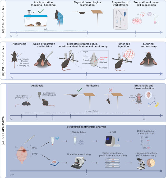
Figure 1: Pre-, intra- and post-operative procedures for the stereotactic intracortical tumor cell injection in mice. Schematic overview of the procedure. (A) The pre-operative phase includes animal acclimatization, health assessment, preparation of the surgical site and the tumor cell suspension. (B) The intra-operative phase involves anesthesia induction, scalp incision, placement in the stereotactic frame, precise craniotomy, injection of tumor cells into the cerebral cortex using stereotactic coordinates, wound closure and recovery from anesthesia. (C) The post-operative phase comprises administration of analgesics, routine monitoring to assess post-surgical recovery, general health status, and development of neurological symptoms (Hanging Wire Test), followed by euthanasia and organ harvesting. A structured postmortem analysis is included to assess metastatic load in fresh frozen (FF) tissue via qPCR and to evaluate histological growth patterns in FFPE samples from a digital slide library. Key procedural stages are illustrated sequentially to emphasize timing and surgical workflow. Image created in BioRender. Please click here to view a larger version of this figure.
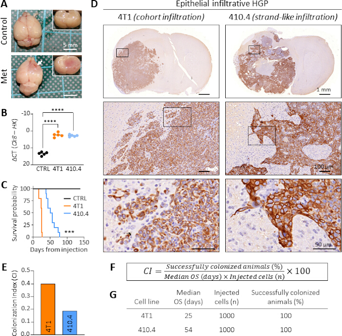
Figure 2: Representative results of metastatic growth in mouse models of brain metastasis following stereotactic injection of 4T1 and 410.4 breast cancer cells. (A) Macroscopic images showing the absence of tumor growth in ECM-injected control Balb/C mice (Control) and the presence of visible metastatic lesions in mice injected with tumor cell-ECM suspensions (Met). (B) Quantification of metastatic burden in ECM-injected control mice (CTRL) and mice with brain metastases derived from 4T1 and 410.4 breast cancer cells. Metastatic load was assessed by qPCR using Ck8 as a tumor marker, with Gapdh and Pgk1 as housekeeping genes (HK). Data are presented as mean values with individual data points. Statistical analysis was performed using one-way ANOVA followed by Dunnett's multiple comparisons test (n = 5; ****P < 0.0001). (C) Kaplan-Meier survival curves comparing overall survival (OS) of mice stereotactically injected with ECM alone (control, black), 4T1 (orange) or 410.4 (blue) tumor cells. Statistical analysis was performed using the Log-rank (Mantel-Cox) test (4T1 vs 410.4; n = 10; ***P < 0.001). (D) Representative histological images of brain metastases derived from 4T1 and 410.4 cells, illustrating differences in growth patterns, including cohort and strand-like infiltrative epithelial HGPs, respectively. Tissue sections were stained with an anti-cytokeratin 8 antibody. (E) Comparative analysis of colonization efficiency using the Colonization Index (CI). CI values of 4T1 (orange) and 410.4 (blue) are shown. (F) Formula used to calculate the CI. (G) Table summarizing the parameters included in the CI for both models. Please click here to view a larger version of this figure.
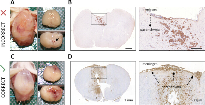
Figure 3: Representative examples of successful and suboptimal stereotactic injection outcomes. (A,C) Macroscopic images showing metastatic growth in different locations: (A) tumor growth in the skull and meninges resulting from an incorrect injection and leakage of tumor cells, and (C) successful parenchymal metastasis following proper intracortical injection. (B,D) Corresponding histological sections confirming (B) intrameningeal tumor growth with subsequent infiltration into the brain parenchyma, and (D) secondary dissemination to the meninges from an established parenchymal metastasis. Tissue sections were stained with an anti-cytokeratin 8 antibody. Please click here to view a larger version of this figure.
Discussion
The stereotactic intracranial injection technique enables the precise and localized delivery of tumor cells or cellular mixtures directly into the cerebral cortex of experimental mice. When combined with a structured postmortem analysis, this model is particularly valuable for studying late stages of brain metastasis, including metastatic outgrowth, secondary intra-organ dissemination, and the potential cause of death. In contrast, systemic injection methods (e.g., tail vein or intracardiac injection) are better suited for investigating the early steps of the metastatic cascade, such as tumor cell survival in the circulation, extravasation, and seeding in distant organs. However, these approaches are not brain-specific and typically result in low and inconsistent rates of brain colonization18,19,20.
Systemic models also show high biological variability, particularly in terms of which brain regions are affected, making therapeutic studies challenging and requiring large animal cohorts. Additionally, animals often develop extracranial metastases, which can lead to early trial discontinuation19. These limitations significantly reduce their usefulness for studying late-stage brain metastasis. Moreover, secondary dissemination and re-colonization are particularly difficult to investigate using systemic models, as distinguishing between primary spread (originating from the primary tumor) and secondary spread (arising from an already established metastasis) is nearly impossible. Furthermore, a structured postmortem analysis, especially for assessing macro-metastatic growth and the HGPs of brain metastases, is limited in these settings.
Compared to ex vivo systems, such as patient-derived organoids (PDOs) and organotypic brain slice (co)-cultures21,22,23,24, the stereotactic injection offers several critical advantages. While ex vivo models preserve aspects of native brain architecture and may include resident and infiltrating immune cells from patients or donors, they lack vascularization, systemic signaling, and the full complexity of the in vivo tumor microenvironment, particularly the further infiltration of T cells or other blood- or bone marrow-derived immune cells23. These limitations restrict their application to short-term studies and simplified settings, such as drug screening or basic metastatic cell-brain interaction assays. In contrast, the stereotactic approach supports long-term studies of tumor progression, immune response, and therapeutic intervention within a living, intact organism. Importantly, the evaluation of neurological symptoms as a humane endpoint allows for a clinically relevant measure of macro-metastatic onset. This makes the model even more reflective of the human disease course, which is especially valuable in translational research. Although it involves the use of animals, raising ethical considerations addressed by adherence to the 3Rs (Replacement, Reduction, and Refinement), this model currently cannot be fully replaced by ex vivo alternatives. Its ability to replicate the full physiological context of brain metastasis makes it indispensable for advancing our understanding of metastatic dynamics and CNS-specific pathophysiology.
Despite its numerous advantages, the stereotactic injection model has some limitations that should be considered. A key concern is the potential leakage of the tumor cell suspension during injection, which can result in misleading growth patterns. In particular, unintended adherence of tumor cells to the meninges may lead to iatrogenic meningeal metastasis8, which does not accurately reflect the natural course of metastatic dissemination in this particular cell line. To ensure data reliability, the growth patterns of a given tumor cell line should be consistently compared across animals, surgical procedures, and experimenters. To minimize tumor cell leakage, we propose using extracellular matrix (ECM), as its gel-like consistency and ability to solidify at body temperature make it a useful tool. However, ECM also presents challenges, including the need for rapid handling during injection to prevent premature solidification. Another consideration in our protocol is that tumor cells are injected in a mixture of ECM and serum-containing media. While FBS enhances tumor cell engraftment by supplying nutrients that are typically limited in the brain, it may also promote inflammation. If this is a concern, serum-free media or balanced salt solutions (e.g., HBSS or PBS) can be used as alternatives.
In addition, stereotactic injection is technically more demanding than other methods, such as intravenous (i.v.) injection. It requires specific training to ensure reproducibility, and the procedure is significantly more time-consuming, typically taking 15-30 min per animal compared to 3-5 min for i.v. injections. This longer duration limits the number of animals that can be processed per session, which may pose a challenge for therapeutic studies requiring large cohorts to achieve statistical power. However, this limitation can be effectively addressed by distributing injections over multiple days, which not only increases experimental flexibility and feasibility but also introduces valuable biological variability, reflecting more realistic inter-individual differences in both tumor cell behavior and host response.
Finally, it is important to note that this model bypasses earlier steps of the metastatic cascade, as previously discussed. The appropriate experimental model should be selected based on the research question and the specific phase of metastasis being studied. For investigating late-stage brain colonization, the stereotactic intracortical injection remains the gold standard.
Disclosures
The authors declare that they have no competing interests.
Acknowledgements
J.A.L. and R.B. received funding from the DFG (TRR305-B03).
Materials
| Name | Company | Catalog Number | Comments |
|---|---|---|---|
| Animal ear punch pliers | LabArt | ||
| Bepanthen® eye and nose ointment | Bayer Vital GmbH | 1578675 | |
| Bone wax | B.Braun | 210434 | |
| Cotton swabs | Hartmann | 143213 | |
| Cultrex Basement Membrane (ECM) Extract | B&D | 3432-005-01 | |
| Disinfectant | B.Braun | 107190 | |
| Drill Bit | Bilaney Consultants GmbH | D #76 | |
| Eye scissors | Hermle | 501 | |
| Hamilton syringe (10µL volume) | VWR | 5491135 | |
| Heating pad | Intergastro | 258122 | |
| Mouse Nase/Tooth Bar Assay (900/1430) | Bilaney Consultants GmbH | DKI 926-B | |
| Noyes eye scissors | Hermle | 545 | |
| Paur 60° tip non noptore Mouse Ear Bars | Bilaney Consultants GmbH | DKI 922 | |
| Scalpel | PFM Medical AG | 17027 | |
| Semkin standard forceps | Hermle | 710 | |
| Seralon polyamide suture (DR-009, USP 7/0, EP 0.5) | Serag-Wiessner GmbH | V0053491 | |
| Stereotaxic Drill -Jacobs Chuck-18000rpm | Bilaney Consultants GmbH | DKI 1471-200-CE-A | |
| Stereotaxic without Rat adaptor | Bilaney Consultants GmbH | DKI 963 % 920 | |
| Syringe Hdder for 5&10µl Hamilton | Bilaney Consultants GmbH | DKI 1772-F1 | |
| TC Needleholders Mayo-Heear 160mm | Hermle | 6425 |
References
- Wu, A., Colón, G. R., Lim, M. Quality of life and role of palliative and supportive care for patients with brain metastases and caregivers: A review. Front Neurol. 13, 806344(2022).
- Ostrom, Q. T., Wright, C. H., Barnholtz-Sloan, J. S. Brain metastases: Epidemiology. Handb Clin Neurol. 149, 27-42 (2018).
- Aizer, A. A., et al. Brain metastases: A society for neuro-oncology (sno) consensus review on current management and future directions. Neuro Oncol. 24 (10), 1613-1646 (2022).
- Wang, B. X., et al. Impacts of egfr mutation and egfr-tkis on incidence of brain metastases in advanced non-squamous nsclc. Clin Neurol Neurosurg. 160, 96-100 (2017).
- Goncalves, P. H., et al. Risk of brain metastases in patients with nonmetastatic lung cancer: Analysis of the metropolitan detroit surveillance, epidemiology, and end results (seer) data. Cancer. 122 (12), 1921-1927 (2016).
- Kim, Y. J., Kim, J. S., Kim, I. A. Molecular subtype predicts incidence and prognosis of brain metastasis from breast cancer in seer database. J Cancer Res Clin Oncol. 144 (9), 1803-1816 (2018).
- Sampson, J. H., Carter, J. H. Jr, Friedman, A. H., Seigler, H. F. Demographics, prognosis, and therapy in 702 patients with brain metastases from malignant melanoma. J Neurosurg. 88 (1), 11-20 (1998).
- Sparrer, D., et al. Primary and secondary metastatic dissemination: Multiple routes to cancer-related death. Molecular Cancer. 24 (1), 203(2025).
- Siam, L., et al. The metastatic infiltration at the metastasis/brain parenchyma-interface is very heterogeneous and has a significant impact on survival in a prospective study. Oncotarget. 6 (30), 29254-29267 (2015).
- Proescholdt, M. A., et al. Metinfilt: A prospective trial highlighting the importance of the histological growth pattern in brain metastases. Translational Oncology. 60, 102480(2025).
- Berghoff, A. S., et al. Invasion patterns in brain metastases of solid cancers. Neuro Oncol. 15 (12), 1664-1672 (2013).
- Blazquez, R., et al. The macro-metastasis/organ parenchyma interface (mmpi) - a hitherto unnoticed area. Semin Cancer Biol. 60, 324-333 (2020).
- Dankner, M., et al. Invasive growth associated with cold-inducible rna-binding protein expression drives recurrence of surgically resected brain metastases. Neuro Oncol. 23 (9), 1470-1480 (2021).
- Blazquez, R., et al. Intralesional tlr4 agonist treatment strengthens the organ defense against colonizing cancer cells in the brain. Oncogene. 41 (46), 5008-5019 (2022).
- Blazquez, R., et al. Pi3k: A master regulator of brain metastasis-promoting macrophages/microglia. Glia. 66 (11), 2438-2455 (2018).
- Blazquez, R., et al. Lef1 supports metastatic brain colonization by regulating glutathione metabolism and increasing ros resistance in breast cancer. Int J Cancer. 146 (11), 3170-3183 (2020).
- Heppner, G. H., Miller, F. R., Shekhar, P. M. Nontransgenic models of breast cancer. Breast Cancer Res. 2 (5), 331-334 (2000).
- Francia, G., Cruz-Munoz, W., Man, S., Xu, P., Kerbel, R. S. Mouse models of advanced spontaneous metastasis for experimental therapeutics. Nature Reviews Cancer. 11 (2), 135-141 (2011).
- Singh, M., Bakhshinyan, D., Venugopal, C., Singh, S. K. Preclinical modeling and therapeutic avenues for cancer metastasis to the central nervous system. Frontiers in Oncology. 7, 220(2017).
- Valastyan, S., Weinberg, R. A. Tumor metastasis: Molecular insights and evolving paradigms. Cell. 147 (2), 275-292 (2011).
- Zhu, L., Miarka, L., Baena, P., Perea-García, M., Valiente, M. Protocol to generate murine organotypic brain cultures for drug screening and evaluation of anti-metastatic efficacy. STAR Protocols. 4 (2), 102194(2023).
- Steindl, A., Valiente, M. Potential of ex vivo organotypic slice cultures in neuro-oncology. Neuro Oncol. 27 (2), 338-351 (2025).
- Blazquez, R., et al. Organotypic 3d ex vivo co-culture model of the macro-metastasis/organ parenchyma interface. Methods Mol Biol. 2764, 165-176 (2024).
- Chuang, H. N., et al. Coculture system with an organotypic brain slice and 3d spheroid of carcinoma cells. J Vis Exp. (80), e50881(2013).
Reprints and Permissions
