Endoscopic en bloc resection for thyroid carcinoma presents significant technical challenges, with current literature lacking comprehensive methodological descriptions. A systematic development and description of the "Sun's Seven-Step Technique" for endoscopic en-bloc thyroidectomy via the chest-breast approach was presented, demonstrating its superior operative efficiency, safety, and clinical feasibility.
Method Article
"Sun's Seven-Step Technique" for Endoscopic En-Bloc Resection of Thyroid Cancer via the Chest-Breast Approach
* These authors contributed equally
In This Article
Summary
Abstract
The oncological radicality and cosmetic outcomes of endoscopic thyroid cancer surgery via the chest-breast approach have been confirmed by numerous studies. The concept of en-bloc resection has been increasingly applied in radical thyroidectomy, whether in open or endoscopic procedures. However, endoscopic en-bloc resection for thyroid cancer presents significant technical challenges, and detailed methodological descriptions remain scarce. To address this, a systematic endoscopic en-bloc resection protocol, termed the "Sun's Seven-Step Technique", was developed. This technique incorporates optimized surgical plane selection, modified operative sequences, and a specialized lymphatic tracer injection method. Compared with the conventional resection group (lobectomy followed by separate lymph node dissection), the en-bloc resection group showed no significant differences in blood loss, lymph node dissection efficacy, or surgical complications; however, it achieved markedly shorter operative times. "Sun's Seven-Step Technique" demonstrates better compliance with oncological radicality principles, facilitates the identification of accidentally resected inferior parathyroid glands, and utilizes preserved tissue integrity to provide mechanical support for exposing lower-level lymph nodes. It served as a valuable reference surgical protocol.
Introduction
The safety, feasibility, and cosmetic outcomes of endoscopic thyroid surgery have been increasingly validated by numerous studies, garnering growing recognition and preference among both surgeons and patients1,2,3. In China, there are mainly three approaches for endoscopic thyroid surgery: the chest-breast approach, transaxillary approach, and transoral approach. Among them, the chest-breast approach has emerged as the most widely adopted technique owing to its broader indications and technically straightforward execution.
However, in endoscopic thyroid cancer surgery, concerns have been raised regarding potential oncological radicality due to inferior visual field limitations inherent in this approach, where the manubrium sterni and clavicular heads may obstruct lower-level lymph nodes, as the endoscopic view progresses from inferior to superior. A previous study4 detailed techniques for addressing inferior visual field limitations inherent to this approach, employing a sequenced procedural order at the time: initial lobectomy followed by gland removal, and then proceeding to subsequent lymph node dissection. With accumulated surgical experience, enhanced familiarity with endoscopic anatomical perspectives, and the widespread adoption of preoperative needle biopsy, the concept of en-bloc resection has gained increasing application in endoscopic radical thyroidectomy5,6,7.
En-bloc resection refers to the removal of the primary lesion, its affiliated organ, and regional lymphatic-adipose tissues as a single intact block, thereby achieving radical resection of the tumor, standardizing the scope of surgery and dissection, ensuring omission-free radical resection, and maximizing patient benefits. Given that endoscopic en-bloc resection was technically more challenging than the conventional sequential approach, its implementation should be based on substantial experience with both traditional endoscopic thyroid cancer surgeries and open thyroid en-bloc resections. Patient selection criteria must also be more stringent. Tumors or suspicious lymph nodes smaller than 25 mm may be considered indications for surgery, whereas cases with extrathyroidal extension, retrosternal suspicious lymph node metastasis, secondary surgery, or previous neck surgery history should be excluded.
The author has routinely performed en-bloc resection in endoscopic thyroid cancer surgery via the chest-breast approach since August 2022. After overcoming the learning curve, a fixed operative pathway and feasible surgical protocol gradually formed, culminating in the "Sun's Seven-Step Technique". This article will further study the feasibility, superiority, and technical strategies of the seven-step en-bloc resection method for thyroid cancer under endoscopy.
Protocol
The protocol adheres to the guidelines of the Human Research Ethics Committee of Chaozhou Central Hospital. No specific ethics approval is required since this treatment was performed in routine clinical care. The consumables and equipment used are listed in the Table of Materials.
1. Operating setting and anesthesia
- Administer general anesthesia via endotracheal intubation (following institutionally approved protocols). Place the patient supine on the operating bed with the lower limbs abducted approximately 30°. Elevate the shoulders using padding to maintain cervical hyperextension. Perform routine disinfection and draping.
- Position the surgeon between the patient's legs. Position the endoscope holder lateral to the patient's right leg and to the left of the surgeon. Have the second assistant stand on one side of the patient.
- Make a 12-mm incision at the 2-4 o'clock position of the right areola. Inject approximately 30 mL of normal saline with adrenaline (1:200,000) using a Veress needle to distend the subcutaneous tissues. Use a specially designed separation rod to create a subcutaneous tunnel extending to the suprasternal fossa.
- Insert a 10-mm trocar and introduce a 10-mm 30° endoscope. Set the CO2 gas pressure to 6 mmHg. Create bilateral 5-mm incisions at the 11 o'clock positions of both areolae. Under endoscopic visualization, insert a 5-mm trocar into each incision, designating the left areolar port as the dominant operating channel.
2. Surgical technique (Sun's Seven-Step Technique)
- Establish the surgical space
- Expose the lower segments of the bilateral sternocleidomastoid muscles (SCM). In the midline of the neck, separate and expose the sternohyoid muscle closely adhering to the deep layer. Continue dissection upward along the surface of the sternohyoid muscle (Figure 1A), and separate the sternocleidomastoid muscle, subcutaneous fat, and anterior cervical vein toward the upper part of the visual field to form the skin flap (Figure 1B).
- Extend the surgical field superiorly to the level of the thyroid cartilage and laterally to the medial borders of the SCM bilaterally, completing delineation of the operative space.
- Expose the thyroid gland and inject the lymphatic tracer agent.
- Make an inferior-to-superior incision through the linea alba at the lower point of the surgical field to expose the thyroid gland. Insert a 7 G silk suture percutaneously from the cervical skin surface to suspend the anterior cervical muscles and retract them laterally. Further separate the anterior capsule of the thyroid gland.
- Using a custom-designed "Z"-shaped long needle, inject 0.1 mL of lymphatic tracer into the thyroid gland (Figure 2A). Apply a gentle digital massage to facilitate tracer dispersion throughout the gland, then wait for approximately 1 min.
- Expose the common carotid artery (CCA) and identify the initial segment of the recurrent laryngeal nerve (RLN)
- Continue lateral and deep dissection of the thyroid capsule until identifying the CCA, which serves as the lateral boundary for central lymph node dissection (CLND).
- Carefully divide the visceral fascia layer by layer along the medial border of the carotid sheath to isolate the initial segment of the RLN located posterior to the carotid sheath (Figure 2B).
- Expose the thymus and identify the inferior parathyroid gland (PTG).
- Continue dissection along the lateral thyroid capsule inferiorly, tracing the CCA downward to expose the innominate artery. Identify the lingual lobe of the thymus anterior to the innominate artery, then trace the thymus to its distal extent to assess exposure of the inferior PTG (Figure 3A).
NOTE: If the inferior PTG cannot be identified at this location, expose the inferior thyroid artery for further exploration (Figure 3B). Preserve the gland in situ whenever possible. If in situ preservation is not feasible, resect and autotransplant the gland into the SCM.
- Continue dissection along the lateral thyroid capsule inferiorly, tracing the CCA downward to expose the innominate artery. Identify the lingual lobe of the thymus anterior to the innominate artery, then trace the thymus to its distal extent to assess exposure of the inferior PTG (Figure 3A).
- Separate the lymph nodes anterior and posterior to the RLN.
- After managing the inferior PTG, retract and dissect the pretracheal lymphatic-adipose tissue toward the affected side (Figure 4A), using the contralateral inferior thyroid vein as an anatomical landmark (Figure 4B). Continue dissection along the established plane extending to the affected tracheoesophageal groove (Figure 4C).
- Enter the plane posterior to the RLN between the RLN and trachea on the affected side (Figure 5A). Expose the prevertebral space (Figure 5B) and dissect the lymphatic-adipose tissue posterior to the RLN.
- Retract the posteromedial tissue of the RLN anterolaterally (Figure 5C). Progressively expose the RLN superiorly while dissecting the surrounding lymphatic-adipose tissue until reaching the inferior thyroid pole, thereby completing the CLND.
- Divide the isthmus, resect the pyramidal lobe, and dissect the prelaryngeal lymph nodes.
- Divide the isthmus anterior to the trachea near the contralateral side, then dissect superiorly to expose the cricothyroid muscle (Figure 6A).
- Resect the pyramidal lobe in the prelaryngeal region and dissect the prelaryngeal lymph nodes.
- Remove the thyroid lobe and complete the operation.
- Retract the inferior thyroid pole and dissected central lymphoadipose tissue superiorly. Place a dark protective strip anterior to the RLN, then dissect along the RLN tunnel superiorly until reaching the nerve's laryngeal entry point.
- At the RLN's laryngeal entry point, identify and preserve the superior PTG in situ on the posterior aspect of the thyroid gland (Figure 6B). Divide the superior pole vessels.
- Remove the specimen, irrigate the surgical field, and place a drainage tube in the thyroid fossa through the chest incision. Maintain negative pressure drainage, then withdraw the trocars and surgical instruments. Suture and close the incisions to complete the surgery (Figure 6C).
3. Postoperative management
- After returning the patient to the ward, continuously monitor the drain tubes and neck condition. Perform routine electrocardiogram monitoring and provide oxygen inhalation for 6 h.
- Administer intravenous and/or oral calcium supplementation to patients with hypocalcemia or hypoparathyroidism.
- Administer routine postoperative oral levothyroxine sodium. Monitor and adjust the thyroxine dosage regularly based on recurrence risk stratification to maintain individualized TSH targets.
Results
Consecutive cases of papillary thyroid carcinoma (PTC) who received endoscopic surgery (lobectomy and CLND) from June 2021 to June 2024 were collected. Cases before July 2022 underwent the conventional resection sequence (thyroid resection followed by lymph node dissection, Group CR), while cases from August 2022 onward underwent en-bloc resection (Group ER). All cases were sourced from the First Affiliated Hospital of Jinan University and Chaozhou Central Hospital, with all procedures performed by the author (Peng Sun). Inclusion criteria: (1) PTC measuring ≤ 25 mm in greatest diameter; (2) cN0 or cN1a, the largest lymph node ≤ 25 mm in diameter. To eliminate the interference of the exploratory process on the research outcomes, the initial 20 cases of en-bloc resection were excluded from the study, as a fixed operative sequence and procedure had not yet been established during the initial exploratory phase. Given the differential surgical complexity between left and right-sided thyroid cancers, only cases undergoing right lobectomy plus right CLND were selected. Left-sided carcinomas, total thyroidectomies, and cases without CLND were excluded. The final cohort comprised 68 cases in Group CR and 79 cases in Group ER. All patients were equally informed about the availability of intraoperative neuromonitoring (IONM) to assist in protecting the RLN, with the decision to use it determined by the patient's financial situation and personal preference. Chi-square and Student's t-tests were employed for intergroup comparisons. Continuous variables are expressed as mean ± standard deviation (SD). Statistical significance was defined as a two-tailed P-value < 0.05 for all analyses.
The demographic and clinical characteristics of patients in Group CR and Group ER are presented in Table 1. There were no statistically significant differences between the two groups in terms of gender, age distribution, tumor size, or the proportion of IONM usage. The comparison of surgical outcomes between the two groups is presented in Table 2. The operation time was significantly shorter in Group ER than in Group CR (112.0 ± 13.6 min vs. 103.3 ± 10.76 min, P < 0.001). No significant differences were observed in intraoperative blood loss, number of dissected lymph nodes, or number of metastatic lymph nodes between the groups (all P > 0.05). In the Group ER, one patient experienced intraoperative loss of neuromonitoring signal and developed postoperative hoarseness, which resolved spontaneously after two months. This was considered a result of thermal injury caused by the harmonic scalpel. No other patients experienced RLN injury (all had normal neuromonitoring signals and postoperative vocal function), hypoparathyroidism, infection, hemorrhage, or chyle leakage. All patients were routinely followed up at postoperative months 1, 3, and 6, with assessments including thyroid function, thyroglobulin, and thyroglobulin antibody levels. Neck ultrasound was performed at 3 months postoperatively and repeated every six months thereafter, along with blood tests. All patients were followed up until June 2025. The mean follow-up time was (40.9 ± 4.5) months in the Group CR and (23.7 ± 6.0) months in the Group ER. No evidence of recurrence was observed in any case.
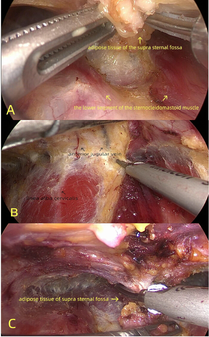
Figure 1: Exposure of the anterior cervical region. (A) Separation along the surface of the sternohyoid muscle. (B) The sternocleidomastoid muscle, subcutaneous fat, and anterior cervical vein were separated and retracted superiorly to widen the visual field. (C) Excision of the suprasternal fossa adipose tissue at the superficial aspect of the flap. Please click here to view a larger version of this figure.
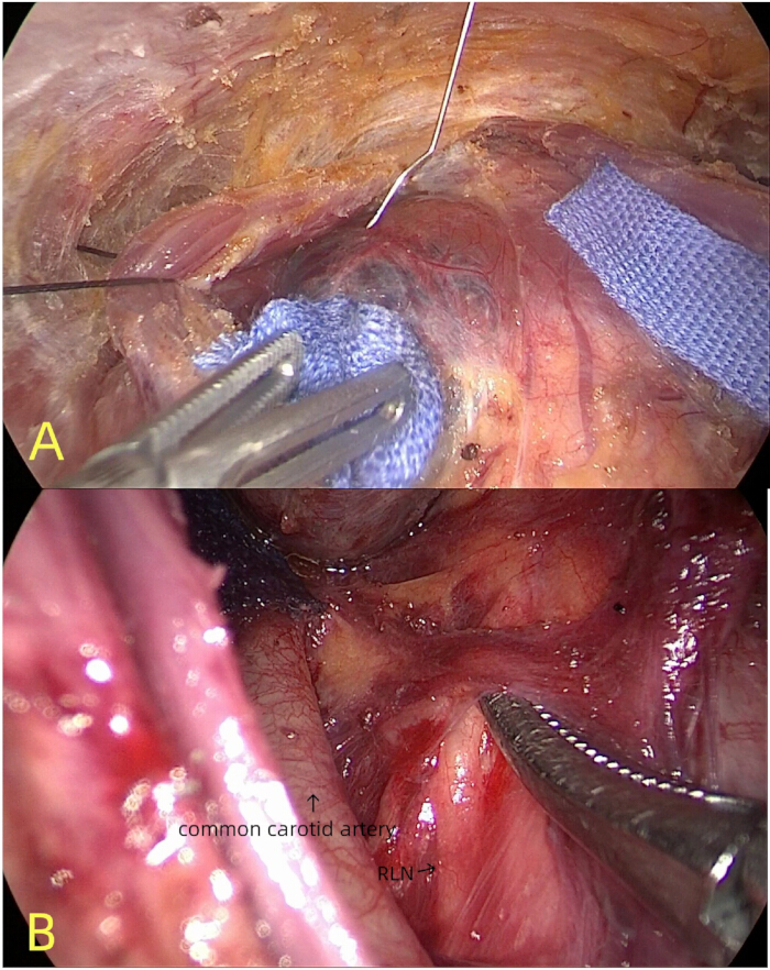
Figure 2: Localization of the recurrent laryngeal nerve (RLN). (A) A "Z"-shaped long needle was used to inject the lymphatic tracer into the thyroid gland. (B) The initial segment of the RLN was identified along the medial border of the common carotid artery (CCA). Please click here to view a larger version of this figure.
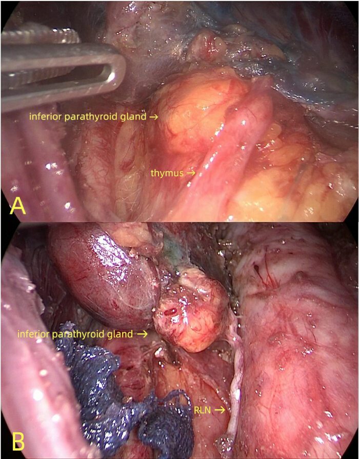
Figure 3. Identification of the inferior parathyroid gland (PTG). (A) The inferior PTG was exposed at the terminal end of the lingual lobe of the thymus. (B) The inferior PTG was exposed near the inferior thyroid artery. Please click here to view a larger version of this figure.
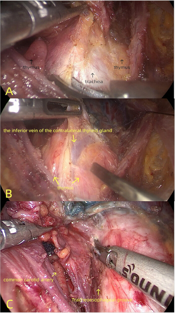
Figure 4: Dissection of the central lymphatic-adipose tissue. (A) The pretracheal lymphatic-adipose tissue was retracted and dissected toward the affected side. (B) Identification of the contralateral thyroid inferior vein, marking the medial boundary of the central lymph node dissection. (C) Exposure of the tracheoesophageal groove. Please click here to view a larger version of this figure.
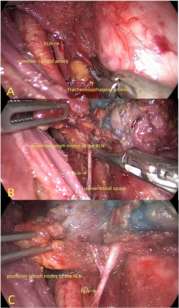
Figure 5: Exposure and mobilization of the RLN and prevertebral space. (A) Entry into the plane posterior to the RLN between the RLN and the trachea on the affected side. (B) Exposure of the prevertebral space. (C) Retraction of the posteromedial tissue of the RLN in an anterolateral direction. Please click here to view a larger version of this figure.
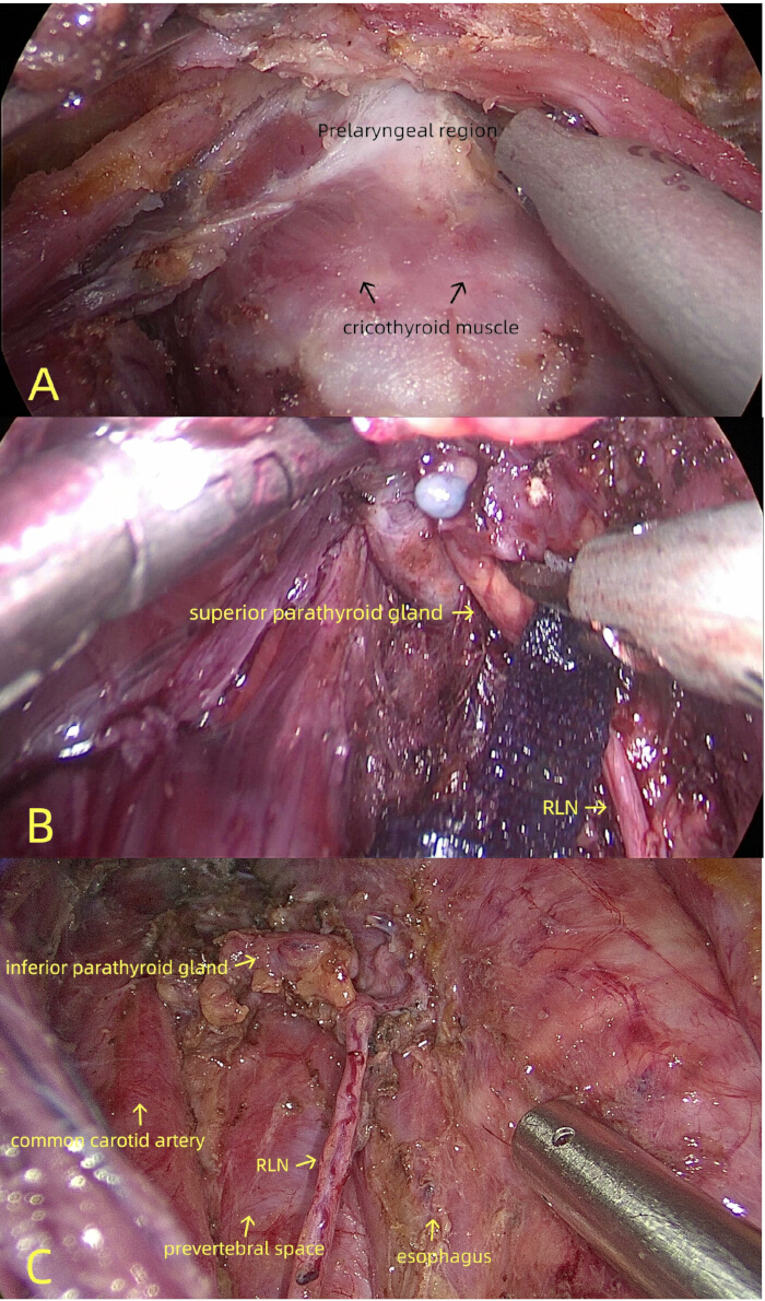
Figure 6: Completion of central lymph node dissection. (A) Removal of the pyramidal lobe and prelaryngeal lymph nodes. (B) Dissection along the posterior thyroid capsule to preserve the superior PTG in situ. (C) Postoperative appearance following right central lymph node dissection. Please click here to view a larger version of this figure.
| Groups | Male/Female | Age (Years) | Tumor size (mm) | IONM(with/without) |
| Group CR (n=68) | 13/55 | 34.7±9.8 | 14.8±8.1 | 61/7 |
| Group ER (n=79) | 15/64 | 35.3±10.1 | 13.9±7.8 | 70/9 |
| P value | 0.984 | 0.585 | 0.834 | 0.831 |
Table 1: Demographic and clinical characteristics of patients. Abbreviation: IONM, intraoperative neuromonitoring.
| Group CR(n=68) | Group ER(n=79) | P value | |
| Intraoperation | |||
| *Operation time(min) | 112.0 ± 13.6 | 103.3 ± 10.76 | 0 |
| *Intraoperative blood loss(mL) | 11.6 ± 6.2 | 13.1 ± 7.1 | 0.658 |
| *No. of dissected lymph nodes | 6.7 ± 4.2 | 7.2 ± 4.2 | 0.721 |
| *No. of metastatic lymph nodes | 2.1 ± 2.1 | 2.6 ± 2.2 | 0.367 |
| Complications | |||
| Temporal RLN palsy | 0 | 1 | - |
| Hypoparathyroidism | 0 | 0 | - |
| Recurrence | 0 | 0 | - |
Table 2: Comparison of surgical results between the two groups. Abbreviation: RLN, recurrent laryngeal nerve. *Values are expressed as mean ± standard deviation.
Discussion
According to the Chinese guidelines for the diagnosis and management of differentiated thyroid cancer (DTC)8, CLND was advocated for PTC while ensuring functional preservation of the RLN and PTG. Consequently, lobectomy plus CLND constitutes the standard surgical approach. With accumulated endoscopic experience and technical refinements, en-bloc resection has been increasingly adopted in endoscopic thyroid surgery5,6,7. However, previous studies predominantly focused on comparative outcomes rather than providing procedural demonstrations and technical specifications. This article details the operative sequence with stepwise technical standards, aiming to establish a practical reference manual for this technique.
Some scholars prefer transaxillary or transoral endoscopic thyroid cancer surgery, questioning the oncological radicality of the chest-breast approach due to inadequate exposure of low-lying central compartment lymph nodes. Previous publication addressed these concerns by presenting surgical outcomes and countermeasures. Two critical innovations were introduced: (1) Surgical Plane Adjustment: Unlike open surgery's subplatysmal dissection, endoscopic dissection proceeds immediately superficial to the anterior cervical muscles, redirecting subcutaneous fat and anterior jugular veins to the flap (superior in endoscopic view). This key maneuver allows complete removal of suprasternal fossa adipose tissue (Figure 1C), resulting in bare anterior cervical muscles devoid of fatty coverage. Though seemingly minor, this step was pivotal for exposing lower-level nodes, unremoved fossa fat would obstruct the endoscopic view, severely compromising low-node visualization. (2) Visceral Fascia Integrity Preservation: Maintain intact visceral fascia during dissection to leverage its "enveloping traction" effect for retracting lower lymphatic-adipose tissue. Analogous to fishing with an unbroken net, this "Net-Trawl Dissection" technique retrieves nodes from visual blind zones. These innovations ensure complete exposure of inferior nodes, achieving open-surgery-equivalent resection boundaries while preventing omissions.
The efficacy of lymphatic tracing in ensuring oncological radicality and parathyroid protection has been validated by multiple studies9,10,11,12. Optimal outcomes depend on achieving homogeneous intraglandular dispersion and uniform staining of the tracer. Excessive injection or tracer leakage compromises surgical efficacy13. Some scholars advocated preoperative ultrasound-guided tracer administration to enhance staining quality14. The development of a custom-engineered "Z"-shaped needle enabled endoscopic depth-controlled tracer injection. This design facilitates homogeneous dispersion within the glandular parenchyma, thereby enhancing lymph node mapping accuracy, improving lymph node retrieval rates, and further guaranteeing oncological radicality and surgical outcomes.
The en-bloc resection sequence fundamentally differs from conventional methods, adhering to the principle: lateral-to-medial, inferior-to-superior progression, prioritizing exposure of the RLN and inferior PTG, with lymph node dissection preceding gland resection. For right-sided procedures, the protocol was as follows: (1) Preserve the isthmus, utilizing its adhesion to the trachea to provide countertraction medially and superiorly. The lateral thyroid capsule was first dissected to expose the CCA and the initial RLN segment. (2) Identify and expose the inferior PTG by tracing the thymus lingula or the inferior thyroid artery branches. (3) At the pretracheal plane, use the contralateral inferior thyroid vein as the medial boundary marker. Dissect laterally along this plane toward the tracheoesophageal groove. (4) Enter the plane posterior to the RLN between the trachea and nerve. Develop the prevertebral space and dissect lymphatic-adipose tissue superiorly along this plane until reaching the inferior thyroid pole. (5) Divide the isthmus, resect the pyramidal lobe, and dissect the prelaryngeal lymph nodes. (6) Mobilize the thyroid lobe and remove the entire specimen containing the gland and lymphatic-adipose tissue.
Given that the RLN traverses through right-sided lymphatic-adipose tissue, dividing the right central compartment into pre-RLN and post-RLN nodal basins, excessive traction during dissection increases RLN injury risk. Consequently, endoscopic right CLND poses greater technical challenges than left-sided procedures15. Some scholars advocated against pursuing complete en-bloc resection during endoscopic right thyroid cancer surgery. Other scholars proposed the "Alar Fascia Theory", recommending initial integrated resection of the pre-RLN lymphatic-adipose tissue along with the thyroid gland, followed by separate dissection of the post-RLN lymph nodes16,17. While acknowledging these perspectives, emphasizing that surgical outcomes supersede rigid adherence to en-bloc principles, this study achieved complete single-stage resection of pre-RLN nodes, post-RLN nodes, and the thyroid lobe in all enrolled right-sided cases. We initiate dissection immediately along the tracheal border, accessing the retro-RLN space from its medial aspect. Upon identification of the prevertebral space within the deep plane, we first resect the lymphatic-adipose tissue posterior to the RLN. Following mobilization, this tissue was retracted laterally beyond the RLN, after which the pre-RLN nodal tissue was dissected. During the procedure, gentle manipulation controls the risk of RLN injury to a level comparable to that of conventional methods.
In this study, the Group ER demonstrated only a shorter operative time compared to the conventional group, potentially due to limited sample size and selection bias. However, based on our operative experience, we summarize the advantages of en-bloc resection as follows: (1) Time Efficiency - Eliminates separate dissection of gland and lymph nodes; (2) Anatomical Preservation - Maintains tissue relationships, facilitating identification of inadvertently resected parathyroid glands; (3) Oncological Radicality - Standardizes resection and dissection boundaries per tumor principles; (4) Mechanical Advantage -En-bloc removal of thyroid with periglandular lymphatic-adipose tissue provides traction-mediated exposure of lower-level lymph nodes. We look forward to future studies with larger sample sizes, longer follow-up periods, prospective designs, and more refined approaches (such as exploring the positive significance of parathyroid protection).
In summary, "Sun's Seven-Step Technique" for endoscopic en-bloc resection of thyroid carcinoma was safe and feasible. Compared with conventional methods, it yields higher operative efficiency and demonstrates certain advantages, potentially offering a valuable technical standard and reference framework for endoscopic en-bloc resection. However, due to its procedural complexity, it is recommended that the technique be performed by experienced surgeons.
Disclosures
The authors declare that the research was conducted in the absence of any commercial or financial relationships that could be construed as a potential conflict of interest.
Acknowledgements
This study was funded by the Scientific Research Project of Chaozhou Health Commission (grant number 2024043).
Materials
| Name | Company | Catalog Number | Comments |
|---|---|---|---|
| Carbon Nanoparticles Suspension Injection | Chongqing Lummy Pharmaceutical Co.,Ltd | National Drug Approval Number H20073246 | Lymphatic tracer |
| Intraoperative nerve monitoring system | Medtronic | CNDA Registration Number: 2023307016 | Intraoperative nerve monitoring |
| Laparoscopic system | STORZ MEDICAL | CNDA-2020-060294 | Endoscope |
| Ultrasonic scalpel | Ethicon Endo-Surgery,LLC | CNDA-SPR-20153010137 | Used for separating skin flaps during surgery and removing the thyroid gland |
References
- Pace-Asciak, P., Russell, J. O., Tufano, R. P. Surgical treatment of thyroid cancer: Established and novel approaches. Best Pract Res Clin Endocrinol Metab. 37 (1), 101664(2023).
- Lu, Q., Zhu, X., Wang, P., Xue, S., Chen, G. Comparisons of different approaches and incisions of thyroid surgery and selection strategy. Front Endocrinol (Lausanne). 17 (14), 1166820(2023).
- Nguyen, V. C., et al. Feasibility of remote-access and minimally invasive video-assisted approaches in lateral neck dissection for papillary thyroid carcinoma: A systematic review and network meta-analysis. Eur J Surg Oncol. 50 (9), 108469(2024).
- Sun, P., Zhan, J., Chong, T. H., Li, J., Wang, C. Endoscopic central lymph node dissection of thyroid cancer via chest-breast approach: Results, indications, and strategies. Surg Endosc. 36 (6), 4239-4247 (2022).
- Xie, Q., et al. Feasibility and safety of modified en-bloc resection in endoscopic thyroid surgery via bilateral areolar approach: Long-term institutional analysis ten years after surgery. Front Endocrinol (Lausanne). 17 (15), 1302510(2024).
- Chen, W., et al. The learning curve for gasless transaxillary posterior endoscopic thyroidectomy for thyroid cancer: A cumulative sum analysis. Updates Surg. 75 (4), 987-994 (2023).
- Sheng, X., Liu, J., Fang, J., Zheng, X., Wang, S. En bloc resection of total thyroid and bilateral central compartment lymph nodes via a gasless transoral approach in papillary thyroid carcinoma. Front Endocrinol (Lausanne). 27 (14), 1130791(2023).
- Guidelines for the diagnosis and management of thyroid nodules and differentiated thyroid cancer (Second edition). Chin J Endocrinol Metab. 39 (3), Chinese Society of Endocrinology. 181-226 (2023).
- Ma, J. J., Zhang, D. B., Zhang, W. F., Wang, X. Application of nanocarbon in breast approach endoscopic thyroidectomy thyroid cancer surgery. J Laparoendosc Adv Surg Tech A. 30 (5), 547-552 (2020).
- Gao, H., et al. Application of nanocarbon tracing technology in thyroid cancer and its relationship with cytotoxic T lymphocyte antigen 4 gene polymorphism. J Nanosci Nanotechnol. 21 (2), 949-954 (2021).
- Liu, Y., et al. Radical resection of differentiated thyroid cancer in elderly patients: Evaluation of the efficacy of the immunocolloidal gold strip method combined with nanocarbon negative imaging tracing technology for parathyroid gland imaging. J Invest Surg. 38 (1), 2447850(2025).
- Lin, D. X., et al. Enhancing parathyroid preservation in papillary thyroid carcinoma surgery using nano-carbon suspension. Sci Rep. 14 (1), 24680(2024).
- Li, W. Infiltration of nanocarbon into the trachea during thyroidectomy surgery: Case series and literature review. Ann Med Surg (Lond). 86 (8), 4338-4343 (2024).
- Wang, Y., et al. Preoperative ultrasound-guided injection of nanocarbon for central lymph node dissection in patients with papillary thyroid carcinoma. Sci Rep. 14 (1), 29185(2024).
- Sun, P., Mak, T. K., Li, J., Wang, C. Endoscopic left central lymph node dissection of thyroid cancer: Safe, feasible, and relatively easy. Surg Innov. 28 (6), 747-753 (2021).
- Shan, J., Jiang, H., Ren, D., Wang, C. Anatomic relationship between right recurrent laryngeal nerve and cervical fascia and its application significance in anterior cervical spine surgical approach. Spine (Phila Pa 1976). 42 (8), 443-447 (2017).
- Phan, T., Lay, J., Scali, F. The alar fascia and danger space: A modern review. Cureus. 14 (12), e32871(2022).
Reprints and Permissions
