This protocol describes a rapid, reproducible organic solvent-based extraction and precipitation method for purifying elastin-like polypeptide (ELP) from Escherichia coli, providing a potentially scalable alternative to conventional ELP purification methods.
Method Article
Efficient Purification of Elastin-Like Polypeptides (ELPs) from E. coli Using an Organic Solvent-based Extraction and Precipitation Method
In This Article
Summary
Abstract
Elastin-like polypeptides (ELP) are engineered biopolymers built from repetitive pentapeptide sequences that mimic motifs found in mammalian tropoelastin. Their unique characteristics make them ideal candidates for a wide array of biomedical applications, ranging from drug and gene delivery to tissue engineering and targeted molecular imaging. Conventional purification approaches from Escherichia coli (E. coli) expression can be ineffective for ELP due to the formation of inclusion bodies. Other methods, such as inverse transition cycling (ITC), utilize the lower critical solution temperature (LCST) properties of ELP to separate it from contaminants such as lipopolysaccharides (LPS), but typically require multiple heating and cooling steps that are time-consuming and can result in low recoveries depending on the sequence, concentration, and molecular weight of the ELP construct. To tackle these challenges, we have developed an organic solvent-based extraction-precipitation workflow that exploits the intrinsic hydrophobicity of ELP to enable rapid, robust, and broadly applicable purification directly from E. coli cell pellets. This method uses polar organic solvents to aid in cell disruption and selectively solubilize ELP in a single step. A subsequent precipitation step effectively removes residual organic solvents, low-molecular-weight impurities, and endotoxins, yielding highly pure ELP with LPS levels below 1 EU/mL in under 3 h. Atomic force microscopy data suggest that ELP-fusion proteins purified in this manner can self-assemble into reverse micelle-like structures that retain fusion protein function. This rapid purification method offers researchers a straightforward and potentially scalable way to purify ELP, creating new possibilities for using ELP and their fusion proteins as flexible building blocks for material and biomedical applications.
Introduction
Elastin-like polypeptides (ELPs) are biopolymers composed of repeating (VPGXG)n motifs1,2,3,4,5,6,7,8,9. The guest residue X in the pentapeptide unit can be any amino acid other than proline. Variations in X can be used to tune the hydrophobicity, thermal transition behavior, and functional characteristics of the ELP construct to create modular biocompatible materials that have been widely explored in various biomedical applications10,11,12 .
While chemical synthesis can be used to produce short peptides, it is poorly suited for high molecular weight ELPs or for retaining the functionality of fusion proteins13,14. For reasons such as these, ELPs are typically expressed recombinantly in E. coli, allowing for sequence-defined production in large quantities. However, this approach can also bring challenges. During their expression in E. coli, ELPs often accumulate within dense inclusion bodies, requiring efficient recovery strategies to extract functional protein15. Moreover, bacterial expression introduces unwanted biomolecules such as host cell proteins, nucleic acids, and lipopolysaccharides (endotoxins) that must be separated from the ELP during purification. Traditional solubilization strategies using detergents (e.g., sodium dodecyl sulphate (SDS) or Triton X-100) or chaotropic agents like urea have met with limited success for our ELP fusion sequences. In our experience, Triton and urea failed to extract ELPs from the pellet phase, while SDS enabled partial solubilization but interfered with downstream affinity purification and was difficult to fully remove from the hydrophobic ELP sequence.
To date, inverse transition cycling (ITC) remains the most widely used purification method for ELPs. This technique exploits their LCST behavior to selectively precipitate and resolubilize the ELP across temperature and salt gradients16. However, with lower molecular weight proteins, ITC is a time-consuming multi-step process that is poorly suited for ELPs trapped in inclusion bodies, requiring multiple ITC cycles that can lead to protein loss at each step7,16.
To overcome these limitations, we adopted an organic solvent-based extraction and precipitation workflow. While Yakhnin et al. first demonstrated a hydrophobicity-driven partitioning strategy for GFP purification using ethanol and salt gradients in 199817, the novel method we developed employs organic solvent blends to directly extract ELP fusion proteins from E. coli cells7. Sweet et al. and Darji et al. adapted this approach to ELP fusions, showing that organic solvent extraction enables rapid recovery of functional ELPs while maintaining structural integrity and bioactivity8,9. This approach uses sonication and organic solvents to lyse bacterial cells and extract ELPs in a single operation, while precipitating most of the host proteins and nucleic acids. Recovery of the ELP-rich organic phase, followed by rapid precipitation to isolate the ELPs from solvents and solubilized impurities, yields highly pure ELPs. This approach is also capable of yielding pure ELP from E. coli pellets with endotoxin levels below 1 EU/mL in under three hours7,8.
Studies to date show that this method is capable of purifying ELP constructs and ELP fusion proteins of varying molecular weight and isoelectric point, without multiple ITC cycles or solubilization steps (Figure 1). Atomic force microscopy (AFM) and transmission electron microscopy (TEM) imaging suggest that ELP-fusion proteins purified in this manner form reverse micelle-like structures (Figure 2), thus helping to preserve the structure and function of ELP fusion domains during organic solvent extraction. This streamlined workflow enables faster and more reliable ELP purification for diverse biomedical applications7,8. Notably, Aayush et al. reported that ELP constructs purified by this method retained epidermal growth factor receptor-binding function18, highlighting the point that this method not only produces pure ELP material, but also maintains the activity of the fusion protein domain.
As a further demonstration of the utility of this method in isolating an ELP-enzyme fusion, we describe a detailed organic solvent-based method to purify an ELP-Intein-Chorismate mutase 2 (ELP-I-Cm2) construct (Figure 3). This method may be useful for rapid purification of other ELPs for drug delivery and nanomaterials applications.
Protocol
1. Materials preparation
- Bacterial strain: Use Escherichia coli BL21(DE3) chemically competent cells as the expression host.
NOTE: They were selected due to their high transformation efficiency and suitability for recombinant protein production. - Expression plasmid: Use pET21(+) vector encoding ELP-I-Cm2 fusion protein with ampicillin-resistance9. Purchase the plasmid vector from addgene. Insert the Cm2 gene using the BsrG I site at the end of the intein.
NOTE: The plasmid DNA sequence for this protein is provided in Supplementary File 1. - Growth media: Terrific Broth (TB) media preparation for 1200 mL media
- Prepare media (Part 1) by adding 16 g of Tryptone, 32 g of Yeast Extract, 16 g of proline, 6.8 mL of glycerol to 1100 mL of ultrapure water. Mix thoroughly to dissolve all components. Adjust the final volume to 1200 mL using ultrapure water.
- Prepare the potassium phosphate buffer (Part 2) by mixing 23.1 g of KH2PO4 and 125.4 g of K2HPO4, and then bring the volume to 1 L with ultrapure water.
- Autoclave the components (Parts 1 and 2) separately. After autoclaving, assemble the media by dividing the TB base into four 300 mL aliquots. Add 33 mL of phosphate buffer to each aliquot under sterile conditions.
- Additives for expression
- IPTG: Perform induction using IPTG at a final concentration of 1.2 mM (e.g., add 396 µL of 1 M IPTG to 330 mL culture).
- Antibiotic: Add ampicillin to a final concentration of 100 µg/mL to ensure plasmid selection.
- Lysis buffer (pH 8.5): Prepare the lysis buffer with 10 mM Tris Base and 22 mM ethylenediaminetetraacetic acid (EDTA). Adjust the pH to 8.5 using HCl or NaOH as required.
- Organic solvents for extraction
- Employ a variety of organic solvents (Table 1) to extract the ELP-I-Cm2 fusion protein from inclusion bodies. Test these solvents both individually and in binary combinations (1:1 vol: vol) to evaluate efficiency and ELP purity.
NOTE: All solvents used were HPLC Grade, except Acetonitrile (Optima grade), Ethanol (USP grade), and 1-Butanol (99% pure).
CAUTION: All solvents listed above are volatile and flammable. They must be handled inside a chemical fume hood using appropriate PPE (lab coat, nitrile gloves, and eye protection). Many types of plasticware are not compatible with organic solvents. Polypropylene plasticware is highly recommended for extraction-precipitation experiments with organic solvents; otherwise, the plasticware to be used should be tested without a valuable sample to ensure that it is appropriate for use. The disposal of solvent waste should follow the procedures outlined in the relevant institutional hazardous waste policies.
- Employ a variety of organic solvents (Table 1) to extract the ELP-I-Cm2 fusion protein from inclusion bodies. Test these solvents both individually and in binary combinations (1:1 vol: vol) to evaluate efficiency and ELP purity.
2. Cell lysis and preparation (Figure 4A)
- Resuspension: Weigh out 1 g of cell pellet and resuspend it in 4 mL of freshly prepared lysis buffer. Vortex gently until the pellet is fully dispersed and mixed with the buffer.
- Enzymatic lysis: Add ~5 mg of lysozyme to assist in cell wall digestion. Allow the suspension to incubate at 4 °C for 1 h. This facilitates partial enzymatic digestion to boost cell lysis.
- Sonication: After incubation, sonicate the sample to complete cell lysis and shear genomic DNA. Use short bursts of microtip probe sonication (350 W device, power level 3) with intervals to avoid overheating (e.g., 5 s on/10 s off, repeated as needed). Sonicate the samples on ice and apply the ultrasonic energy until a uniform lysate free of visible particulates is obtained, typically for about 5-8 min.
NOTE: After sonication is complete, store a 100 μL aliquot of the sample (or the lysate) for ELP expression analysis to monitor purification progress before and after the extraction and precipitation steps. - Centrifugation and expression analysis.
- Clarify the lysate by centrifugation at 10,000 g for 10 min at 20 °C, then carefully separate the supernatant (soluble fraction) from the pellet (insoluble/inclusion-body fraction).
- Assess ELP expression in both fractions by SDS-PAGE: withdraw an aliquot (e.g., 10-20 µL) of the supernatant and resuspend an equivalent pellet mass in lysis buffer (50 mM Tris-HCl pH 8.0, 150 mM NaCl, 1 mM EDTA) to the same volume; mix each with 4x Laemmli buffer to a final 1x, heat at 95 °C for 5 min, and load equal amounts to enable direct comparison.
- If the target ELP is predominantly soluble, proceed with the organic-solvent extraction using the clarified supernatant. If it is predominantly insoluble (inclusion bodies), discard the supernatant and continue with the inclusion-body workflow (washing/solubilization and extraction) by processing the pellet by organic solvent extraction.
- Pellet washing and drying (only for ELP in inclusion bodies): Invert the centrifuge tubes to allow gravity-assisted drainage of any residual lysis buffer. Once visible liquid has drained (~1-2 min), leave the pellet to air dry at room temperature for 5-10 min, ensuring it is free of excess moisture before proceeding to organic extraction.
3. Organic extraction (Figure 4B)
NOTE: Each ELP has its own preferred solvent condition for yielding high-purity extractions (e.g., AC performance in Figure 1). Therefore, an initial screen of 28 organic solvents and their combinations should be tested, and the isolates analyzed by PAGE to identify the optimal solubilization efficiency conditions for the construct of interest.
- Solvent screening. Screen the individual solvents shown in step 1.6 and their 1:1 (v/v) binary solvent mixtures. Evaluate additional mixtures (e.g., 1:2, 2:1, 4:1) to enhance the effectiveness of the process.
NOTE: At present, there are no guidelines for rationally selecting the most effective solvents or combinations, so a screening campaign is needed to select from solvents that have previously met with success in solubilizing hydrophobic protein aggregates. The final selection of the organic solvent extractant to be used for purification should be based on SDS-PAGE analysis of the isolates obtained from this screen. - Extraction procedure
- Solvent addition: Add 4 mL of organic solvent or solvent mixture to the air-dried pellet/supernatant obtained after lysis. For mixed combinations, ensure precise volumetric ratios (e.g., 2 mL of each for a 1:1 combination).
- Vortexing: Vortex the suspension vigorously for 1 min to ensure thorough mixing and penetration of solvent into the pellet.
- Incubation (Retention time): Allow the suspension to incubate at 20 °C for 5 min, ensuring sufficient interaction time for extracellular proteins to aggregate and sediment while the ELPs solubilize in the organic phase.
- Centrifugation: Centrifuge the mixture at 13,000 × g for 10 min at 20 °C to separate solubilized proteins from residual insoluble debris.
- Supernatant collection: Carefully collect the supernatant without disturbing the pellet. The supernatant contains the solubilized ELP, while the pellet contains host cell proteins, nucleic acids, and lipids.
- Volume measurement: Record the volume of the recovered supernatant precisely. Care should be taken to ensure the supernatant is free from any pellet particles. This measurement is critical for the downstream precipitation step.
4. Protein precipitation
- Anti-solvent addition: Add acetone or acetonitrile to the organic supernatant at 2.33 times the measured volume of the supernatant.
- Incubation: Incubate the mixture at 20 °C for 5-7 min.
- Centrifugation: Centrifuge at 10,000 g for 10 min at 20 °C.
- Pellet handling: After centrifugation, carefully discard the supernatant and air dry the sample by leaving the tube uncapped under a gentle nitrogen gas stream for 10-15 min, or incubate by placing uncapped centrifuge tubes upside down on lint-free wipes in a fume hood for 1 h at 20 °C (do not apply heat).
5. Protein pellet handling and resuspension
- Resuspension: Following precipitation and air-drying, resuspend the protein pellet in 50 µL of phosphate-buffered saline (PBS). Gently pipette up and down to ensure the pellet is fully dissolved.
- Storage: Store the resuspended protein at -20 °C for short- to medium-term use. Avoid repeated freeze-thaw cycles to maintain protein functional integrity.
6. Validation via SDS-PAGE
- Sample preparation: Mix the purified protein with 4× SDS-PAGE loading buffer in a 3:1 ratio and vortex briefly to ensure homogeneity.
- Electrophoresis conditions: Load onto a 15% SDS-PAGE gel for ELP-I-Cm2 and run at 100-120 V until the tracking dye reaches the bottom.
NOTE: The % crosslinking in the gel should be based on the size of the ELP construct of interest - Staining and visualization: Stain the gel using Instant Blue Coomassie stain or other suitable stain for 2 h at 20 °C. Destain by rinsing with deionized water until a clear background is achieved. Test the protein isolated in activity assays to validate that activity retention has occurred after the organic solvent extraction-precipitation purification workflow.
Results
After going through the final phases of the protocol, the next step is visualization of the extraction-precipitation outcome by SDS-PAGE analysis and Coomassie blue staining. When there is a lysis step prior to organic extraction, a prominent ELP band should be apparent. In the case of ELP-I-Cm2, this band appears at 100 kDa (Figure 3A).
Screening of different organic solvent combinations demonstrated variable extraction efficiencies (Figure 3B), with every combination producing different concentrations and purities. We select the best combinations for the extraction-precipitation based on the purity and functional assay of the fusion protein for use with subsequent larger-scale ELP purifications of that construct. The AG (isopropanol:acetonitrile) and BG (butanol:acetonitrile) 1:1 mixtures produced the highest protein recovery (2.2-2.6 g/L) compared to the other combinations for the case of ELP-I-Cm2.
To determine whether the purified ELP remains functional after organic extraction, a fusion protein activity assay should be performed. In the case of ELP-I-Cm2, a Cm2 enzyme activity assay was conducted (Figure 2C). This assay measures the conversion of chorismate into prephenate in the presence of the Cm2 enzyme, with subsequent conversion into phenylpyruvate (Figure 2D). Phenylpyruvate production is tracked by measuring the increase in absorbance at 320 nm as a direct indication of the reaction extent. The protein extracted with the AG solvent mix retained about 80% of the activity compared to ELP-I-Cm2 isolated by the standard ITC method; the BG mix retained around 50% activity. As expected, the V40 control, which has a similar molecular weight but lacks the Cm2 enzyme, showed almost no activity.
In summary, the ELP-I-Cm2 results suggest that a combination of a lysis step before extraction with the appropriate solvent mix (e.g., AG or BG) is effective in isolating pure ELP-I-Cm2 that retains much of its enzymatic activity, making this approach a viable alternative to ITC purification.
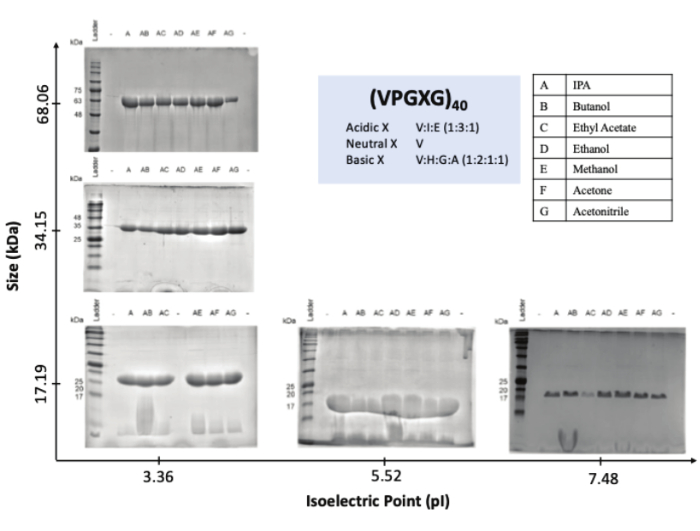
Figure 1: Organic solvent extraction and precipitation of elastin-like polypeptides from E. coli generates purified constructs in a manner that is independent of molecular weight and isoelectric point. This figure has been adapted with permission from Aayush, A., Darji, S., Estes, K. M., Yeh, E., Thompson, D. H. Development of an elastin-like polypeptide-based nucleic acid delivery system targeted to EGFR+ bladder cancer cells using a layer-by-layer approach. Biomacromolecules. 25 (9), 5729-5744 (2024)18. Copyright © 2024 American Chemical Society. Please click here to view a larger version of this figure.
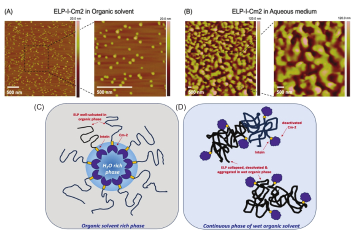
Figure 2: Representative images of the ELP-I-Cm2 observed by atomic force microscopy (AFM) in organic solvent mix AG (1:1 isopropanol and acetonitrile) and aqueous medium. (A) Organic solvent mix AG (1:1 isopropanol and acetonitrile) and (B) Aqueous medium. Differences in enzyme activity of ELP-I-Cm2 in organic versus aqueous environments likely arise from distinct microphase arrangements, as visualized by AFM. (C) In organic solvent, Cm2 is retained within a hydrated reverse micellar core surrounded by solvated ELP chains, supporting activity by preserving structure and enabling substrate access. (D) In contrast, limited solvent compatibility causes ELP collapse and aggregation into amorphous assemblies, leading to structural disruption of Cm2 and reduced catalytic activity. This figure has been reprinted with permission from Darji S., Aayush, A., Estes, K. M., Strock, J. D., Thompson, D. H. Unravelling the mechanism of elastin-like polypeptide-enzyme fusion stabilization in organic solvents. Biomacromolecules. 25 (1), 272-281 (2024)9. Copyright © 2024 American Chemical Society. License Number: 6137271264771 Please click here to view a larger version of this figure.
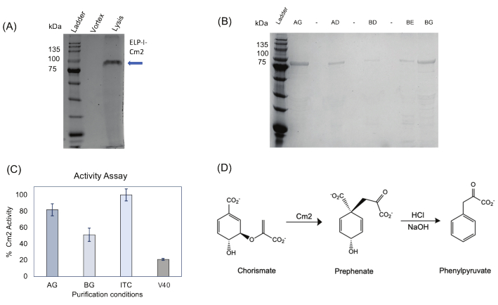
Figure 3: Representative images of the ELP-I-Cm2 protein expression and purification via SDS-PAGE analysis, followed by its activity assay results. (A) SDS-PAGE electrophoresis analysis to showcase the comparison between E.coli pellet masses that are just vortexed versus a single-step cell lysis followed by organic extraction. (B) SDS-PAGE electrophoresis analysis of ELP-I-Cm2 proteins in different organic solvents. The AG and BG combinations showed maximum protein extraction. To ensure reproducibility, ELP-I-Cm2 was purified using organic extraction from at least three biological replicates. (C) Activity assay of ELP-I-Cm2 from organic extraction (AG and BG) in comparison to proteins extracted by standard ITC alone (positive control), while the negative control, V40 showing low activity (each condition had n = 3 replicates). (D) The Cm2 activity assay tracks the reaction by measuring the increase in absorbance at 320 nm, corresponding to the formation of phenylpyruvate product, as shown. This figure has been reprinted with permission from Darji S., Aayush, A., Estes, K. M., Strock, J. D., Thompson, D. H. Unravelling the mechanism of elastin-like polypeptide-enzyme fusion stabilization in organic solvents. Biomacromolecules. 25 (1), 272-281 (2024)9. Copyright © 2024 American Chemical Society. License Number: 6086020722974 Please click here to view a larger version of this figure.
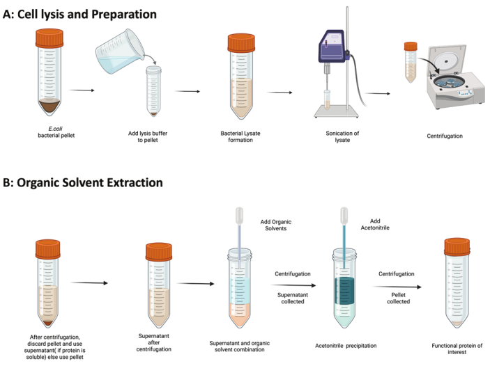
Figure 4: Workflow for organic solvent-based extraction and precipitation of ELP fusion proteins. (A) Cell lysis and preparation. E. coli bacterial pellets expressing the protein of interest are suspended in lysis buffer to generate a bacterial lysate. The lysate is disrupted by sonication and then followed by centrifugation to separate soluble and insoluble fractions. (B) Organic Solvent Extraction. The soluble fraction (or pellet if the protein is insoluble) is mixed with organic solvents to extract the target protein. After centrifugation, the protein-containing supernatant is collected and precipitated with acetonitrile. The final centrifugation yields a pellet containing the purified protein of interest. Figure created with BioRender.com Please click here to view a larger version of this figure.
| Label | Solvent |
| A | Isopropanol |
| B | Butanol |
| C | Ethyl Acetate |
| D | Ethanol |
| E | Methanol |
| F | Acetone |
| G | Acetonitrile |
Table 1: Organic solvents employed to extract the ELP-I-Cm2 fusion protein from inclusion bodies.
Supplementary File 1: The plasmid DNA sequence for the ELP-I-Cm2 protein. Please click here to download this File.
Discussion
ITC has previously been used for non-chromatographic purification of ELP19; however, it can be a time-consuming method and low-yielding process. In the methods detailed above, we describe an approach for the rapid purification of an ELP-I-Cm2 fusion protein, demonstrating how the organic solvent-based extraction-precipitation method can be effectively used for the purification of a wide variety of ELP and ELP fusions. The organic solvent protocol is not only faster than ITC, but also much more effective for shorter ELP sequences that can challenge the ITC method. It may also be a more scalable alternative than ITC for industrial applications. This approach efficiently removes impurities, including nucleic acids, LPS, metabolites, and host cell proteins at each step of the workflow. Previous work from our lab has shown that this method is effective across a wide range of ELP constructs, regardless of sequence length or isoelectric point (Figure 1), making it a promising alternative to ITC7,9,18. Unlike ITC, which relies heavily on the thermoresponsive behavior of ELPs and requires multiple heating and cooling cycles, this method simplifies the process into a single-step partitioning strategy with high purity and reproducibility.
To assess the activity of proteins purified using this method, we performed enzymatic assays using the ELP-I-Cm2 fusion protein. Cm2 catalyzes the conversion of chorismate to prephenate, which is subsequently converted into the UV-active product phenylpyruvate in the presence of HCl. The purified protein retained catalytic activity comparable to its ITC-purified counterpart9. This highlights the important finding that organic solvent extraction of ELP constructs can quickly purify samples while preserving the function of the fusion protein.
Suitable activity assays have been performed using other fusion proteins, such as ELP-CryS96 (chaperone) and ELP-EGF (EGFR binding), both of which demonstrated preserved activity after purification9,18. These findings highlight the capacity of the organic solvent extraction-precipitation method for purifying ELP fusions while maintaining their biochemical functionality across different ELP sequences.
During purification, we encountered challenges with certain ELP constructs. Specifically, low molecular weight ELPs tended to aggregate into inclusion bodies, whereas higher molecular weight ELPs were more frequently found in the soluble fraction. As a result, the extraction protocol described here should be adapted accordingly, such that soluble ELPs are treated with organic solvents and isolated from the supernatant, while ELPs forming inclusion bodies should be extracted directly from the pellet.
To better understand the structural behavior of ELPs in different environments, we previously examined their morphology using atomic force microscopy and transmission electron microscopy. In the aqueous phase, ELPs formed amorphous aggregates, whereas in the organic solvent phase, we consistently observed uniform ~50 nm spherical features, suggestive of a reverse micelle-like architecture (Figure 2). In this working model, the polar Cm2 fusion protein is sequestered within an aqueous-rich interior compartment, while the hydrophobic ELP segments interact with the surrounding bulk organic solvent9. This configuration provides a rationale for the surprising observation that bioactivity (e.g., Cm2 enzyme activity) is preserved even after exposure to large quantities of organic solvent during the extraction-precipitation process.
The reversed micelle model also helps explain why maintaining a precise water content during extraction is critical. The presence of excess water can disrupt the formation of reverse micelle structures and compromise extraction efficiency. This may partly explain the variability observed between biological replicates and highlights the need for tight control of solvent ratios and hydration levels throughout the protocol. Additionally, it is essential to control the incubation time following the addition of acetone or acetonitrile. Extended incubation beyond the optimal window may negatively affect protein solubility and folding, possibly leading to aggregation and loss of functionality and/or yield. Careful attention to timing, hydration, and solvent handling is, therefore, key to ensuring reproducibility and functional recovery of ELP-fusion proteins.
Disclosures
The authors have nothing to disclose.
Acknowledgements
We thank the Purdue University Institute for Cancer Research and the NIH CCSG program (CA23168) for support of this work.
Materials
| Name | Company | Catalog Number | Comments |
|---|---|---|---|
| 2-Mercaptoethanol | Bio-Rad | 161-0710 | SDS-PAGE Component |
| 30% Acrylamide/Bis-acrylamide | TCI | A3218 | SDS-PAGE Component |
| Acetone | Fisher Scientific | A949 | Organic Solvent |
| Acetonitrile | Fisher Scientific | A996 | Organic Solvent |
| Ampicilin | Gold Bio- Gold Biotechnology | A-301-25 | Antibiotic |
| APS (Ammonium Persulfate) | Bio-Rad | 1610700 | SDS-PAGE Component |
| Butanol | Fisher Scientific | L13171 | Organic Solvent |
| EDTA | Fisher Bioreagents | BP118-500 | Lysis Buffer Component |
| Ethanol | Decon Labs | UN1170 | Organic Solvent |
| Ethyl Acetate | Fisher Scientific | E195 | Organic Solvent |
| Glycerol | Research Products International | G22020-0.5 | Terrific Broth (TB) Media Component |
| Hydrochloric Acid | Fisher Scientific | A144SI-212 | Lysis Buffer Component |
| Instant Coomasie stain | Abcam | ab119211 | SDS-PAGE Component |
| IPTG | Gold Bio- Gold Biotechnology | 12481C25 | Protein Expression Component |
| Isopropanol | Fisher Scientific | A451-1 | Organic Solvent |
| K2HPO4 (Potassium Phosphate Monobasic) | Wards' Science | 470302-246 | Part 2 Buffer Component |
| KH2PO4 (Potasium Phosphate Dibasic) | Wards' Science | 470302-254 | Part 2 Buffer Component |
| L-Proline | Research Products International | P5200-500.0 | Terrific Broth (TB) Media Component |
| Lysozyme | Gold Bio- Gold Biotechnology | L-040-1 | Lysis Buffer Component |
| Methanol | Fisher Scientific | A452 | Organic Solvent |
| Native Sample Buffer | Bio-Rad | 161-0738 | SDS-PAGE Component |
| Protein Ladder | Gold Bio- Gold Biotechnology | P008-500 | SDS-PAGE Component |
| Resolving Gel Buffer | Bio-Rad | 1610798 | SDS-PAGE Component |
| Sodim Dodecyl Sulfate (SDS) | Thermo Scientific | J18220-36 | SDS-PAGE Component |
| Sodium Hydroxide | Fisher Scientific | S318-500 | Lysis Buffer Component |
| Stacking Gel Buffer | Bio-Rad | 1610799 | SDS-PAGE Component |
| TEMED | Bio-Rad | 1610801 | SDS-PAGE Component |
| Tris Base | Fisher Bioreagents | BP-152 | Lysis Buffer Component |
| Tryptone | Research Products International | T60065-1000.0 | Terrific Broth (TB) Media Component |
| Yeast Extract | Research Products International | Y20025-1000.0 | Terrific Broth (TB) Media Component |
References
- Despanie, J., Dhandhukia, J. P., Hamm-Alvarez, S. F., MacKay, J. A. Elastin-like polypeptides: therapeutic applications for an emerging class of nanomedicines. J Control Release. 240, 93-108 (2016).
- MacEwan, S. R., Chilkoti, A. Applications of elastin-like polypeptides in drug delivery. J Control Release. 190, 314-330 (2014).
- Jenkins, I. C., Milligan, J. J., Chilkoti, A. Genetically encoded elastin-like polypeptides for drug delivery. Adv Healthc Mater. 10 (13), e2100209(2021).
- Ju, Y., et al. Molecular targeting of immunosuppressants using a bifunctional elastin-like polypeptide. Bioconjug Chem. 30 (9), 2358-2372 (2019).
- Nettles, D. L., Chilkoti, A., Setton, L. A. Applications of elastin-like polypeptides in tissue engineering. Adv Drug Deliv Rev. 62 (15), 1479-1485 (2010).
- Meyer, D. E., Chilkoti, A. Purification of recombinant proteins by fusion with thermally responsive polypeptides. Nat Biotechnol. 17 (11), 1112-1115 (1999).
- VerHeul, R., Sweet, C., Thompson, D. H. Rapid and simple purification of elastin-like polypeptides directly from whole cells and cell lysates by organic solvent extraction. Biomater Sci. 6 (4), 863-876 (2018).
- Sweet, C., Aayush, A., Readnour, L., Solomon, K. V., Thompson, D. H. Development of a fast organic extraction-precipitation method for improved purification of elastin-like polypeptides that is independent of sequence and molecular weight. Biomacromolecules. 22 (5), 1990-1998 (1990).
- Darji, S., Aayush, A., Estes, K. M., Strock, J. D., Thompson, D. H. Unravelling the mechanism of elastin-like polypeptide-enzyme fusion stabilization in organic solvents. Biomacromolecules. 25 (1), 272-281 (2024).
- Urry, D. W. Physical chemistry of biological free energy transduction as demonstrated by elastic protein-based polymers. J Phys Chem B. 101 (51), 11007-11028 (1997).
- Guo, Y., Liu, S., Jing, D., Liu, N., Luo, X. The construction of elastin-like polypeptides and their applications in drug delivery system and tissue repair. J Nanobiotechnology. 21, 418(2023).
- Yi, A., Sim, D., Lee, S. -B., Sarangthem, V., Park, R. -W. Application of bioengineered elastin-like polypeptide-based system for targeted gene delivery in tumor cells. Biomater Biosyst. 6, 100050(2022).
- Clement, H., et al. Comparison between the recombinant expression and chemical synthesis of a short cysteine-rich insecticidal spider peptide. J Venom Anim Toxins Trop Dis. 21, 19(2015).
- Miranda, L. P., Alewood, P. F. Accelerated chemical synthesis of peptides and small proteins. Proc Natl Acad Sci U S A. 96 (4), 1181-1186 (1999).
- Eweje, F., et al. Self-assembling protein nanoparticles for cytosolic delivery of nucleic acids and proteins. Nat Biotechnol. , (2025).
- Hassouneh, W., Christensen, T., Chilkoti, A. Elastin-like polypeptides as a purification tag for recombinant proteins. Curr Protoc Protein Sci. 6 (11), 1-16 (2010).
- Yakhnin, A. V., Vinokurov, L. M., Surin, A. K., Alakhov, Y. B. Green fluorescent protein purification by organic extraction. Protein Expr Purif. 14 (3), 382-386 (1998).
- Aayush, A., Darji, S., Estes, K. M., Yeh, E., Thompson, D. H. Development of an elastin-like polypeptide-based nucleic acid delivery system targeted to EGFR+ bladder cancer cells using a layer-by-layer approach. Biomacromolecules. 25 (9), 5729-5744 (2024).
- Christensen, T., et al. Fusion order controls expression level and activity of elastin-like polypeptide fusion proteins. Protein Sci. 18 (7), 1377-1387 (2009).
Reprints and Permissions
