This method outlines the development of an in vitro tick blood-feeding system incorporating a 3D-printed chamber and a laboratory-fabricated silicone membrane. The system enables artificial feeding of ticks, allowing for easy sampling at various feeding stages and utilizing low volumes of blood, providing an invaluable tool for studying ixodid ticks.
Method Article
Tick Blood Feeding through an Artificial Membrane on a 3D-Printed Chamber in Ixodid Tick Research
In This Article
Summary
Abstract
This work presents a detailed protocol for a tick feeding system through an artificial silicone membrane utilizing a 3D-printed chamber created from high-resolution photopolymer resin. The method includes the design of the feeding chamber, preparation of the silicone membrane, assembly of the system, and optimized conditions for tick feeding. Ticks are one of the leading vectors of disease in the United States and worldwide. Tick research is essential for developing effective strategies to prevent and reduce the incidence of tick-borne illnesses. As obligate hematophagous ectoparasites, ticks must feed on blood to survive and thus transmit disease, making blood-feeding a fundamental process when investigating vector-host interactions, pathogen transmission, and the biology, physiology, and anatomical morphology of ticks. Tick feeding systems with a silicone membrane have been widely utilized and refined over the past several decades. However, many existing systems are tailored to a single tick species, life stage, or specific set of conditions. The tick feeding system described in this protocol has been successfully used to obtain both partially fed and fully engorged specimens from several of the most important tick vector species in the United States, including Amblyomma americanum, A. maculatum, Ixodes scapularis, and Rhipicephalus sanguineus adults. The system has shown success in initiating feeding of Dermacentor variabilis adults and nymphal A. americanum, with the highest feeding rates (>85%) observed in adult A. americanum. Consequently, the feeding chamber developed in this study is adaptable for use with a low volume of different blood, down to 100 µL, which is crucial in pathogen transmission and toxicological studies. The transparent lid with minimal height of the chamber facilitated the observation of ticks over tick tick-feeding duration.
Introduction
Ticks are obligate blood-feeding ectoparasites that can carry and transmit a wide range of pathogens, including bacteria, viruses, and parasites that cause disease to animals and humans worldwide1,2,3,4. As the number one disease vector in the United States, second only to mosquitoes in the rest of the world2,3,5, several tick species can transmit pathogen-associated diseases, while other species are also known to induce non-communicable diseases such as alpha-gal syndrome (AGS, also known as red meat allergy), which has been increasingly reported worldwide in recent years6,7,8,9,10,11,12,13,14,15,16. Research aimed at understanding tick biology, physiology, genetics, and vector competence is essential for developing strategies to reduce the economic and public health burden of both communicable and non-communicable tick-borne diseases. Studying tick-host interactions often requires partially fed or fully engorged ticks to address specific research questions. This can be achieved through live animal feeding, allowing ticks to complete their full life cycle17. While the use of live animals for tick feeding is highly effective for developing and maintaining tick colonies18,19, it can be challenging to implement and may not be applicable to every research hypothesis due to host availability limitations and facility regulations. Most importantly, the successful implementation of in vitro tick feeding systems helps reduce the use of live animals, which is a significant goal in research under the “Animal Use Alternative (3Rs)” framework (replace, reduce, refine)20.
There are numerous advantages of using artificial membrane tick feeding systems in research, providing broad opportunities to address a wide range of scientific questions. These can be broadly summarized in three groups; (1) assessment of the effect on host blood variation and vector-host interactions, using different blood source, such as tick alpha-gal production during feeding21, (2) evaluation of acaricide toxicity through delivery of compounds with the blood22 and the recovery of tick secreted compounds during bloodfeeding23, (3) evaluation of vector competence, through the delivery of pathogens by natural acquisition during blood feeding24,25,26,27, among others. Furthermore, the nature of the artificial membrane feeding system allows large-scale testing of various treatments and concentrations without introducing host-related variations, at a small scale and volume, reducing the need for live animals.
Tick feeding systems through artificial membranes have been widely used in research and to support tick-colony rearing19,28,29, in some cases enabling completion of the tick’s long life cycle30,31. Many protocols on the optimization of the tick feeding systems and their materials have been reported for different tick species32,33,34. Nonetheless, it is well known that in vitro tick feeding can be highly variable depending on system conditions, the materials used, and, most importantly, tick species and stages34. Furthermore, many systems rely on large volumes of blood, such as continuous-flow systems27, which are highly effective but can be costly due to the significant blood volumes required. This protocol details an artificial membrane tick feeding system that has been shown to be successful with the adult stages of five different tick disease vector species in the United States. This includes Amblyomma americanum, A. maculatum, Ixodes scapularis, Rhipicephalus sanguineus, and Dermacentor variabilis.
Protocol
Tick feeding experiments were performed using commercially available defibrinated bovine blood (Hemostat Laboratories, CA, USA) and defibrinated human blood (Bioivt, CA, USA), which collects and processes blood in accordance with Institutional Animal Care and Use Committee (IACUC) and Institutional Review Board (IRB)-approved protocols and guidelines. No live vertebrate animals were used for tick feeding in this study. All ticks used in this protocol were purchased from the Oklahoma State University tick rearing facility. The reagents and the equipment used are listed in the Table of Materials.
1. Tick feeding chambers
- Design the chambers using a free 3D design Software to fit on a 35 mm x 10 mm Petri dish. Printed parts (Figure 1A) sizes are as follows: chamber body (part i) 25 mm (inner diameter) x 20 mm (high), with 3 mm wall thickness at the base and 6 mm wall thickness at the top lid ring (part ii) 30 mm (inner diameter) x 5 mm (high) and 3.5 mm wall thickness and outer ring (part iii) 31 mm (inner diameter) x 5 mm (high) and 3 mm wall thickness.
NOTE: The lid ring includes four circular 3 mm (diameter) x 2 mm (height) openings to the top to place magnets. The top of the chamber also has the same size openings for attaching the magnets (Figure 1B). The 3-D model design should be saved as an .stl file, the most common 3-D printing file format. Files containing the 3D design used in this protocol will be provided upon request. - Print chambers using a 10" 14K LCD 3D Printer with gray or white photopolymer resins (Figure 1A) or another high-resolution 3D printer/services that prints 3-D models from a .stl file.
NOTE: This protocol used the University of Arizona’s printing services and resins. Using high-resolution resins ensures the creation of detailed and durable chambers. - Using gloves, ensure there are no resin residues on the parts before assembly by washing and, if needed, gently sanding with fine sandpaper (180 to 200 grit). Ensure that the flat bottom of part i (Figure 1A) is smooth. This ensures proper adhesion of the silicone membrane, reducing the risk of leakage.
- Expose all dry pieces to UV light for 10 min to ensure a complete resin polymerization.
NOTE: Most 3-D printing services that use toxic resins for printing use UV light to cure the resin (hardening the models and removing resin toxicity); however, it occasionally remains “not fully cured”. A non-fully cured model will remain slightly sticky to the touch and may carry unpolarized resin, which can be toxic or cause irritation if handling. Extra UV light exposure will ensure complete resin hardening. - Glue the 3 mm x 2 mm round magnets to the slits on the top of the chambers and lid rings using clear epoxy glue. Allow to dry overnight at room temperature (20–25 °C/minimum of 16 h). Ensure that the poles of the magnets are toward one direction to match the attraction of the poles on the magnets in the upper part of the lid ring (Figure 1A, part ii).
- Assemble all pieces for the model to ensure proper fit of all components, as shown in Figure 1A–C.
2. Membrane preparation
- Place a 30 cm x 30 cm piece of plastic wrap on a clean surface and ensure it is wrinkle-free by stretching it on a flat surface and securing the edges with tape (Figure 2Ai).
- Place 4–5 single sheets of lens cleaning paper over an extended piece of plastic/cling wrap spaced 5 cm apart (Figure 2Aii).
- Prepare the silicone membrane using the components and quantities provided below.
NOTE: The quantities are sufficient to prepare 4–5 (7 cm × 12 cm) membranes, each accommodating up to 5–6 feeding chambers, for a total of 20–30 chambers. - Prepare the silicone mixture by combining 15 g of silicone clear caulk (20–25 Shore A hardness) and adding 4.5 g of silicone oil (30% of the total silicone), and 2.9 g of hexane (19.3% of the total silicone). Place the ingredients in a plastic weighing boat and gently mix using a spatula (Figure 2Aiii).
NOTE: Ensure minimal bubble formation by mixing ingredients gently. - Place 2–3 g of silicone mixture along the smaller edge of the lens paper to secure it to the plastic wrap. Allow 5–10 s for the silicone to seep through the lens paper (Figure 2Aiv).
- Evenly spread the silicone mixture along the lens paper sheet using a straight-edge instrument, such as a lid, ruler, or a spatula. Ensure that the object used covers the full area of the lens paper (Figure 2Av). This ensures silicone coverage in a single stroke, preventing membrane ruffling and ripples at the object edge.
- Ensure that all silicone mixture is used in the membrane by returning the spreading tool to the starting position for spreading. As some silicone mixture will be retained on the spreading tool, returning the tool to collect it will ensure all the silicone is used to avoid gaps (Figure 2Avi).
NOTE: Membrane should appear uniformly translucent without ripples (Figure 2B). - Prepare membranes of different thicknesses initially by adding more or less silicone mixture, as the thickness cannot be measured until the membranes are dry.
- Let the membranes air-dry overnight at room temperature (20–25 °C/minimum of 16 h).
- Measure membrane thickness using a digital outside micrometer measuring tool. Different membrane thicknesses are used for different adult tick species, ranging from short mouth parts (70 µm membrane thickness) to longer mouthparts (200 µm membrane thickness).
- Remove all parts from previously assembled chambers and apply silicone glue to the bottom rim of the chambers. For precise glue placement, use a 1 ml syringe and fill it with silicone glue (Figure 2Ci).
- Place a 200 µL pipette tip on the tip of the syringe (instead of the needle) for glue application (Figure 2Cii) and screw it onto the end of the syringe, as with a needle tip (Figure 2Ciii). This will allow fine glue application to the bottom rim of each chamber (Figure 2Civ).
- Place the chamber over the membrane (Figure 1, part i) with a weighted object to ensure settling. Allow to sit overnight at room temperature (20–25 °C/minimum of 16 h) (Figure 2D,E).
NOTE: Use any flat weighted object. This protocol used a 2.5 kg piece of acrylic (Figure 2E). - Cut the membrane excess from the chambers using curved scissors (Figure 3A).
- Before using the chamber for tick feeding, remove any remaining plastic wrap (where the membrane is prepared (step 2.1) (Figure 3B).
- Assemble the chamber parts back together, include a 35 mm x 10 mm Petri dish at the bottom, and use the lids of the dishes as the lids of the chambers by inverting them and placing them between the magnets (Figure 3C,D).
NOTE: Use the lids inverted, as the lid holder ring is an exact fit for the lid if inverted (see part descriptions in Figure 3C,D). - Soak the chamber membrane in distilled water using the bottom portion of 35 mm x 10 mm Petri dishes. Let the membrane sit in water overnight at room temperature (20–25 °C/minimum of 16 h). Since the membrane is initially hydrophobic, soaking it in water before adding blood will help reduce the formation of trapped bubbles.
NOTE: Absence of bubbles confirms that hydrophobicity has been removed.
3. Tick feeding and blood preparation
- Make 2–3 small breathing holes in the lids of every chamber (from the 35 mm x 10 mm Petri dishes) (part iv in Figure 1A) mentioned in step 2.16 (Figure 3E). Use a heated metal scalpel or a thin metal spatula to poke the breathing holes in the lid.
NOTE: Breathing holes allow humidity to be internalized within the chamber housing. - Supplement the blood with gentamicin sulfate at a final concentration of 50 µg/mL. Add antibiotics only to the blood intended for use within 1 week and store at 4 °C. Keep bovine blood (or any other animal blood) at 4 °C before and after adding antibiotics.
- Place 2–3 mL of blood in a clean 35 mm x 10 mm Petri dish, then place the chamber on it. Adjust chamber height to increase or reduce the amount of blood used per chamber by rotating the adjustment ring (part iii from Figure 1A). Adjust chamber height, leaving a 1–2 mm space from the bottom of the Petri dish to the membrane. See magenta arrows in Figure 4A for reference.
NOTE: The adjustment ring design of the chambers allows for the control of the volume of blood used. i.e., a larger space between the bottom of the Petri dish and the membrane will require a larger volume to fill the space. - Place unfed ticks inside chambers in equal numbers of males and females. Ensure that both sexes are always included in the chamber, as this can affect feeding rates (Figure 4B shows 4 males and 4 females of adult A. americanum inside the chamber). A maximum of 10–15 ticks per chamber (for the adult stage) is used for easier handling.
NOTE: When working with immature stages, the number of ticks is higher (20–50 nymphs per chamber, not presented in this protocol). - Place chambers in covered dry baths at 37 °C ± 1 °C with a humidity source (i.e., water cups) that maintains a relative humidity of 63% ± 2% (Figure 4C) under a 12:12 h light-dark photoperiod.
NOTE: These requirements may vary depending on the tick species and laboratory conditions; try other humidity settings if necessary and evaluate the best setup for the laboratory.
4. Replacing blood and chambers
- Prepare two squirt bottles with the solutions to be used for rinsing the membrane between blood changes. In one bottle, add 0.005% nystatin solution in distilled water; in the second bottle, add distilled water.
- After 12–16 h, prepare new Petri dishes with fresh blood and place them in the dry bath at 37 °C before placing the chamber.
- While the fresh blood is warming, remove the chamber from the used blood and rinse the underside of the membrane with distilled water for 3–5 s, then with 0.005% nystatin solution for 3–5 s to prevent fungal contamination. Rinse the membrane again with distilled water to remove any remaining nystatin residue.
NOTE: This process takes less than 5 min and does not affect the feeding of ticks attached to the membrane on the upper surface. - Discard all used blood and rinsing water into a container containing 0.5% Sodium hypochlorite solution (10% bleach). Blood waste is discarded in accordance with the institutional safety protocol.
- After rinsing, place the chamber in the new Petri dish with blood.
NOTE: Use chambers for up to 2–3 months before washing, provided there is no contamination and no leaks. Chambers and membranes become dirty and are at risk of contamination beyond 3 months of use without washing. This results in reduced tick feeding rates. Reuse the chambers for up to 1.5 years (~10 washing cycles). - Reuse chambers by placing them in 0.5% sodium hypochlorite solution (10% bleach) for up to 30 min, then washing with dish soap. Do not expose the chambers to bleach for prolonged periods (>30 min), as the smell reduces attachment rates and can weaken resin integrity over time.
NOTE: Prolonged bleach exposure makes the chamber flexible when wet and brittle when it dries, leading to breakage. This makes the chamber not last longer than 4–5 wash cycles based on repeated observations. Continuous use of the same chamber without washing (up to 3 months) results in faster tick attachment (1–3 days after placement in the chamber) compared to a recently washed chamber (often >7 days to attach). This is likely due to the smell of frass and other aggregation pheromones from ticks feeding in the chamber, which can stimulate tick feeding35,36.
Results
Membrane thickness for each tick species.
Highest tick feeding rates were observed at the following membrane thickness specifications for each adult tick species: Amblyomma americanum 100–200 µm; A. maculatum 100–250 µm; Rhipicephalus sanguineus 70–100 µm; Ixodes scapularis <150 µm, Dermacentor variabilis <150 µm. All tick species were maintained under the same conditions of humidity and temperature during feeding (63% RH and 37 °C) (Table 1).
Ticks and blood sources used in this protocol
All ticks were obtained from the Oklahoma State University tick rearing facility and maintained at >90% relative humidity at 20 °C, under a 12:12 h light-dark photoperiod. All ticks were fed on defibrinated bovine or human blood as part of a research project investigating alpha-gal (αGal) levels in various tick species. This study compared the effects of feeding on different blood sources within a broader project of alpha-gal syndrome (AGS) conducted by the research group. Most tick specimens described here were dissected prior to full engorgement to conduct αGal quantification analyses in partially fed ticks (2–5 days of feeding). Only a subset of Amblyomma americanum ticks (those not required for αGal quantification) were allowed to feed to repletion. Although detailed comparisons between bovine-fed and human-fed tick feeding rates are not presented here, it is notable that full engorgement (i.e., feeding success) was achieved more in ticks fed on bovine blood. Among the ticks that were allowed to remain in the feeders for extended periods (n = 35), 15 reached full engorgement, representing a 42.8% feeding success rate. Of these, 9 were fed on bovine blood and 6 on human blood (60% bovine-fed compared to 40% human-fed).
Tick feeding rates vs. tick feeding success rates
Tick feeding rates in this study were determined based on the acquisition of partially fed ticks (2–5 days of feeding), whereas feeding success was defined as full engorgement, typically indicated by a change in alloscutum coloration of the female (data available only for Amblyomma americanum). Male feeding rates were defined as continuous feeding for more than 2 days (2–5 days). Overall feeding rates were calculated by combining data from ticks fed on bovine and human blood and calculating percentages of ticks achieving 2–5 days of feeding or full engorgement from ticks placed in the chambers. Ticks that did not attach or died after <2 days of attachment were considered for the calculations of feeding rates. Although quantitative data on the influence of blood source on tick feeding rates are not presented, this observational evidence suggests that, for most tick species, attachment and feeding rates do not differ substantially between bovine and human blood during the 2–5-day feeding period. An exception was observed for Rhipicephalus sanguineus, which exhibited lower feeding rates on human blood between days 3 and 5 compared to bovine blood. Feeding rates for all tick species that achieved partial feeding and full engorgement using this in vitro tick-feeding system are summarized in Table 1.
Amblyomma americanum female weight and feeding stage classification
Partially fed female A. americanum were classified based on days of feeding as: (1) unfed, (2) 2–3 days partially fed, (3) 4–5 days partially fed, and (4) engorged (Figure 5). Engorged feeding stage included ticks at the onset of the rapid engorgement phase (with weights ranging from 48.9 mg to 109 mg) and fully engorged ticks (with weights ranging from 272.7 mg to 348.7 mg).
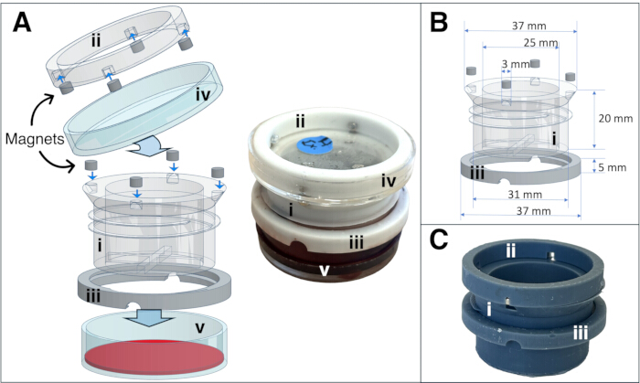
Figure 1: Tick feeding chamber - 3D model and printed assembly with membrane. Exploded 3D model and final assembled chamber displaying individual components: (i) chamber body (tick housing), (ii) lid holder ring, (iii) height adjustment ring, (iv) 40 mm x 10 mm Petri dish lid, and (v) 40 mm x 10 mm Petri dish bottom with blood (A). Assembly of the 3D printed model and individual part sizes shown in mm (B). Assembled chamber printed in gray 8K-resolution resin (C). Please click here to view a larger version of this figure.
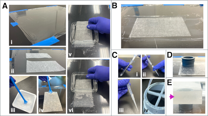
Figure 2: Preparation of the silicone membrane and chamber assembly. (i) A 30 cm × 30 cm plastic wrap secured with tape, ensuring no visible wrinkles, (ii) individual sheets of lens cleaning paper placed on the wrap, spaced approximately 5 cm apart, (iii) gentle mixing of the silicone components with a spatula using circular motions, (iv) each sheet of lens paper secured by applying 1–2 g of the silicone mixture along its edges, (v) the silicone mixture evenly spread across the lens paper using a straight-edged instrument larger than the paper (image shows the edge of a glass lid), (vi) note the reduced amount of silicone mixture during spreading, the retained silicone on the tool is returned and redistributed to ensure uniform coverage (A). Properly spread membranes showed no air bubbles or gaps (B). Preparation of the syringe for glue application: (i) a 1 mL syringe filled with silicone glue, (ii) a 200 µL pipette tip fitted into the syringe tip, (iii) the pipette tip screwed in place to ensure a leak-proof seal, and (iv) the placement of glue on the underside rim of part i (C). The chamber placed on the dried membrane (D), and a weighted object (a thick acrylic block weighing 2.5 kg; magenta arrow) placed on top to ensure firm adhesion (E). Please click here to view a larger version of this figure.
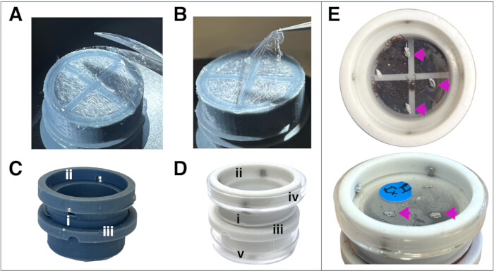
Figure 3: Final steps in chamber and membrane preparation before use. Trimming of edges of the membrane surrounding the chamber using curved small scissors (A), removal of plastic wrap before the chamber is used (B), final model assembly (C), placement of the chamber in a 40 mm x 10 cm Petri dish containing water, and use of the Petri dish llid as the chamber lid (D) (numbers i-v indicate parts as specified in Figure 1A), and chamber lids displaying breathing holes (indicated by magenta arrowheads) (E). Please click here to view a larger version of this figure.
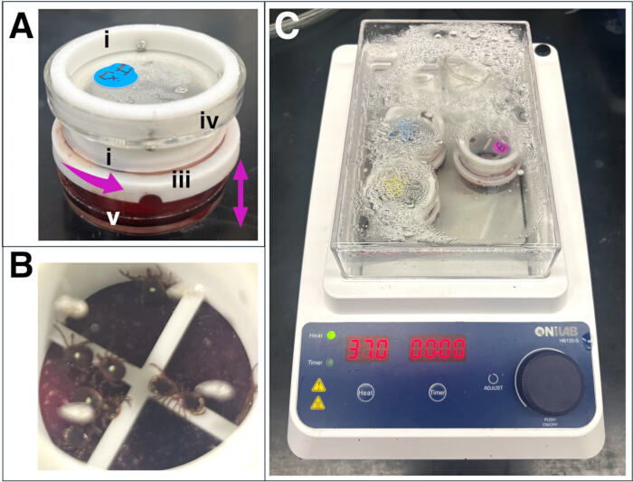
Figure 4: Blood feeding setup and tick placement. Chamber placed in a blood-filled dish, with the height adjustment ring highlighted (magenta arrows) (A). Interior view of the chamber housing containing an equal number of male and female ticks (B). Tick chambers positioned in dry baths with a humidity source, maintaining 63% ± 2% relative humidity (RH) and a temperature of 37 °C ± 1 °C (C). Please click here to view a larger version of this figure.
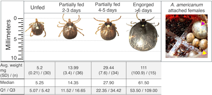
Figure 5: Amblyomma americanum size and weight variation during feeding. Unfed tick size and average weight of partially fed ticks are shown. The images represent typical examples corresponding to the average weight recorded at each feeding interval. The accompanying table presents average weights, standard deviations, the number of ticks analyzed, median, and quartiles 1 and 3 for each feeding time point. Ticks fed for 2–3 days showed a weight range from 7.4 mg (minimum) to 19.2 mg (maximum). Ticks fed for 4–5 days ranged from 20.6 mg to 44.8 mg. Ticks at the onset of rapid engorgement phase and fully engorged ticks weighed between 48.9 mg and 348.7 mg. Image of attached A. americanum females 4-5 days fed female (yellow arrowhead), and fully engorged female (magenta arrowhead). Please click here to view a larger version of this figure.
| Tick species and sex | Number of ticks fed/ Total ticks placed in chambers (Number of ticks dead) | Feeding rate (ticks attached 2 -5 days) | Average weight (mg) (SD) | Membrane thickness used (μm) |
| A. americanum females | 85/97 (12) | 87.60% | 37.3 (54.40)* | 100 - 200 |
| A. americanum males | 19/22 (2) | 86.30% | Not recorded | 100 - 200 |
| A. maculatum females | 45/66 (21) | 68.20% | 122.4 (141.65) | 100 - 250 |
| A. maculatum males | 36/47 (11) | 76.60% | Not recorded | Not recorded |
| R. sanguineus females | 34/60 (26) | 56.60% | Not recorded | 70 - 100 |
| I. scapularis females | 15/23 (8) | 65.20% | Not recorded | <150 |
| *Feeding success rate evaluated in a subset of bovine fed Aa adult ticks is (n=35) was 42.8% | ||||
Table 1: Tick feeding rates and average weights.
Supplementary Figure 1: Low-volume (100 µL) feeding of the individual ticks already attached to a site of the 3D chamber. A small PCR tube attached to the bottom of the artificial membrane with silicone glue is filled with 100 µL of blood.Please click here to download this file.
Discussion
Tick artificial membrane feeding systems are highly effective tools for tick research and are extensively used in many research laboratories as viable alternatives to live animal feeding28,29. The primary value of these systems lies in their ability to deliver or recover various agents through the blood meal, including pathogens, acaricides, tick-derived compounds, and other modulatory molecules22,23,25,27. The optimization of artificial membrane feeding systems could facilitate the development of sustainable tick colony rearing, thereby reducing reliance on live animals.
The in vitro tick feeding system outlined in this protocol has been particularly successful with adult Amblyomma americanum and Ixodes scapularis. Tick feeding success rates are defined as the proportion (or percentage) of ticks that successfully complete feeding to full engorgement. However, partially fed ticks (typically 3–4 days post-feeding initiation) are often sufficient to address specific experimental hypotheses for most tick research laboratories studying the tick biology or physiology. In this artificial membrane feeding system protocol, and to address specific research objectives in the alpha-Gal (αGal) project, the terms “tick feeding rate” were used to refer to the acquisition of partially fed ticks (2–5 days of feeding) and “tick feeding success” to refer to full engorgement in A. americanum. The highest feeding rates were in A. americanum females (87.6%), followed by A. americanum males (86.3%), A. maculatum females (68.2%), A. maculatum males (76.6%), I. scapularis females (65.2%), and R. sanguineus females (56.6%), while A. americanum feeding success rate was 42.8% (Table 1). Partially fed female A. americanum were classified based on days of feeding in 4 categories (feeding phases), modified from previous publications as follows21: (1) unfed, (2) 2–3 days partially fed, (3) 4–5 days partially fed, and (4) engorged (Figure 5). Engorged feeding stage included ticks at the onset of the rapid engorgement phase (with weights ranging from 48.9 mg to 109 mg) and fully engorged ticks (with weights ranging from 272.7 mg to 348.7 mg) (Figure 5). This artificial membrane tick-feeding system has enabled multiple studies within the same research group and collaborators by allowing the generation of partially fed ticks on bovine blood, which were subsequently used to infest mice and test the transmission hypothesis of alpha-gal syndrome (AGS), a tick-bite–induced red meat allergy, and for an N-glycomics study11,21,37. The convenience of the individual setup enabled the use of different blood sources, and the clear lid and minimal height of the chamber facilitated visualization of the ticks during the feeding period under a stereomicroscope with sufficient working distance.
Recent modifications in humidity (from water bath, 90% RH, to dry bath, 63% ± 2% RH), temperature (from 37 °C to 35 °C and back to 37 °C), and membrane thickness (Table 1) have enabled the successful feeding of multiple tick species. While temperature and relative humidity (RH) are important variables to control in in vitro tick feeding, membrane thickness and strength are critical factors ensuring successful feeding across different tick species33,38. It is essential to consider that ticks with short mouthparts, such as Rhipicephalus sanguineus and Dermacentor variabilis, will require thinner membranes than those with long mouthparts39, such as Amblyomma spp and Ixodes spp. The highest tick feeding rates were in Amblyomma americanum and A. maculatum at a membrane thickness of 100–200 µm; Rhipicephalus sanguineus at thicknesses between 70–100 µm; Ixodes scapularis at thicknesses <150 µm, and Dermacentor variabilis <150 µm (Table 1). A key factor to note in artificial membrane tick feeding is that fluctuations in feeding rates still occur. In this system, a slight change in temperature, i.e., from 37 °C to 35 °C and back to 37 °C after a couple of days, often enhances or restores tick feeding rates. Furthermore, switching from dry bath to water bath yields similar results for Amblyomma spp. and Ixodes scapularis, however, this effect was not observed for Rhipicephalus sanguineus, which feeds more effectively at 63% ± 2% RH in dry baths.
Other artificial tick feeding systems, such as those adapted from the five-part continuous flow system developed by Ueti, M. et al. (USDA-ARS) and later modified for additional tick species by Yamasaki, Y. et al., have demonstrated high efficiency, yielding consistently high tick feeding success rates27,40. However, studies that require frequent access to the system for the dissection of partially fed ticks make these setups less suitable, as the repeated opening and closing of the tick housing chamber can be cumbersome. The 3D-printed chamber described in this protocol features a magnetic sure-lock lid and individual Petri dishes, allowing easy handling and visual inspection of tick feeding without disturbing the setup. The transparent lid allows visual monitoring without disturbance and securely attaches to the chamber via embedded magnets as opposed to the most common tick feeding systems, such as those optimized by the original design from Kröber and Guerin22,29, where individual visualization is not possible without removing the feeding chamber from the blood as all feeding units are integrated into a single plate and where the methods used to prevent ticks from escaping do not allow for visualization. Moreover, this in vitro tick-feeding system can be further refined to deliver smaller, site-specific volumes of blood. For instance, once female ticks are attached at defined feeding sites, the bottom Petri dish could be replaced with miniature reservoirs, such as the upper portion of a 100 µL PCR tube, containing less than 100 µL of blood positioned directly beneath the attachment area (Supplementary Figure 1). This low-volume feeding approach would enable detailed studies on pathogen acquisition and transmission, as well as investigations into the effects of tick toxicants and host-derived antibodies. This protocol has been demonstrated to be successful in obtaining partially fed ticks from various adult tick species. For the purposes of this study, egg-laying and full engorgement rates were not evaluated in this protocol; however, further optimization is being conducted with the goal of obtaining tick progeny, which could be achieved with Amblyomma americanum and Ixodes scapularis based on preliminary observations. Perhaps the most innovative feature of this system is its flexibility for small-scale experimental manipulations, enabling controlled variation of treatments and environmental conditions, while also supporting colony rearing applications.
In vitro tick feeding is a valuable tool for researchers studying tick biology, physiology, and vector competence. However, artificial membrane tick feeding is a time-consuming and effort-demanding task. This protocol, featuring a 3D-printed chamber with a sure-lock system and a simple laboratory-crafted membrane, offers a straightforward setup once the chambers are printed and membranes are prepared, allowing for extended use. Additionally, by adjusting membrane thickness, the system can be effectively adapted to different tick species. Further optimization, including chamber size and membrane thickness, can enable its application across various tick life stages.
Disclosures
The authors declare no material financial interests that relate to the research described in this paper.
Acknowledgements
The authors would like to acknowledge the diligent work of the undergraduate students and workers, Kara Golden, Sydney Ingham, and Omayma Jaddou Najma, who assisted with tick feeding and setup of the tick chamber and membrane.
Materials
| Name | Company | Catalog Number | Comments |
|---|---|---|---|
| 1mL Luer-Lok Syringe | BD | 309628 | Luer-Lok Tip |
| Advanced Clear Silicone | General Electric | 2810435 | Clear silicone caulk. This caulk forms a water-impermeable barrier after curing. |
| Clear Epoxy glue | JB Weld-Clear | B009EU5ZM0 | Amazon product number |
| Clear food wrap (cling seal) | Glad | CLO00020 | |
| Defibrinated Bovine blood | Hemostat Laboratories | DBB100 | |
| Defibrinated Human blood | BioIVT | ||
| Disposable spatulas | LevGo, Inc. | 17211 | Amazon product number |
| Dry bath incubators | ONiLAB Scientific | HB120-S | Amazon product number. Heating block is inverted to obtain a flat surface. |
| Gentamicin sulfate salt | Millipore Sigma | G3632-5G | |
| Lens Cleaning Paper | Tiffen | EK1546027T | |
| Micropipette tips, 200 μL | Biotix uTIP | 63300057 | For syringe. Silicone caulk applicator |
| Nitrile gloves | Inspire | COBALT-OG | Amazon product number |
| Nystatin | Fisher BioReagents | BP2949-5 | |
| Petri dish 35 mm x 10 mm | Falcon | 351008 | |
| Phrozen Sonic MEGA 8K V2 Resin Printer | Phrozen | B0F9NBXVVT | Build volume of 320 x 175 x 300 mm. Can be purchased through Amazon if in-house printing is preferred |
| Pro 8K photopolymer Resin Aqua Gray | Phrozen | Through University of Arizona | Low Shrinkage. High-quality & extremely intricate 3D models with 8K resolution. |
| Round magnets 3mm x 2 mm | Mealos | B08NZTN426 | Amazon product number |
| RPG photopolymer Resin Smoke White | Phrozen | Through University of Arizona | High-quality & extremely intricate 3D modelsGreat flex with impressive bend. |
| Silicone oil | Millipore Sigma | 378321-250ml | |
| Tinkercad free 3D software design | Tinkercad | N/A | https://www.tinkercad.com |
| Weighing boats (100 mL capacity) | Pura Ponta | mdo-azoc-1030 |
References
- De La Fuente, J., Estrada-Pena, A., Venzal, J. M., Kocan, K. M., Sonenshine, D. E. Overview: Ticks as vectors of pathogens that cause disease in humans and animals. Front Biosci. 13, 6938-6946 (2008).
- Gubler, D. J. In vector biology, ecology and control. Atkinson, P. W. , Springer. Netherlands, Dordrecht. 39-62 (2010).
- Sonenshine, D. E., Roe, R. M. In biology of ticks. 1, Oxford University Press. Cary. 3-17 (2013).
- A global brief on vector-borne diseases. , World Health Organization. 1-54 (2014).
- Mullen, G. R., Durden, L. A. In medical and veterinary entomology. , Academic Press. Amsterdam. 517-558 (2002).
- Choudhary, S. K., et al. Tick salivary gland extract induces alpha-gal syndrome in alpha-gal deficient mice. Immun Inflamm Dis. 9 (3), 984-990 (2021).
- Van Nunen, S. A., O'Connor, L. R., Clarke, L. R., Boyle, R. X., Fernando, S. L. An association between tick bite reactions and red meat allergy in humans. Med J Aust. 190 (9), 510-511 (2009).
- Fischer, J., et al. Spatial distribution of alpha-gal in Ixodes ricinus – A histological study. Ticks Tick Borne Dis. 11 (5), 101506(2020).
- Hamsten, C., et al. Identification of galactose-α-1,3-galactose in the gastrointestinal tract of the tick Ixodes ricinus; possible relationship with red meat allergy. Allergy. 68 (4), 549-552 (2013).
- Chinuki, Y., Ishiwata, K., Yamaji, K., Takahashi, H., Morita, E. Haemaphysalis longicornis tick bites are a possible cause of red meat allergy in Japan. Allergy. 71 (3), 421-425 (2016).
- Maldonado-Ruiz, L. P., Boorgula, G. D., Kim, D., Fleming, S. D., Park, Y. Tick intrastadial feeding and its role on IgE production in the murine model of alpha-gal syndrome: The tick “transmission” hypothesis. Front Immunol. 13, 1-11 (2022).
- Commins, S. P., et al. Delayed anaphylaxis, angioedema, or urticaria after consumption of red meat in patients with IgE antibodies specific for galactose-α-1,3-galactose. J Allergy Clin Immunol. 123 (2), 426-433 (2009).
- Commins, S. P., Jerath, M. R., Cox, K., Erickson, L. D., Platts-Mills, T. Delayed anaphylaxis to alpha-gal, an oligosaccharide in mammalian meat. Allergol Int. 65 (1), 16-20 (2016).
- Crispell, G., et al. Discovery of alpha-gal-containing antigens in North American tick species believed to induce red meat allergy. Front Immunol. 10, 1056(2019).
- Park, Y., et al. Alpha-gal and cross-reactive carbohydrate determinants in the N-glycans of salivary glands in the lone star tick, Amblyomma americanum. Vaccines. 8 (1), 95(2020).
- Sharma, S. R., Karim, S. Tick saliva and the alpha-gal syndrome: Finding a needle in a haystack. Front Cell Infect Microbiol. 11, 680264(2021).
- Sonenshine, D. E. In biology of ticks. Sonenshine, D. E., Roe, R. M. Vol 1, New York. 51-66 (1991).
- Narasimhan, S., et al. Laboratory management of mammalian hosts for Ixodes scapularis host–pathogen interaction studies. Comp Med. 74 (4), 235-245 (2024).
- Allan, S. A. Tick rearing and in vitro. feeding. Biology of ticks. Sonenshine, D. E., Roe, R. M. , Oxford University Press. New York. 445-473 (2013).
- Costa-Da-Silva, A. L., Carvalho, D. O., Kojin, B. B., Capurro, M. L. Implementation of artificial feeders in hematophagous arthropod research cooperates with the vertebrate animal use replacement, reduction and refinement (3Rs) principle. J Clin Res Bioeth. 5 (2), 1-3 (2014).
- Maldonado-Ruiz, L. P., et al. High levels of alpha-gal with large variation in the salivary glands of lone star ticks fed on human blood. Sci Rep. 13 (1), 21409(2023).
- Kröber, T., Guerin, P. M. An in vitro. feeding assay to test acaricides for control of hard ticks. Pest Manag Sci. 63 (1), 17-22 (2007).
- Stone, B. F., Commins, M. A., Kemp, D. H. Artificial feeding of the Australian paralysis tick, Ixodes holocyclus, and collection of paralysing toxin. Int J Parasitol. 13 (5), 447-454 (1983).
- Bonnet, S., Liu, X. Laboratory artificial infection of hard ticks: a tool for the analysis of tick-borne pathogen transmission. Acarologia. 52 (4), 453-464 (2012).
- Oliver, J. D., et al. Infection of immature Ixodes scapularis (Acari: Ixodidae.) by membrane feeding. J Med Entomol. 53 (2), 409-415 (2015).
- Vimonish, R., et al. Isolation of infectious Theileria parva sporozoites secreted by infected Rhipicephalus appendiculatus ticks into an in vitro. tick feeding system. Parasit Vectors. 14 (1), 616(2021).
- Vimonish, R., et al. Quantitative analysis of Anaplasma marginale acquisition and transmission by Dermacentor andersoni fed in vitro. Sci Rep. 10 (1), 470(2020).
- Kröber, T., Guerin, P. The tick blood meal: From a living animal or from a silicone membrane. Altex. 24, 39-41 (2007).
- Kröber, T., Guerin, P. M. In vitro feeding assays for hard ticks. Trends Parasitol. 23 (9), 445-449 (2007).
- Waladde, S. M., Ochieng, S. A., Gichuhi, P. M. Artificial-membrane feeding of the ixodid tick, Rhipicephalus appendiculatus, to repletion. Exp Appl Acarol. 11 (4), 297-306 (1991).
- Khoo, B., Cull, B., Oliver, J. D. Tick artificial membrane feeding for Ixodes scapularis. J Vis Exp. (189), e64553(2022).
- Krull, C., Böhme, B., Clausen, P. H., Nijhof, A. M. Optimization of an artificial tick feeding assay for Dermacentor reticulatus. Parasit Vectors. 10 (1), 60(2017).
- Garcia Guizzo, M., et al. Optimizing tick artificial membrane feeding for Ixodes scapularis. Sci Rep. 13 (1), 16170(2023).
- Rochlin, I., et al. Optimization of artificial membrane feeding system for lone star ticks, Amblyomma americanum (Acari: Ixodidae), and experimental infection with Rickettsia amblyommatis (Rickettsiales: Rickettsiaceae). J Med Entomol. 61 (2), 442-453 (2023).
- Barré, N., Garris, G., Lorvelec, O. Field sampling of the tick Amblyomma variegatum (Acari: Ixodidae.) on pastures in Guadeloupe: Attraction of CO and/or tick pheromones and conditions of use. Exp Appl Acarol. 21 (2), 95-108 (1997).
- Norval, R., Peter, T., Sonenshine, D., Burridge, M. Responses of the ticks Amblyomma hebraeum and A. variegatum. to known or potential components of the aggregation-attachment pheromone. III Aggregation. Exp Appl Acarol. 16 (3), 237-245 (1992).
- Park, Y., et al. Alpha-gal and cross-reactive carbohydrate determinants in the N-glycans of salivary glands in the lone star tick, Amblyomma americanum. Vaccines. 8 (1), 1-18 (2020).
- Trentelman, J. J., Kleuskens, J. A., Van De Crommert, J., Schetters, T. P. A new method for in vitro feeding of Rhipicephalus australis (formerly Rhipicephalus microplus.) larvae: A valuable tool for tick vaccine development. Parasit Vectors. 10 (1), 153(2017).
- Kemp, D. H., Stone, B. F., Binnington, K. C. In physiology of ticks. Obenchain, F. D., Galun, R. , Pergamon. 119-168 (1982).
- Yamasaki, Y., Singh, P., Vimonish, R., Ueti, M., Bankhead, T. Development and application of an in vitro. tick feeding system to identify Ixodes tick environment-induced genes of the Lyme disease agent, Borrelia burgdorferi. Pathogens. 13 (6), 1-13 (2024).
Reprints and Permissions
