We present an efficient, optimized protocol for Agrobacterium-mediated transformation of an asexual amphibious Brassicaceae species, Rorippa aquatica, without resource-intensive sterile tissue culture procedures. Utilizing a visual transformation marker and shoot regeneration pipeline successfully generated near clonal transformants, thereby advancing the future studies of plant's adaptation to the underwater environment.
Method Article
Efficient Regeneration-based Agrobacterium-Mediated Transformation of an Asexual Amphibious Brassicaceae Species, Rorippa aquatica
In This Article
Summary
Abstract
Stable genetic transformation of plants mediated by Agrobacterium tumefaciens has been a powerful tool for fundamental plant biology research as well as plant biotechnologies that directly impact crop improvement. Simple and effective transformation pipelines, such as the floral-dipping method for Arabidopsis, have drastically advanced large-scale analyses of gene function. However, such a pipeline cannot be applied to plant species that do not undergo sexual reproduction. Rorippa aquatica is an emerging amphibious Brassicaceae model species exhibiting heterophylly, drastic changes of leaf and plant form in terrestrial or underwater environments. R. aquatica exclusively propagates vegetatively. The optimized pipeline presented here provides efficient and reliable Agrobacterium-mediated R. aquatica transformation that does not require sterile tissue culture. Leaf cuttings are briefly immersed in Agrobacterium suspended in transformation buffer containing a binary vector that expresses a visual marker, RUBY, under the constitutive promoter. Subsequently, the inoculants are grown on a water-soaked filter paper in a growth chamber. Then, regenerating plantlets with RUBY red pigmentation are separated and potted in soil. Those initial T1 regenerants (R1-T1) express red sectors. Through cutting out the red sector and repeating the regeneration steps, a large number of transgenic siblings are produced in the next R2-T1 generation, some of which are nearly uniformly red; indicative of RUBY expression. This simple transformation pipeline can be adopted for R. aquatica and possibly other vegetatively propagating plant species to introduce genes of interest to investigate their functions in the adaptation to terrestrial vs. aquatic environments.
Introduction
Stable genetic transformation, i.e., introduction and expression of genes of interest to a given plant species, is central for basic plant science and plant biotechnology. Agrobacterium tumefaciens-mediated transformation is by far the most widely used means of plant genetic engineering for its reliable generation of stable transformants1. The development of a binary vector system, in which the T-DNA cloning vector and disarmed vir helper plasmid are separated into two entities, revolutionized Agrobacterium-mediated transformation1,2. For the model plant Arabidopsis thaliana, a simple, cost-effective floral dipping transformation procedure enabled high-throughput generation of clonal (hemizygous) transformants for functional genetics and genomics3,4. However, such an approach is not feasible for plant species that predominantly or exclusively propagate vegetatively.
Amphibious plants exhibit a unique ability to thrive in both terrestrial and aquatic (underwater) environments5. Rorippa aquatica is an amphibious Brassicaceae species that exhibits heterophylly. In a terrestrial environment, R. aquatica develops round leaves with epidermis composed of pavement cells and stomata. In contrast, their aquatic leaves exhibit morphologies that are well adapted to the underwater environment: highly dissected needle-like leaves with epidermis devoid of cuticulated pavement cells or stomata6,7. Phylogenetically, R. aquatica is closely related to Arabidopsis thaliana; both belong to the same family, Brassicaceae6. Together with the availability of whole genome sequence data and high sequence conservation of coding genes between R. aquatica and A. thaliana, R. aquatica serves as an emerging amphibious model plant to study the molecular basis of adaptation to land and water8. Indeed, recent physiological and gene expression studies suggest that rewiring of the light and hormone signaling pathways is critical for the heterophylly of R. aquatica7,8.
In order to test the molecular functions of candidate genes and pathways, genetic transformation is absolutely essential. R. aquatica propagates vegetatively in nature8,9. Therefore, unlike A. thaliana, Agrobacterium floral dip transformation cannot be applied to R. aquatica. Remarkably, R. aquatica easily regenerates whole plantlets from detached leaves without exogenous supplementation of phytohormones10. Briefly, within a few days of leaf segment detachment, cell proliferation becomes active in the vascular tissue at the proximal end of the cut surface. Within a week, both roots and shoots regenerate, eventually producing young plantlets (rosette plantlets). Individual plants can be separated and grown to maturity10. This spontaneous whole plantlet regeneration process can readily be utilized for a genetic transformation of R. aquatica.
The optimized pipeline reported here enables efficient and reliable Agrobacterium-mediated transformation of R. aquatica by harnessing its ability for spontaneous regeneration after leaf detachment. For an easy selection of transformants without the need for costly sterile culture processes, a non-invasive visual selection marker RUBY was adopted, a synthetic vector with three genes leading to the biosynthesis of betalain11. By optimizing bacterial culture conditions, inoculation steps and incubation processes of leaf cuttings for regeneration, reliably transgenic R. aquatica plants were produced to maturity. Nearly all of the initial transformants at the R1-T1 generation exhibit red sectors, indicating that they are heterogeneous, genetic mosaics. By cutting out the RUBY+ red leaf sectors and repeating the regeneration processes, generation of entirely red rosette plants, thus nearly clonal, R. aquatica transformants at the subsequent R2-T1 generation is possible. This transformation pipeline can be readily applied to introduce/express foreign genes of interest into R. aquatica, thereby accelerating the molecular genetic studies of heterophylly and plant adaptation to extreme environments.
Protocol
The reagents and the equipment used are listed in the Table of Materials.
1. Preparation of plant materials
- Excise and cut, entirely lobed, mature healthy leaves into 1-2 cm pieces from two-month-old, mature mother plants (Figure 1A).
- Line a plant propagation tray (20 x 30 cm Perma Nest Trays) with two layers of autoclaved folded paper towels. Saturate paper towels with sterile water, so that there is a thin layer of water on top.
- Place cuttings adaxial side up with space around each explant to avoid overlapping (Figure 1A).
- Lightly mist cuttings and tightly cover the tray in plastic wrap.
- Place trays in the growth chamber at 25 °C-28 °C with 16 h light and 8 h dark period under humid conditions and a light intensity of 50 µmol/m2/s.
- Inspect and maintain clean, humid propagation trays 2-3 times a week.
- Add sterile water to maintain a thin layer of water over saturated paper towels.
- Replace plastic wrap that has lost adhesion and/or shows signs of contamination (e.g., visible water turbidity).
- Using either micro-scissors or scalpel, carefully excise regenerated, individual healthy rosette plantlets that bear both a shoot and a root between 14-21 days after establishment (Figure 1B).
NOTE: Careful excision is required to obtain a single rosette plantlet with shoot and root intact. Rorippa rosette plantlets proliferate densely along the intersection of the leaf explant and the midrib, complicating the identification of individual rosette plantlets. A rosette plantlet's shoot grows adaxially, whereas roots grow beneath the leaf surface, complicating visibility of the whole rosette plantlet (Figure 1B). - Transplant individual rosette plantlets into pots containing a moist soil medium (2:1:1 ratio of potting mix: Vermilculite: Perlite) supplemented with a slow-release fertilizer (Figure 1B).
- Water with tap water amended with 0.25 g/L of a granulated NPK fertilizer and cover with a humidity dome or plastic wrap to maintain humid conditions.
- Place trays in the growth chamber under prior conditions (25 °C-28 °C with 16 h light and 8 h dark period under humid conditions and a light intensity of 50 µmol/m2/s)
NOTE: It is critical to preserve moist soil conditions. Maintain a thin layer of water beneath the potted transplants.
2. Agrobacterium cultures for transformation
- Streak A. tumefaciens strain GV3101 harboring the Ti-plasmid pMP90 (pTiC58DT-DNA) that contains the RUBY vector on a 30 mL LB plate containing rifampicin (0.03 mg/mL), gentamycin (0.025 mg/mL), and spectinomycin (0.05 mg/mL) and grow at 28° C for 48-72 h until single colonies are evident.
- Select a single colony and inoculate 5 mL of MG/L broth amended with biotin and prior selective antibiotics (Table 1) and incubate in a 28 °C rotary shaker at 200 rpm for 16 h.
- Transfer inoculum into a conical flask with 250 MG/L broth and incubate in a 28 °C rotary shaker at 200 rpm for 16 h.
- Ensure that the liquid culture has a minimum OD600 of 1.2 using a spectrophotometer. Pellet the bacteria by centrifugation at 3396 x g for 10 min at room temperature (approximately 22 °C). Amend the inoculum broth with 200 µM Acetosyringone (AS).
NOTE: Aliquots of filter-sterile AS are prepared in DMSO at a working concentration of 100 mM and stored at -20 °C. Inoculum broth should be made fresh. AS is added directly before the centrifugation step to minimize degradation. Media descriptions and components are listed in Table 1. - Avoiding direct disturbance of the bacterial pellet, discard the supernatant and resuspend the pellet in inoculum broth.
- Hand swirl the flask so that the solution has an OD600 of approximately 0.8.
- Incubate inoculum on a shaker at 90 rpm for 2.5 h at ambient temperature (approximately 22 °C).
- Record pertinent conditions (Culture and Inoculum OD600, AS concentration and surfactant quantity, inoculation period, and number of experimental units required for transformation efficiency.
NOTE: Optimal bacterial and resuspension culture concentrations are essential for maximizing transformation efficiency while minimizing excessive bacterial growth. Before centrifugation, ensure the bacterial culture has a minimum OD600 of 1.2; resuspend to an inoculum OD600 of 0.8. Amendments to the induction broth are optimized to enhance transformation without compromising regenerant viability. The recommended concentrations are 150-225 µM for AS and 0.005% for surfactant.
CAUTION: Dispose of all transgenic bacteria and plants in designated biohazardous waste containers.
3. Transformation of R. aquatica leaf cuttings
- While the inoculum resuspension culture undergoes incubation, select several 2-month-old stock plants that exhibit healthy leaves with entire margins.
- Prepare a plant propagation tray (20 x 30 cm Perma Nest Trays) with 2 layers of autoclaved paper towels. Fold paper towels. Saturate paper towels with sterile water, so that there is a thin layer of water on top.
NOTE: Establish a negative control tray in which uninoculated explants undergo identical environmental conditions. - Ensure that the lab bench is equipped with 100-500 mL ddH2O, sterile containers appropriate for the anticipated volume of resuspended inoculum, a micropipette, and tips.
- Preceding the termination of the incubation phase, quickly excise ten leaves at the petiole base from the selected stock plants. Place explants in a Petri dish with sterile water.
NOTE: It is critical to use freshly excised tissue for leaf inoculation to avoid leaf desiccation. - After 2.5 h, pipette 50 µL surfactant into 1 L resuspension solution (0.005% surfactant concentration) and gently swirl flask to mix (Figure 2A). Ensure accessibility of a propagation tray to place the explants after inoculation (Figure 2B).
- Place the leaf abaxial side up and cut perpendicularly along the midrib into pieces 1 cm long with a scalpel (Figure 2C).
- Immediately place freshly cut leaves into a 50 mL tube, suitable for the rotating tube mixer, to serve as inoculation explants.
- Pour inoculum into the explant tube at a volume to saturate all material and then agitate in a rotary mixer for 10 min at room temperature for inoculation (Figure 2D,E).
- Remove all inoculated leaves from the solution with forceps and directly place them in the prepared propagation tray.
- Separate and spread soaked explants so that they are partially but not fully immersed in the thin layer of water atop the saturated paper towels (Figure 2F).
CAUTION: Maintain aseptic conditions and avoid any contact with surfaces or tools used during Agrobacterium procedures prior to their disinfection. - Seal each tray with plastic wrap and incubate in the dark at ambient (22° C) temperature for three days.
- Upon completion of the dark period, ensure trays are sealed with transparent plastic wrap and amend the trays with ddH2O to maintain the moisture level.
- Move inoculation trays to a growth chamber under vegetative propagation conditions.
CAUTION: Consolidate inoculum waste in a waste container, treat it with a 10% bleach solution, and dispose of it according to biosafety guidelines.
4. Regeneration of rosette plantlets and establishment of the R1-T1 generation
- Inspect inoculation trays 2-3 times a week to ensure clean and moist conditions.
- Add ddH2O using a squeeze bottle. If available, mist explants using a spray bottle before resealing trays.
- Maintain high humidity by replacing plastic wrap that has lost adhesion or shows signs of contamination (e.g., visible water turbidity).
- Visually inspect regenerated rosette plantlets for transplant suitability characteristics. Ideal regenerants will exhibit an intact shoot with multiple leaves and roots at least 2-3 mm in length (Figure 2G).
NOTE: Transgenic rosette plantlets typically regenerate more slowly, usually reaching transplant suitability 21-28 days after being placed into the growth chamber. - Excise and transplant intact transgenic rosette plantlets under the same conditions as WT clonal regenerants.
- (Optional) Record the number of regenerated transformants and correlate with their transformation conditions to assess efficiency.
NOTE: Exercise extra caution during selection. Transgenic regenerants are often small and easily overlooked. Carefully inspect both adaxial and abaxial leaf surfaces using forceps to maximize yield and minimize damage. - Excise rosette plantlet, while avoiding touching any transgenic leaves. Trim any excess foliage immediately surrounding the transgenic leaf. Transplant one plant per pot.
- Quickly fertilize, water, and mist each plant and place under a plastic dome to retain humidity (Figure 2G,H).
- Label and record each transgenic seedling.
- Return explants that contain transformants that lack shoots, roots, or appear too small to transplant to the propagation tray for continued screening. Transplant additional transformants when growth is sufficient .
- Mist and water transplants daily for the first week. Ensure that there is always water in the tray.
- Remove and dispose of any transplants that have senesced or no longer exhibit RUBY pigment expression.
CAUTION: Dispose of all transgenic materials according to institutional biosafety regulations.
5. Subsequent regeneration and establishment of the near-clonal R2-T1 generation
- Once rosette plantlets develop into large, healthy plants (2 months), excise expanded RUBY-expressing leaves from a single seedling (Figure 2H).
NOTE: While RUBY-expressing leaves may appear uniformly red, they are more commonly chimeric. Both phenotypes are suitable for propagation. Proceed with standard vegetative propagation methods in the subsequent steps. - Divide transformed leaf pieces and place them in a propagation tray. Clearly label each propagation tray with the corresponding seedling donor ID.
CAUTION: Do not mix leaves from multiple seedlings in the same tray. Each seedling represents a unique transgenic event and must be tracked as a separate generation. - Transplant regenerated R2-T1 plants (14-28 days post propagation). Assign each plant a new identifier reflecting its second status. Continue standard after transplant care as previously described (Figure 2I,J).
Results
Parameters of A. tumefaciens transformation were optimized to successfully produce RUBY-expressing transgenic, R. aquatica plants, bypassing the sterility-based limitations while taking advantage of its rapid asexual regeneration rate (Figure 1 and Figure 2). After leaf piece explants were inoculated and exposed to a subsequent 72 h dark treatment, regenerated plantlets began growing within 2 weeks under the described conditions. Regenerated plantlets exhibiting chimeric RUBY sectors that were large enough to transplant were found between 3-5 weeks after inoculation (Figure 3) and were somewhat smaller than those non-expressing rosette plantlets. No RUBY regenerated plantlets were found on the control, wild-type trays, confirming this phenomenon was disparate from R. aquatica physiology. Bacterial culture broth type, resuspension inoculum broth type, infiltration method, and inoculation period did not differ significantly in their ability or rate of generating transformants (Table 2 and Table 3). Treatment factors that positively affected transformation efficiency included increased AS concentration and auxin exposure treatment prior to leaf explant inoculation. An increased surfactant concentration negatively affected the yield and was found unsuitable.
R version 4.4.212,13,14,15 was used to perform Poisson General Linear Mixed Model (Poisson GLMM) analysis of rosette plantlet count data. However, in Experiment D (Table 2D), where RUBY-expressed regenerants were examined for a 15 min inoculation amended with 150 µM AS or 225 µM AS, the model violated assumptions due to under dispersion, and a Negative Binomial GLMM was applied instead.
The effects of different bacterial broth and culture conditions on transformation efficiency were carefully examined. Among the five experimental replicates, use of Miller's LB broth or transformation-specific, MG/L broth resulted in transformants expressing RUBY under near identical parameters and did not differ significantly in their effects (Table 2A). Means of RUBY expressed regenerated rosette plantlets from individual leaf pieces, consisting of 15-60 explants per treatment, varied from 1.61 ± 0.71 regenerants produced from experiments that utilized LB and non-MS based culture resuspension broth (Control Induction Broth, Table 1) and 3.23 ± 1.13 regenerants from those experiments featuring MG/L and the Optimized Induction Broth. Despite the lack of a significant difference, due to subjective observations, the MG/L culture and Optimized Induction Broth (Table 1) were selected for future optimization experiments.
Next, we tested factors affecting bacterial load and the ability to access meristematic plant tissue. Among eight replicates (Table 2B), inoculated explants with increased surfactant concentration of 0.01% exhibited extreme stress one month later, resulting in small, largely nonviable regenerants. There appeared to be no regenerated transformants among those replicates, though the explants' stressed regenerant phenotype was maroon, brown, and yellow pigmentation, so there may have been potential transformants. Regardless, the nonviability of the regenerants validated maintaining a 0.005% surfactant concentration.
Two mechanistic techniques, agitation or vacuum infiltration, were compared in their effect to introduce the desired DNA into the host plant. The control infiltration method, where explants were submerged in 50 mL of resuspended inoculum and rotated in a tube mixer, was successful in producing RUBY-expressed regenerants across the listed treatments. Applied vacuum infiltration produced RUBY-expressed regenerants as well, but did not differ in its transformation efficiency compared to the control method (p = 0.478, Table 2D). Roughly 2-5 RUBY-expressed regenerants per propagation tray were observed, whether inoculated via vacuum infiltration or physical agitation.
The amended Acetosyringone (AS) concentration of bacterial inoculum clearly differed in its effect on transformation efficiency. Increasing AS concentration from 150 µM to 225 µM significantly improved the production of RUBY expressing transgenic explants (p = 0.000309, 0.000458; Table 3C,D). Among the four experiments, each treatment consisted of a tray with 40 identically treated inoculated explants. Those inoculated at the higher concentration averaged nearly 5 RUBY expressed regenerants as opposed to 1 transformant per control AS tray (Table 3C). Furthermore, the AS effect appeared to be separate from the inoculation duration periods.
Among the three experiments with 15 min inoculation durations, featuring explants amended with 225 µM AS, a mean of 7.28 ± 4.13 RUBY-expressed regenerants was found, which was clearly greater than the 150 µM treatments, where 2.03 ± 1.32 regeneration rate (Table 3D). There appeared no clear effect on time interval among 150 µM AS control treatments (p = 0.676) for which the mean 10 min inoculation rate was 1.48 ± 0.954 compared to 1.84 ± 1.230 regenerants among 15 min inoculations (Table 3B). Similarly, 10 min, 225 µM-treated inoculants regenerated roughly 5 RUBY expressed rosette plantlets per treatment, which was not clearly different from 15 min inoculant treatments, where 6-7 regenerants for the treatment tray were indicated (Table 3A).
Furthermore, the plant hormone auxin, which aids plant regeneration in general, was tested. Exposing freshly excised leaf pieces to a pulse auxin treatment prior to inoculation had a clear positive effect on increased RUBY regeneration compared to those in ddH2O (p = 0.00969, Table 2C). When placed in ddH20 amended with 285 µM indole-3-acetic acid (IAA), explants exhibited a mean 6.67 ± 1.490 transformant rate per treatment compared to the auxin-free treatment, which averaged a 2.00 ± 0.816 regeneration rate.
The immediately regenerated transgenic line was designated as the R1-T1 generation (Figure 2), with individual regenerants as lines. All initial R1-T1 rosette plantlets exhibited RUBY expression in the form of chimeric leaves, with a variety of chimeric patterns. Most commonly 1-3 plantlet leaves expressed distinct chimeras where RUBY-expressed and unexpressed tissue diverged at the midrib.The majority of one-month-old R1-T1 seedlings maintained chimeric expression (Figure 3A), but a small number of entirely RUBY-expressing seedlings were found.
Asexually propagated RUBY-pigmented chimeric sectors from R1-T1 seedling leaf pieces (Figure 3B) regenerated either fully RUBY or chimeric R1-T1 rosette plantlets within 2-4 weeks post excision (Figure 3C). After one month, most R2-T1 seedlings displayed predominantly RUBY leaves, with some showing minor unexpressed chimeric regions (Figure 3D,E).
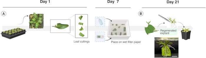
Figure 1: Vegetative propagation protocol and timeline. Representative images illustrate the production pipeline used to regenerate asexual rosette plantlets. (A) Excising mature, healthy, entire lobed leaves from established mother plants and cutting them into 2-3 pieces. (B) Placing explants in a propagation tray and maintaining them under specified conditions. After 3 weeks, the rosette plantlets were excised from the explants and transplanted into the soil media. Below is an actual photo of the regenerated rosettes still attached (pink arrows) to the mother leaf cutting. These plantlets are at the optimal stage for transplantation. Scale bar: 10 mm. Please click here to view a larger version of this figure.
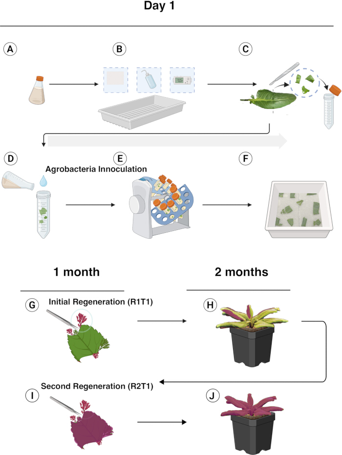
Figure 2: Transformation of R. aquatica leaf cuttings and regeneration of transformants expressing RUBY transcripts. Graphical overview of the transformation procedure and production of transformed regenerants. (A) Obtaining bacterial inoculum. (B) Preparing a propagation tray with sterile filter paper and water. (C) Excising leaves into explant pieces. (D) Immersing explants in inoculum. (E) Agitating in a rotary mixer for 20r/1 min for 10 min. (F) Spreading saturated explants in the tray, covering with a plastic dome, and incubating under dark conditions for 72 h. (G,H) After 1 month, excising intact transformed regenerants from Agrobacterium-exposed explants and transplanting to establish mature plants. (I,J) Repeating the propagation step (G,H) to produce a near clonal R2-T1 generation. Please click here to view a larger version of this figure.
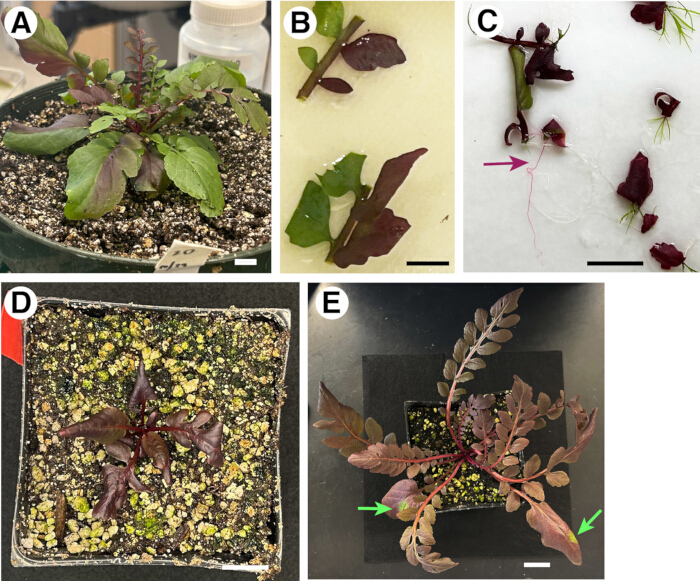
Figure 3: Asexually propagated generations of a RUBY transgenic line. (A) Mature R1-T1 seedling exhibiting chimeric leaves bearing RUBY expression. Scale bar: 1 cm. (B) Two of twenty explants freshly placed in a propagation tray from the 3-month-old seedling shown in (A). The explants exhibit a chimeric pattern of RUBY separated at the midrib. Scale bar: 1 cm. (C) Regenerated R2-T1 rosette plantlets with varying degrees of chimeric RUBY expression, one month after excision, prior to transplant. Regenerated RUBY positive roots (marron arrow). Scale bar: 2 cm. (D) R2-T1 seedling shown 1 month after transplanting, exhibiting RUBY expression. Scale bar: 2 cm. (E) Mature R2-T1 seedling 3 months after transplant exhibiting small sectors (green arrows). Scale bar: 2 cm. Please click here to view a larger version of this figure.
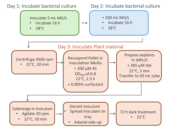
Figure 4: Flowchart of the optimized pipeline. Please click here to view a larger version of this figure.
Table 1: Media Formulations Please click here to download this Table.
Table 2: Effect of four optimization parameters on the transformation efficiency of R. aquatica. Please click here to download this Table.
Table 3: Effect of inoculum acetosyringone concentration on transformation efficiency ofR. aquatica. Please click here to download this Table.
Discussion
In this study, we established a simple, direct regeneration-based Agrobacterium-mediated stable transformation of R. aquatica, a Brassicaceae amphibious plant that solely propagates asexually10,16. A thorough transformation pipeline of vegetatively propagated amphibious plants has not been established, nor has R. aquatica's practical transformability been utilized with a visual marker. Notably, this pipeline does not require a laborious, sterile plant tissue culture process or specialized plant growth incubators. As such, this protocol can be widely adopted to diverse experimental settings from scientific research laboratories to the classroom.
Among the different inoculation and incubation conditions tested, a few factors made a significant improvement in transformation efficiency. Notably, a slightly higher acetosyringone (AS) concentration as well as indole-3-acetic acid (IAA) concentration positively affected the transformation efficiency (Table 1 and Table 2). AS is a phenolic compound that attracts Agrobacterium, and it has been demonstrated to improve plant transformation efficiency in Arabidopsis17. The phytohormone auxin is central for plant cell reprogramming. A rapid increase of auxin-related gene expression was observed during the wound-induced regeneration10. For this reason, one could presume that exogenously applied auxin (IAA) during explant inoculation increases the regeneration efficiency. However, differences in the observed number of regenerated plantlets from three trials were not observed (57 7.55 vs. 59.67 5.51 for control vs. treatment). Therefore, auxin treatment may likely aid the transformation process rather than regeneration itself.
A non-ionic surfactant, Alkoxylated trisilane (known as Silwet L-77), greatly increases the transformation efficiency of Arabidopsis via the floral-dip method3, and as such, it is widely used among the plant biology community. There was minimal plant stress observed among explants amended with Silwet L-77 at 0.005% (the concentration recommended for floral dip). When this dose was doubled (0.01%), R. aquatica leaf cuttings were severely damaged, resulting in the recovery of nearly no transformants (Table 2B). During Arabidopsis floral dipping, the use of surfactants reduces the surface tension of inoculants and enhances access to floral buds. In contrast, as an amphibious plant species, R. aquatica develops smooth leaves devoid of trichomes or thick cuticles5. Increased surfactant caused leaf tissue damage, indicating that factors other than surfactant should be prioritized for transformation efficiency in non-pubescent leaf tissues. Surfactant is more relevant for pubescent phenotype, where it facilitates tissue penetration. After integrating all the tested parameters, a pipeline with the most optimal conditions is provided as a flow chart (Figure 4).
Utilization of a visual marker, RUBY, is instrumental to this transformation pipeline. RUBY is a synthetic reporter encoding three coding genes (CYP76AD1, DODA, and Glucosyl transferase) that synthesize a bright red pigment, betalain11. When the RUBY expression was observed in R1-T1 regenerants, it exhibited expression as red sectors, indicating that they are most likely chimeric (Figure 2 and Figure 3). A previous study revealed that R. aquatica regeneration originates from a vascular cambium of a cut leaf section10. Strikingly, R1-T1 regenerants occasionally exhibit a chimeric boundary along the midvein, with half of the leaf blade red and the other half green (Figure 3A,B). Re-regeneration from RUBY sectors occasionally yielded fully red plantlets with red roots (Figure 3C, maroon arrow), which were then grown into uniformly red R2-T1 plants (Figure 3D). No adverse effects of RUBY overexpression on R. aquatica growth and development were observed (Figure 3). In support, no discernible differences were observed in the size and shape of R1-T1 rosette leaves with RUBY sectors (e.g., Figure 3B). It is possible that any strong, constitutive promoter-driven transgene might interfere with normal plant growth and physiological response.
On some occasions, these 'near clonal' RUBY plants developed green sectors (Figure 3E, green arrows), suggestive of reversion or transgene silencing. This indicates that having a visible marker like RUBY is indispensable for ectopic expression or silencing (e.g., RNAi) of a gene of interest for future molecular, mechanistic studies. One can introduce a transgene of interest to the RUBY cassette and observe the phenotypes in the red, RUBY sectors. In conclusion, harnessing the leaf cutting-induced direct reprogramming and a non-invasive visible marker, a simple, efficient transformation pipeline is optimized for R. aquatica, a vegetatively-propagating Brassicaceae. While it is out of the scope of this protocol, future understanding of the exact origin of the regeneration of transformed R. aquatica might provide insight into cellular reprogramming in plants.
Disclosures
We declare that we have no conflicts of interest.
Acknowledgements
We thank Prof. Yunde Zhao for advice on the vector design to modify the existing RUBY plasmids; Dr. Shuka Ikematsu and Prof. Seisuke Kimura for advice on R. aquatica handling; Dr. Issey Takahashi for help in graphics. This work is supported by the start-up funds and endowment from the Johnson & Johnson Centennial Chair of Plant Cell Biology from The University of Texas at Austin, and the Howard Hughes Medical Institute to K.U.T. Figure 1 and Figure 2 were created in BioRender by P.E.G. and modified in Adobe Illustrator by K.U.T.
Materials
| Name | Company | Catalog Number | Comments |
|---|---|---|---|
| 1000 µL Low Retention LTS Filtered Pipet Tips | Rainin | 30389213 | |
| Acetosyringone | Sigma-Aldrich | D134406-1G | |
| Bacto Yeast Extract, Technical | Fisher Scientific | DF0886-17-0 | |
| BD Bacto Agar | Fisher Scientific | 214030 | |
| BD Difco LB Broth, Miller | Midland Scientific | DIFCO 244610 | |
| BD Bacto Dehydrated Tryptone | Fisher Scientific | DF0123-17-3 | |
| Biotin lyophilized powder | Sigma-Aldrich | B4501-100MG | |
| Clear Domes for PermaNest Trays | Grower's Solution | GD221 | |
| Clear Plastic Wrap | Thomas Scientific | 22-305654 | |
| Coarse Vermiculite | Bectine | ||
| di-Potassium hydrogen phosphate anhydrous | Sigma-Aldrich | 105109 | |
| Disposable Petri Dishes 100 x 15 mm | VWR International | 25384-342 | |
| D-Mannitol | Sigma-Aldrich | M9546-250G | |
| Ethanol, 200 proof | Fisher Scientific | 04-355-720 | |
| Falcon 50 mL Conical Centrifuge Tubes | Fisher Scientific | 14-432-22 | |
| Folded paper towels 9.3” x 10.5” | Kimberly-Clark | 2920 | |
| GENESYS 30 Visible Spectrophotometer | Thermo Scientific | 840-277000 | |
| Gentamicin solution | Sigma-Aldrich | G1272-100ML | |
| L-Glutamic acid monosodium salt hydrate | Sigma-Aldrich | G1626-100G | |
| Magnesium chloride | Sigma-Aldrich | M8266-100G | |
| Magnesium sulfate, Heptahydrate | Sigma-Aldrich | M9546-250G | |
| MaxQ 6000 Incubator Shaker | Thermo Scientific | SHKE6000 | |
| MES hydrate | Sigma-Aldrich | M8250-100G | |
| Murashige and Skoog Basal Salt Mixture (MS), powder | Sigma-Aldrich | M5524-50L | |
| MX-RL-pro: Rotisserie Tube Rotator | Scilogex | 824222019999 | |
| myo-Inositol | Sigma-Aldrich | I7508-100G | |
| Percival Growth Chamber | Percival | AR36L3 | |
| Perlite Planting Soil Additive | PVP Industries | PVP105408 | |
| Perma-Nest Plant Trays 8''x12'' | VWR International | 470150-476 | |
| pH meter Seven Compact 5220 | Mettler Toledo | 30019028 | |
| Protected Scalpel | Fisher Scientific | 02-688-79 | |
| Rifampicin | Sigma-Aldrich | R3501-5G | |
| Smart Release Fertilizer Plant Food | Osmocote | B0071CZTBA | |
| Sodium chloride | Fisher Scientific | S271-3 | |
| Spectinomycin dihydrochloride pentahydrate | Sigma-Aldrich | S4014-5G | |
| Spray Bottles with Pump Vaporizer | Thomas Scientific | 1141V43 | |
| Square 3.5" GAGE pots | Growers Nursery Supply Inc. | 03GA-0350S | |
| Sucrose | Sigma-Aldrich | S0389-5KG | |
| Sunshine #4 Professional Growing Mix Soil | Sun GRO Horticulture | 1341CFL0028P S | |
| Surfactant- Silwet L-77 | Fisher Scientific | NC1791615 | |
| Vacuum Desiccator | The Lab Depot | 55207 | |
| Water Soluble All Purpose Plant Food | Miracle-Gro | 160101 |
References
- Gelvin, S. B. Agrobacterium-mediated plant transformation: the biology behind the "gene-jockeying" tool. Microbiol Mol Biol Rev. 67 (1), 16-37 (2003).
- Hellens, R., Mullineaux, P., Klee, H. Technical Focus: A guide to Agrobacterium binary Ti vectors. Trends Plant Sci. 5 (10), 446-451 (2000).
- Clough, S. J., Bent, A. F. Floral dip: A simplified method for Agrobacterium-mediated transformation of Arabidopsis thaliana. Plant J. 16 (6), 735-743 (1998).
- Zhang, X., Henriques, R., Lin, S. S., Niu, Q. W., Chua, N. H. Agrobacterium-mediated transformation of Arabidopsis thaliana using the floral dip method. Nat Protoc. 1 (2), 641-646 (2006).
- Koga, H., Ikematsu, S., Kimura, S. Diving into the water: Amphibious plants as a model for investigating plant adaptations to aquatic environments. Annu Rev Plant Biol. 75 (1), 579-604 (2024).
- Nakayama, H., et al. Regulation of the KNOX-GA gene module induces heterophyllic alteration in North American lake cress. Plant Cell. 26 (12), 4733-4748 (2014).
- Ikematsu, S., et al. Rewiring of hormones and light response pathways underlies the inhibition of stomatal development in an amphibious plant Rorippa aquatica underwater. Curr Biol. 33 (3), 543-556.e4 (2023).
- Sakamoto, T., et al. A chromosome-level genome assembly for the amphibious plant Rorippa aquatica reveals its allotetraploid origin and mechanisms of heterophylly upon submergence. Commun Biol. 7 (1), 431(2024).
- Flora of North America: Volume 7: Magnoliophyta: Dilleniidae, Part 2. , Oxford University Press Inc. 493-506 (2010).
- Amano, R., et al. Molecular basis for natural vegetative propagation via regeneration in North American Lake Cress, Rorippa aquatica (Brassicaceae). Plant Cell Physiol. 61 (2), 353-369 (2020).
- He, Y., Zhang, T., Sun, H., Zhan, H., Zhao, Y. A reporter for noninvasively monitoring gene expression and plant transformation. Hortic Res. 7 (1), 152(2020).
- Hartig, F. DHARMa: Residual Diagnostics for Hierarchical (Multi-Level / Mixed) Regression Models. R package version 0.4.7. , (2024).
- Lenth, R. emmeans: Estimated Marginal Means, aka Least-Squares. R package version 1.10.7. , (2025).
- McGillycuddy, M., Bolker, B. M., Popovic, G., Warton, D. I. Parsimoniously fitting large multivariate random effects in glmmTMB. J Stat Software. 112 (1), 1-19 (2025).
- Bates, D., Mächler, M., Bolker, B. M., Walker, S. C. Fitting linear mixed-effects models using lme4. J Stat Software. 67 (1), 1-48 (2015).
- Nakayama, H., Sinha, N. R., Kimura, S. How do plants and phytohormones accomplish heterophylly, leaf phenotypic plasticity, in response to environmental cues. Front Plant Sci. 8, 1717(2017).
- Sheikholeslam, S. N., Weeks, D. P. Acetosyringone promotes high efficiency transformation of Arabidopsis thaliana explants by Agrobacterium tumefaciens. Plant Mol Biol. 8 (4), 291-298 (1987).
Reprints and Permissions
