We present a simple, effective, and reproducible protocol for Agrobacterium-mediated transformation of Phytophthora palmivora, which is also applicable to P. capsici.
Method Article
A Simplified Method for Agrobacterium-mediated Transformation of Phytophthora palmivora
In This Article
Summary
Abstract
Phytophthora is an oomycete genus that contains numerous destructive plant pathogens, among which is the broad-host-range species P. palmivora. Functional genomics of Phytophthora has been constrained by the limited availability of robust, easy-to-implement transformation methods. Among the available approaches, Agrobacterium-mediated transformation (AMT) is particularly attractive because it requires minimal specialized equipment and often produces stable transformants with single-copy gene insertions. Most AMT protocols for fungal and oomycete transformation use minimal medium (MM) salts in Agrobacterium induction and co-cultivation media. Preparing these media typically involves making and mixing multiple stock solutions, with some containing numerous components at different concentrations. The process is time-consuming and error-prone, therefore leading to inconsistent transformation success. Here, we present a streamlined, simple, and reproducible AMT protocol for P. palmivora that uses commercially available Murashige and Skoog (MS) basal medium to prepare the Agrobacterium induction and co-cultivation media. This protocol has also been successfully applied to transform P. capsici and may be applied to other Phytophthora spp. as well.
Introduction
Phytophthora, a genus of oomycetes, includes many of the world's most destructive plant pathogens, responsible for severe economic losses1,2. Among them, Phytophthora palmivora is a broad-host-range pathogen that infects many economically important crops, including cacao and papaya3. It is the most widespread causal agent of black pod rot in cacao, a tropical perennial tree that produces the major component of the multibillion-dollar chocolate industry4,5,6. Understanding the molecular pathogenicity mechanisms of this destructive pathogen is critical for designing novel, effective disease-control strategies, and genetic transformation is an essential tool to enable such investigations.
Several methods have been developed to transform Phytophthora spp., including microprojectile bombardment7, polyethylene glycol and calcium dichloride (PEG/CaCl2)-mediated protoplast transformation8,9,10,11, zoospore electroporation12,13,14, and Agrobacterium-mediated transformation (AMT)15,16. Among these approaches, AMT offers key advantages as it typically generates single-copy gene integration15,16 and it is relatively easy to perform without requiring specialized equipment.
A protocol for efficient transformation of P. palmivora using AMT has been previously developed16. With this method, we successfully transformed P. palmivora with multiple constructs17,18. This method utilizes minimal medium (MM) salt-based Agrobacterium induction and co-cultivation media, which are commonly used for fungal and oomycete transformation15,16,19. Although the MM-based transformation protocol was shown to be effective in generating transformants, preparing multiple stock solutions and combining them to make media can be a task that is both time-consuming and prone to error.
Here, we present a much simplified method for AMT of P. palmivora by using commercial Murashige and Skoog (MS) basal medium to replace the MM salt-based medium components in the Agrobacterium induction and co-cultivation media16. This method eliminates the need to prepare multiple chemical stock solutions, making the process less time-consuming and highly reproducible. Moreover, this approach appears to yield higher transformation efficiency compared to the previously described method16, and is also applicable to P. capsici.
Protocol
1. Preparation of stock solutions, media, and other materials required for the transformation
- Antibiotic stock solutions
- Prepare stock solutions of kanamycin (50 mg/mL), G418 (15 mg/mL), and cefotaxime (100 mg/mL): Dissolve appropriate amounts of the antibiotics in water, filter-sterilize the solutions, prepare 1-mL aliquots, and store them at -20 °C.
- Prepare stock solution of rifampicin (15 mg/mL): Dissolve the appropriate amount of rifampicin in DMSO, prepare 1-mL aliquots, wrap them in foil, and store them at -20 °C.
NOTE: Ensure to filter-sterilize chemical solutions prepared in DMSO using syringe filters made of DMSO-compatible materials, such as Nylon or PTFE (Teflon).
- Media for Agrobacterium growth
- Luria-Bertani (LB) liquid media: To prepare 250 mL of media, add 6.25 g LB powder to 250 mL of water, autoclave the solution, and store it at room temperature (RT).
- LB agar media: To prepare 250 mL of media, add 6.25 g LB powder and 3.75 g agar to 250 mL of water, autoclave the solution, and store at RT. Add appropriate antibiotics to the melted LB agar before making the plates. Store the plates supplemented with antibiotics at 4 °C.
- Media for P. palmivora growth and transformation
- 10% V8 agar: To prepare 500 mL, add 50 mL of V8 original 100% vegetable juice to 450 mL of water. Then add 0.5 g of CaCO3 to the suspension and stir to mix it well. Autoclave the solution and pour it into plates.
NOTE: Add appropriate antibiotics when needed. Store the plates supplemented with antibiotics at 4 °C. - Half-strength Murashige and Skoog (½ MS) liquid medium: Add 2.15 g of Murashige and Skoog basal medium (MS) to 950 mL of water, adjust pH to 5.6 with 1 N KOH, and add water up to 1 L. Use directly to make ½ MS agar (step 1.3.3), autoclave the remaining ½ MS liquid, and store it at RT.
- ½MS agar for Agrobacterium-zoospore co-cultivation agar (AZCA): To prepare 600 mL of AZCA, mix 567 mL of ½ MS liquid from step 1.3.2 with 9 g agar. Autoclave the solution and store it at RT.
- 50% Glycerol: Mix 50 mL glycerol with 50 mL of water, autoclave the solution, and store it at RT.
- 1M MES (pH 5.6): To prepare 200 mL, add 39.048 g MES Hydrate to 160 mL of water, then adjust the pH to 5.6 with NaOH solution. Next, add water up to 200 mL, filter-sterilize the solution, and store it at RT under dark conditions.
NOTE: MES will dissolve gradually as the pH increases. Initially, use 10 N NaOH to bring the pH close to 5.6, which will almost dissolve the powder. Then, add 1 N NaOH to bring the pH exactly to 5.6. - 20% Glucose: Dissolve 10 g glucose in 40 mL of water, then add water up to 50 mL, filter-sterilize the solution, and store it at RT.
- 200 mM Acetosyringone (AS): Dissolve 40 mg 4'-Hydroxy-3’,5’-dimethoxyacetophenone in 1 mL DMSO (see NOTE under 1.1.2) and cover the solution with foil. Make it fresh on the day of transformation.
- Agrobacterium induction media (AIM): To prepare 40 mL of media, add 400 µL of 50% glycerol, 400 µL of 20% glucose, 1.6 mL of 1 M MES (pH 5.6), 40 µL of 200 mM AS to 37.6 mL of ½ MS liquid (pH 5.6) solution (prepared in step 1.3.2) in a sterile 50 mL tube. Mix the solution well, cover the tube with foil, and use it fresh within several hours.
- Agrobacterium-zoospore co-cultivation agar (AZCA): To prepare 600 mL of the agar, add 6 mL of 50% glycerol, 3 mL of 20% glucose, 24 mL of 1 M MES (pH 5.6), 600 µL of 200 mM AS to autoclaved and melted 567 mL ½ MS agar (pH 5.6) (prepared in step 1.3.3). Pour the solution into plates and cover them with a black cloth to protect from light.
NOTE: Add the chemical solutions to melted ½ MS agar (step 1.3.9) when the temperature is 55-60 °C. Turn off the lights in the hood during steps 1.3.7-1.3.9 as AS is light-sensitive. Perform these three steps on the day of transformation. - Plich agar: To prepare 1 L of the agar, in 950 mL of water, add 0.5 g KH2PO4, 0.25 g MgSO4·7H2O, 1 g L-asparagine, 5 g glucose, 0.5 g yeast extract, 10 µL thiamine (100 mg/mL in water, stored at 4 °C), 10 mg β-sitosterol (dissolve in 3 mL ethanol before adding). Stir the solution to dissolve all the ingredients, then add water to make the volume up to 1 L. Divide the solution into 500 mL portions and add 7.5 g agar to each 500 mL. Autoclave the solution, and add appropriate antibiotics when the temperature drops to 55 °C, then pour it into plates.
- Hybond N+ membrane: Cut the membrane with the protective sheets into pieces of 5 cm x 6 cm, wrap the pieces in aluminum foil, and autoclave them. While placing the autoclaved membrane on AZCA media, handle it with sterile forceps, and discard the protective sheets.
- 10% V8 agar: To prepare 500 mL, add 50 mL of V8 original 100% vegetable juice to 450 mL of water. Then add 0.5 g of CaCO3 to the suspension and stir to mix it well. Autoclave the solution and pour it into plates.
2. Culturing P. palmivora
- Seven to eight days before the transformation, grow P. palmivora papaya isolate P116 on freshly made 10% V8 agar plates by placing one agar plug of a previously grown Phytophthora culture (or from the stock) onto the center of the Petri dish.
NOTE: To obtain a higher concentration of zoospores, it is important to use freshly prepared unclarified 10% V8 agar. - Incubate the plates at 25 °C under a 12-h light/12-h dark cycle for 7-8 days until the transformation.
3. Preparation of Agrobacterium tumefaciens
- Three days before the transformation, streak A. tumefaciens strain EHA105 carrying pCB301TOR-GFP16 on an LB agar plate supplemented with rifampicin (15 µg/mL) and kanamycin (50 µg/mL). Incubate the plate at 28 °C for 2 days.
NOTE: The initial preparation of Agrobacterium (step 3.1) can be done a week before the transformation, and the plates can be stored at 4 °C after the 2-day incubation period. - On the day before transformation, take a small amount of Agrobacterium cells and spread them on a new LB agar plate supplemented with antibiotics (as described in step 3.1) using the bottom and flat base of a sterile 1.5 mL microcentrifuge tube. For easier spreading, add 20 µL LB liquid media to the center of the LB agar plate, mix the bacterial cells with the LB media, and then spread them evenly across the plate. Incubate the plate at 28 °C overnight for about 16-18 h.
- Scoop the cells grown overnight with a 1 mL pipette tip and resuspend them in 1 mL Agrobacterium induction medium (AIM) by pipetting up and down. Dilute the cells to OD600 = 0.4 with AIM in a sterile 50 mL tube covered with foil. Make a minimum of 5 mL to be used for co-incubation with P. palmivora zoospores (step 5.1).
NOTE: An OD600 ranging from 0.1 to 0.8 would allow successful transformation. OD600 = 0.4 is routinely used as this concentration produced the highest transformation efficiency when minimal medium (MM) salt-based Agrobacterium induction and co-cultivation media were used for transformation16. - Incubate the bacteria at RT under dark conditions for 1.5-2 h on a platform shaker with gentle agitation (approximately 70 rpm).
NOTE: It is recommended to keep the hood lights off during step 3.3 and to loosen the cap of the 50 mL tube during incubation (step 3.4).
4. Releasing the zoospores
- After 1 h of the Agrobacterium tumefaciens incubation (step 3.4), flood a 7-8 day old P. palmivora plate with 10 mL water pre-cooled at 4°C, remove the air bubbles on the media surface by gently tapping with a pipette tip, and incubate the plate at 4 °C for 15 min, followed by 15 min at RT under light.
- Gently transfer the released zoospores (about 7-8 mL) to a 15 mL sterile centrifuge tube with a pipette without disturbing the mycelia and sporangia.
- To measure the zoospore concentration, transfer some of the zoospore suspension (e.g., 20 µL) to a 1.5 mL microcentrifuge tube and dilute it 10 times with water (180 µL H2O for 20 µL zoospores). Vortex the tube for 1 min to encyst the zoospores for easy counting, and then count the number of zoospores under a microscope using a hemacytometer. Multiply the counted number by a factor of 10 to determine the concentration of the zoospore suspension prepared in step 4.2.
NOTE: Zoospore concentration is measured for calculating the transformation efficiency. We typically use concentrations ranging from 1.5 × 10⁶ to 5 × 10⁶ zoospores per mL, with transformation success.
5. Co-incubation of Agrobacterium and zoospores
- Add 5 mL of Agrobacterium (prepared in step 3.4) to 5 mL of zoospore suspension (prepared in step 4.2) into a sterile 50 mL tube covered with foil. Gently mix and incubate the tube at RT for 2 h in the dark. Keep the lid loose and do not shake the tube during the co-incubation.
- During the co-incubation period (step 5.1), prepare the AZCA plates (step 1.3.9). When the medium solidifies, place one piece of sterilized Hybond N+ membrane (prepared in step 1.3.11) onto each AZCA plate and keep the plates in the dark.
- After 2 h of the Agrobacterium and zoospore co-incubation (step 5.1), spread 300 µL of the mixture onto each Hybond N+ membrane placed atop the AZCA plates (step 5.2) with a pipette tip, leaving the margins free from the liquid.
- Let the mixture air-dry for about 10 min in the hood by leaving the Petri dishes uncovered. Cover the plates as soon as the solution on the membrane stops flowing around.
NOTE: During steps 5.1-5.4, keep the hood lights off. If necessary, use lights while adding and spreading the mixture on the membranes (step 5.3). - After air-drying, incubate the plates at 25 °C in the dark for 2 days.
6. Selection of G418-resistant transformants
- After 48 h of co-incubation (step 5.5), transfer the Hybond N+ membranes upside-down using sterilized forceps to Plich agar plates supplemented with G418 (30 µg/mL) and cefotaxime (100 µg/mL). While placing the membranes on the plates, do not drag them on the medium surface.
NOTE: The concentration of G418 for selection of transformants for each P. palmivora isolate must be determined, as different levels of sensitivity are observed for different isolates. To do this, place a 0.5-cm-diameter mycelium plug at the center of Plich agar plates supplemented with a series of G418 concentrations in 5 µg/mL increments starting from zero. Prepare three replicate plates for each concentration. Incubate the plates at 25 °C for 7 days. Identify the lowest G418 concentration that completely inhibits the growth of the Phytophthora isolate and use it for the selection of transformants. - Use the flat base of a 1.5 mL microcentrifuge tube to gently press and slide over the surface of the membrane to make sure that it is in close contact with the medium. Observe the underside of the plates to check the presence of air bubbles between the membrane and medium. Gently squeeze out the air bubble, if any, using the flat base of the same microcentrifuge tube.
- Incubate the plates under a 12-h light/12-h dark cycle at 25 °C for 2-3 days.
- After 3 days of incubation, remove the membranes using sterile forceps and continue incubating the plates under the same conditions. The membranes can be removed after 2 days if there are visible colonies appearing on the plates.
NOTE: The transformants typically appear within 1-3 days after membrane removal, but some colonies may appear later. To yield more transformants, monitor the plates for about one week after removing the membranes. - Transfer the transformants onto 10% V8 agar plates with the same concentration of G418 or a concentration reduced by 30%.
Results
By following this protocol, we conducted two independent experiments to transform P. palmivora using A. tumefaciens EHA105 carrying pCB301TOR-GFP16. As expected, co-incubation of zoospores with Agrobacterium yielded G418-resistant colonies on Plich agar plates supplemented with 30 μg/mL G418, whereas no colonies were observed on control plates without infection with Agrobacterium (Figure 1A). The number of G418-resistant transformants per 107 zoospores produced from these two experiments was 55 and 50, respectively (Table 1), which is nearly double the highest number (27 ± 5) previously obtained using the earlier protocol16.
True transformants were verified by two methods: First, observation of GFP fluorescence using a fluorescence microscope, and second, PCR amplification of a 550 bp fragment of the EGFP gene using Taq 2X Master Mix (NEB) with the primer pair EGFP-F2 (5'- GACGTAAACGGCCACAAGTTC-3') and EGFP-R2 (5'-GGGTGCTCAGGTAGTGGTTG-3'). In experiment (Exp) 1, 15 out of 18 G418-resistant transformants, and in Exp 2, 17 out of 21 transformants exhibited GFP fluorescence (Table 1, Figure 1B). For the seven G418-resistant transformants that did not display fluorescence from both experiments, three were confirmed to be true transformants by PCR (Figure 1C). In total, the rate of true transformants was estimated as 90%.
In addition, we were also able to successfully transform a P. capsici squash isolate, PC2024-14sz, using A. tumefaciens EHA105 carrying pCB301TOR-GFP, following the protocol with a minor modification in the preparation of zoospore suspensions (the incubation period at 4 °C after flooding a 7-day-old P. capsici plate with pre-cooled water was extended to 30 min). The number of G418 (30 µg/mL)-resistant transformants per 107 from a single experiment was 13. Nine transformants were observed under a fluorescence microscope, and all produced fluorescence (Supplementary File 1), suggesting that they are true transformants.
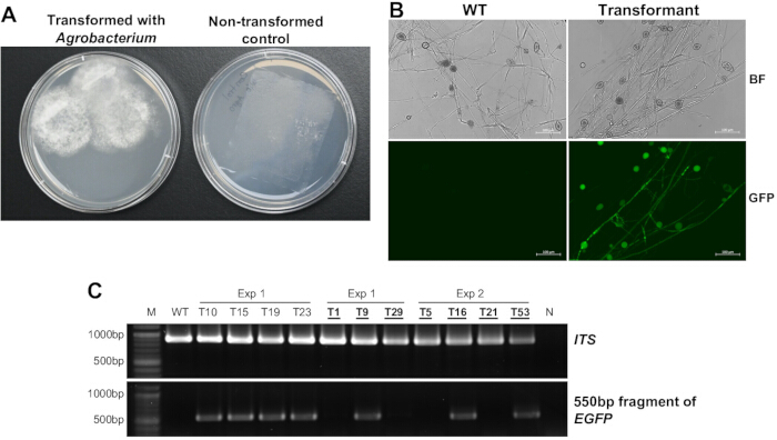
Figure 1: Transformation of P. palmivora using A. tumefaciens EHA105 carrying pCB301TOR-GFP. (A) Appearance of G418-resistant P. palmivora transformants on Plich agar supplemented with 30 µg/mL G418. (B) Detection of GFP expression under a fluorescence microscope in a representative transformant and P. palmivora wild-type (WT) strain. Images were taken under both bright-field (BF) and GFP fluorescence channels. (C) PCR amplification of EGFP from four transformants with detectable GFP fluorescence and seven without GFP fluorescence. Universal primer pair ITS4/ITS520 was used to amplify the internal transcribed spacer (ITS) region to check DNA integrity. Transformants without GFP fluorescence are highlighted in bold, underlined text. Abbreviations: M = NEB 100 bp DNA ladder; WT = wild type P. palmivora P1 isolate; N = negative control PCR using H2O as the template. Please click here to view a larger version of this figure.
| Experiment | OD600 of Agrobacterium | Zoospore concentration (/mL) | Zoospore volume (mL) | Number of G418-resistant transformants | Number of transformants with detectable GFP signal (Detected / Observed) | Number of G418-resistant transformants/ 107 zoospores |
| 1 | 0.4 | 1.8 x 10⁶ | 4.3 | 43 | 15/18 | 55 |
| 2 | 0.4 | 2.9 x 10⁶ | 4.6 | 67 | 17/21 | 50 |
Table 1: Efficiency of Agrobacteria-mediated transformation of P. palmivora using A. tumefaciens EHA105 carrying pCB301TOR-GFP.
Supplementary File 1: GFP expression under a fluorescence microscope in wild-type (WT) P. capsici and a representative transformant generated using A. tumefaciens EHA105 carrying pCB301TOR-GFP. Images were taken under both bright-field (BF) and GFP fluorescence channels. Please click here to download this file.
Discussion
This protocol utilizes half-strength Murashige and Skoog basal medium (½ MS) to replace components of minimal medium (MM) salts that are commonly used in Agrobacterium induction and co-cultivation media for oomycete and fungal transformation. Preparing MM salt-based media requires making multiple stock solutions from numerous individual chemicals and then combining these solutions19. ½ MS also contains many chemical components; however, as MS is widely available commercially as a premixed powder, medium preparation is simplified to dissolving the powder in water. This modification significantly reduces media preparation time and minimizes the potential for human error. In addition, the protocol improves transformation efficiency, yielding a greater number of transformants compared with the previously described AMT method16. It is also effective for transforming P. capsici and is likely applicable to other Phytophthora species. Further supporting its potential broader applicability, MS-based medium has previously been used for co-culturing Agrobacterium tumefaciens strains expressing AtVIP1 (Arabidopsis thaliana VirE2-interacting protein 1) and P. infestans zoospores, enabling successful transformation of P. infestans21.
It is important to note that this protocol may generate a low percentage of false positives. Among 39 G418-resistant transformants that were tested by GFP fluorescence observation or PCR, four showed neither fluorescence nor amplification of EGFP (Table 1, Figure 1C). As partial integration of T-DNA during Agrobacterium-mediated transformation is not uncommon22,23, it is possible that only the portion of the T-DNA containing the NPTII gene from plasmid pCB301TOR-GFP16 was integrated into the P. palmivora genome. Alternatively, the false positives may result from insufficient contact between the membrane and the selective medium, allowing escape from G418 selection. Ensuring that the membrane stays in full contact with the medium (step 6.2) is therefore essential.
This protocol may be further simplified. For example, MES in Agrobacterium induction media (AIM) and Agrobacterium-zoospore co-cultivation agar (AZCA) can potentially be omitted. MES is commonly used as a buffering agent in biochemistry and molecular biology experiments to maintain a stable pH; however, preparing a 1 M MES stock solution at pH 5.6 takes a sufficient amount of time and adds to the cost. In AMT of plants, such as sweet basil24 and papaya25, similar MS-based Agobacterium induction and co-cultivation media without MES were used, suggesting MES may not be essential for P. palmivora transformation. Further experiments are needed to confirm that MES omission does not compromise transformation efficiency.
Overall, this simplified protocol provides a practical and accessible platform for routine genetic manipulation of P. palmivora, P. capsici, and potentially other oomycetes. It supports a broad range of fundamental studies with some having translational potentials, including fluorescent labeling of pathogens to track infection dynamics, tagging proteins to examine subcellular localization, and functional analyses through targeted changes in gene expression or protein-coding sequences. When coupled with CRISPR-based genome editing, the protocol is expected to accelerate the dissection of pathogenicity mechanisms and identification of key virulence factors in these destructive pathogens. The resulting insights will, in turn, enable the development of novel, mechanism-based disease control strategies.
Disclosures
The authors declare no conflicts of interest.
Acknowledgements
The study is supported by the National Science Foundation (NSF Award No. 2418799) and National Institute of Food and Agriculture (NIFA Award No. 2023-67013-42262).
Materials
| Name | Company | Catalog Number | Comments |
|---|---|---|---|
| 1.5 mL Microcentrifuge Tubes | VWR | 525-1126 | |
| 4'-Hydroxy-3',5' dimethoxyacetophenone | TCI | D2666 | Acetosyringone (AS) |
| 50 mL Tube | Greiner Bio-one | 227261 | |
| Absolute Ethanol | Fisher BioReagents | BP28184 | |
| Agar | Fisher BioReagents | BP1423-500 | |
| Aluminium foil | |||
| Hybond-N+ membrane (30 cm × 3 m) | Cytiva | RPN303B | |
| Calcium carbonate | Sigma-Aldrich | 239216-500G | CaCO3 |
| Cefotaxime sodium salt | Thermo Scientific Chemicals | AAJ6269006 | |
| D-(+)-Glucose | Sigma-Aldrich | G8270-1KG | |
| DNeasy PowerLyzer Microbial Kit | Qiagen | 12255-50 | |
| Forceps | |||
| Fluorescence microscope | Zeiss | ZEISS Axioscope 5 fluorescence microscope with filter set 38 HE | |
| G418 sulfate | Thermo Scientific Chemicals | AC329400010 | |
| Glycerol | Sigma-Aldrich | 911046-1L | |
| Hemacytometer (Bright-Line) | Hausser Scientific | ||
| Kanamycin sulfate | Santa Cruz Biotechnology | sc-257635 | |
| L-Asparagine anhydrous | MP Biomedicals | ICN10079425 | |
| LB Broth, Miller | Fisher BioReagents | BP1426-2 | |
| Magnesium sulfate heptahydrate | Sigma-Aldrich | 63138-250G | MgSO4·7H2O |
| MES Hydrate | Sigma-Aldrich | M2933-100G | |
| Murashige and Skoog Basal Medium | Sigma-Aldrich | M5519-50L | |
| Petri Dish (100 mm x 15 mm) | VWR | 25384-342 | |
| Potassium dihydrogen phosphate | Sigma-Aldrich | 1370391000 | KH2PO4 |
| Rifampicin | Santa Cruz Biotechnology | sc-200910 | |
| Taq 2X Master Mix | NEB | M0270L | |
| Thiamine hydrochloride | Sigma-Aldrich | T4625-5G | |
| UV-Visible Spectrophotometer | Fisher Scientific | 14-385-355 | |
| V8 Original 100% Vegetable Juice | Campbell Soup Company | ||
| Vortexer | Fisher Scientific | 14-955-163 | |
| Water | Molecular Biology Grade, purified using Mill-Q water purification system | ||
| Yeast Extract | ACROS | 611805000 | Currently available through Thermo Scientific Chemicals |
| β-Sitosterol | Sigma-Aldrich | 85451-100G |
References
- Kamoun, S., et al. The top 10 oomycete pathogens in molecular plant pathology. Mol Plant Pathol. 16 (4), 413-434 (2015).
- Jones, J. D., Staskawicz, B. J., Dangl, J. L. The plant immune system: From discovery to deployment. Cell. 187 (9), 2095-2116 (2024).
- Tocafundo, F., et al. Direct and cross-pathogenicity of Phytophthora palmivora against cacao, papaya, and peach palm. Trop Plant Pathol. 46 (6), 664-673 (2021).
- Morales-Cruz, A., et al. Independent whole-genome duplications define the architecture of the genomes of the devastating West African cacao black pod pathogen Phytophthora megakarya and its close relative Phytophthora palmivora. G3 (Bethesda). 10 (7), 2241-2255 (2020).
- Ali, S., et al. PCR-based identification of cacao black pod causal agents and identification of biological factors possibly contributing to Phytophthora megakarya's field dominance in West Africa. Plant Pathol. 65 (7), 1095-1108 (2016).
- Marelli, J. -P., et al. Chocolate under threat from old and new cacao diseases. Phytopathology. 109 (8), 1331-1343 (2019).
- Cvitanich, C., Judelson, H. S. Stable transformation of the oomycete, Phytophthora infestans, using microprojectile bombardment. Curr Genet. 42 (4), 228-235 (2003).
- Judelson, H. S., Tyler, B. M., Michelmore, R. W. Transformation of the oomycete pathogen, Phytophthora infestans. Mol Plant Microbe Interact. 4 (6), 602-607 (1991).
- Wang, W., et al. an oxathiapiprolin-resistance gene, functions as a novel selection marker for Phytophthora transformation and CRISPR/Cas9 mediated genome editing. Front Microbiol. 10, 2402(2019).
- Dai, T., et al. An improved transformation system for Phytophthora cinnamomi using green fluorescent protein. Front Microbiol. 12, 682754(2021).
- Fang, Y., Cui, L., Gu, B., Arredondo, F., Tyler, B. M. Efficient genome editing in the oomycete Phytophthora sojae using CRISPR/Cas9. Curr Protoc Microbiol. 44 (1), 21A.1.1-21A.1.26 (2017).
- Evangelisti, E., Yunusov, T., Shenhav, L., Schornack, S. N-acetyltransferase AAC (3)-i confers gentamicin resistance to Phytophthora palmivora and Phytophthora infestans. BMC Microbiol. 19 (1), 265(2019).
- Dong, L., et al. Establishment of the straightforward electro-transformation system for Phytophthora infestans and its comparison with the improved PEG/CaCl2 transformation. J Microbiol Methods. 112, 83-86 (2015).
- Huitema, E., Smoker, M., Kamoun, S. A straightforward protocol for electro-transformation of Phytophthora capsici zoospores. Methods Mol Biol. 712, 129-135 (2011).
- Vijn, I., Govers, F. Agrobacterium tumefaciens mediated transformation of the oomycete plant pathogen Phytophthora infestans. Mol Plant Pathol. 4 (6), 459-467 (2003).
- Wu, D., Navet, N., Liu, Y., Uchida, J., Tian, M. Establishment of a simple and efficient Agrobacterium-mediated transformation system for Phytophthora palmivora. BMC Microbiol. 16 (1), 204(2016).
- Gumtow, R., Wu, D., Uchida, J., Tian, M. A Phytophthora palmivora extracellular cystatin-like protease inhibitor targets papain to contribute to virulence on papaya. Mol Plant Microbe Interact. 31 (3), 363-373 (2018).
- Pettongkhao, S., Navet, N., Schornack, S., Tian, M., Churngchow, N. A secreted protein of 15 kDa plays an important role in Phytophthora palmivora development and pathogenicity. Sci Rep. 10 (1), 2319(2020).
- Minz, A., Sharon, A. Electroporation and Agrobacterium-mediated spore transformation. Molecular and Cell Biology Methods for Fungi. Sharon, A. , Humana Press. Totowa, NJ. 21-32 (2010).
- Bellemain, E., et al. ITS as an environmental DNA barcode for fungi: An in silico approach reveals potential PCR biases. BMC Microbiol. 10 (1), 189(2010).
- Wang, L., et al. A modified Agrobacterium-mediated transformation for two oomycete pathogens. PLoS Pathog. 19 (4), e1011346(2023).
- Wu, E., et al. Optimized Agrobacterium-mediated sorghum transformation protocol and molecular data of transgenic sorghum plants. In Vitro Cell Dev Biol Plant. 50 (1), 9-18 (2014).
- Sun, L., et al. TDNAscan: A software to identify complete and truncated T-DNA insertions. Front Genet. 10, 685(2019).
- Navet, N., Tian, M. Agrobacterium-mediated transformation of sweet basil (Ocimum basilicum). Bio Protoc. 10 (22), e3828-e3828 (2020).
- Hasley, J., Dinulong, R. -J., Adhikari, A., Christopher, D., Tian, M. Genome editing of papaya using both Cas9 and Cas12a. bioRxiv. , (2025).
Reprints and Permissions
