RNA secondary structure has primarily been observed in mature RNA with structure probing methods. Co-transcriptional Structure Tracking sequencing (CoSTseq) unifies nuclear run-on, which has been used to study Polymerase position on nascent RNA, with structure probing. CoSTseq thereby enables observation of RNA secondary structure in RNA under active transcription.
A subscription to JoVE is required to view this content. Sign in or start your free trial.
Method Article
Comparative RNA Structure Analysis of Nascent and Mature Transcripts in Saccharomyces cerevisiae
* These authors contributed equally
In This Article
Summary
Abstract
During transcription, nascent RNA starts base pairing as it exits RNA polymerases (Pols). This base pairing permits the formation of RNA structures that critically influence gene expression at the level of RNA processing, translation, and stability. Established methods to study RNA secondary structure are limited to mature transcripts, while little is known about folding states. Moreover, the relatively lower abundance (< 1%) and transient nature of nascent RNA complicate its isolation and characterization. Co-transcriptional Structure Tracking (CoSTseq) leverages transcriptional run-on with biotin-NTP and dimethyl sulfate (DMS) probing to simultaneously acquire Pol position and base pairing status of nascent transcripts. In Saccharomyces cerevisiae, CoSTseq yields the sequence and structural information near the 3'-end of nascent RNAs transcribed by any of the three RNA Pols. During transcriptional run-on, the biotin-NTP incorporated at the active site effectively stalls Pols. Then, treatment with DMS methylates unpaired A, C, and U nucleotides. Subsequent biotin enrichment and cDNA synthesis with a template-switching reverse transcriptase enables paired-end sequencing and the computation of DMS reactivities as a function of Pol position. CoSTseq is readily performed side-by-side with DMS probing (DMS-MaPseq), enabling capture of the folded mature transcript as well. Here, a detailed protocol is presented for parallel CoSTseq and DMS-MaPseq, including transcriptional run-on, library preparation, and data analysis.
Introduction
RNA can fold into secondary and tertiary structures due to base pairing within RNA molecules, and these structures can further be influenced by proteins that act as chaperones to guide RNA folding1. RNA structure can be highly dynamic, where cellular RNAs can conform to a range of structures defined by the thermodynamic landscape to generate ensembles of possible RNA conformations2. Dynamic conformational changes have the potential to affect gene regulation and expression3,4. Conversely, RNA can also adopt highly favored structures that are tightly related to its function, such as tRNAs, small nuclear RNAs, and rRNA. While these examples highlight mature RNAs with strong regulatory roles, eukaryotic RNA processing steps often co-occur with transcription, including pre-mRNA splicing, 3'-end cleavage, and RNA modification. Likewise, RNA folding can occur before maturation of the transcript, as discussed elsewhere5.
While the sequence of RNA can encode secondary structure, accurate determination often requires experimental validation in vitro or in vivo. The advent of structure-specific chemical modification techniques like DMS-MaPseq (dimethyl sulfate mutational profiling with sequencing) has paved the way for determining cellular RNA secondary structure from living cells6. DMS is introduced to cells and specifically methylates the Watson-Crick base-pairing faces of adenosine, cytosine, and, to a smaller degree, uridine, which are accessible only when unpaired. Following modification by DMS, a high-fidelity and highly processive reverse transcriptase, such as Thermostable Group II Intron Reverse Transcriptase (TGIRT)7, can be used to recode modified bases as mutations in the final prepared cDNA library. These mutations can then be used to infer the accessibility of RNA at the nucleotide level. While a powerful technique, DMS-MaPseq and related methods have primarily been used to study mature RNA that is already fully folded6.
Several methods have been developed for studying nascent RNA regulation and processing. Global run-on sequencing (GROseq) was developed to overcome the limitations of RNA-seq, which enables steady-state measurement of RNA levels in cells and thus provides information that averages across transcription, RNA processing, and degradation processes that affect RNA levels. GROseq utilized bromouridine for nuclear run-on, where an RNA Polymerase (Pol) could add nucleotides to nascent RNA at the resolution of tens of bases8. Precision run-on sequencing (PROseq) was built on this methodology to map RNA Pols on nascent RNA at base-pair resolution by utilizing biotin-NTPs. Here, a biotinylated-NTP is added where Pol was last detected at the 3'-end of the newly synthesized RNA9.
Recently, Schärfen et al. developed and utilized Co-transcriptional Structure Tracking (CoSTseq), which leverages and adapts biotin run-on and DMS probing to enable RNA structure determination of nascent transcripts in S. cerevisiae10. CoSTseq enables determination of RNA secondary structure during active transcription; this protocol details how nuclear run-on is combined with DMS-probing during CoSTseq experiments. Given that CoSTseq requires careful handling during sample preparation, as well as close attention to data quality when proceeding with CoSTseq analysis, this protocol details best practices for generating suitable libraries for paired-end sequencing on the Illumina platform. The CoSTseq protocol simultaneously yields nascent and mature RNA, the latter of which can be analyzed as a DMS-MaPseq dataset to afford rigorous comparison between nascent and mature RNA (Figure 1). Experimentally, access to reagents needed for maintaining yeast cultures and conducting nuclear run-on and DMS probing is required, as detailed in the protocol below. Nascent RNA yield is critical to the success of CoSTseq library prep and data analysis; thus, growth conditions established by appropriate incubators and growth media are critical to securing sufficient starting material. Finally, to conduct computational analysis of CoSTseq data, access to a High-Performance Computing (HPC) cluster is ideal.
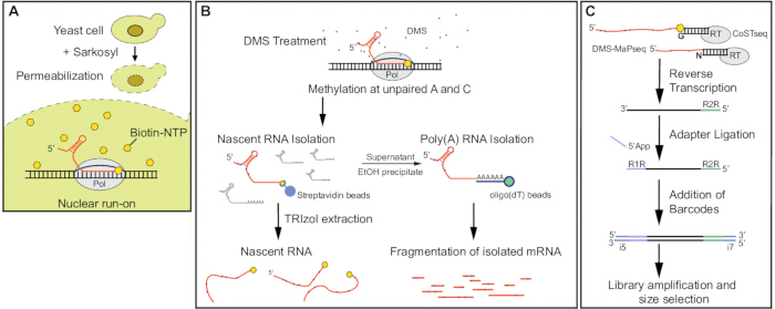
Figure 1: Overview of combined CoSTseq and DMS-MaPseq library preparation from the same biological sample. (A) Permeabilization of the yeast cell wall and nuclear membrane with sarkosyl enables biotin-NTP incorporation at the 3'-end of nascent RNA during a nuclear run-on reaction. (B) DMS methylates unpaired A and C residues (sometimes U) on nascent and mature RNA in the permeabilized cell, and total RNA is obtained by phenol/chloroform extraction. Nascent RNA is then purified from the total RNA by streptavidin-conjugated magnetic beads; TRIzol extraction releases the nascent RNA from the beads. In parallel, the bead supernatant containing polyadenylated mRNAs can be EtOH precipitated; from this material, mRNA can be isolated by oligo(dT) bead pulldown. Poly A+ mRNAs are fragmented before library preparation. (C) A template-switching reverse transcriptase is used to separately generate cDNA from nascent or mature RNA, followed by ligation of 5' and 3' adapters to introduce an N7 UMI and barcodes (see Figure 2 for details of CoSTseq). Note that DMS-MaPseq and CoSTseq reverse transcription steps will use distinct heteroduplex adapters with overhangs complementary to biotin-NTP used in CoSTseq and N overhangs used in DMS-MaPseq. Minimal PCR cycles are used for amplification to limit bias in the library, and size selection is applied to remove adapter dimers. Please click here to view a larger version of this figure.
Access restricted. Please log in or start a trial to view this content.
Protocol
NOTE: Before beginning, make sure all the buffers listed in Table 1 are prepared. All the reagents should be prepared in nuclease-free water. Filter sterilization of buffers is recommended when using non-molecular biology grade chemicals/reagents for buffer preparation. For sections 1 through 4, prepare the following in advance:
1. Preparation of materials and reagents
- Prepare a 10% sarkosyl (v/v) solution at least 1 day in advance to allow sufficient time for complete dissolution. Filter-sterilize the homogeneous solution with a 0.22 µm filter. On the day of the experiment, use the 10% sarkosyl to make a 0.5% sarkosyl solution, and leave it on ice until use.
- Pre-cool ultracentrifuges to 4 °C. In the fume hood, set the thermomixer to 30 °C and pre-warm an aliquot of 2.5x Structure probing buffer. This thermomixer will be used for DMS treatment later.
- In the fume hood, set a heat block to 65 °C and warm 650 µL of acid phenol: chloroform per reaction for RNA extraction.
- Mix DTT with 2.5x transcription buffer to a final concentration of 2 mM. Freshly prepare and pre-cool the quench and wash solutions for immediate use following the DMS treatment (see Table 1).
- Aliquot 40 µL of 20% SDS into tubes for yeast lysis prior to RNA extraction. Prepare a 100 mM Zinc Chloride (ZnCl2) solution and filter-sterilize prior to use.
- Generate deletion, depletion, or knock-in yeast strains that may be required to address any specific research questions about nascent RNA base pairing in comparison to the wild-type strain of budding yeast. Always freshly revive the strains by streaking on an appropriate yeast agar plate (YPD, dropout, or antibiotic plates). Start a liquid culture from a single colony the day before starting the experiment. For more instructions on growth and manipulation of yeast strains, please refer to Schärfen et al., 2025.
2. Preparation of yeast cells for CoSTseq
- Setting up a preculture: Grow S. cerevisiae strain BY4741 in 50 mL of YPAD media until the cells reach mid-log phase (Optical Density (OD) at 600 nm = 0.6) at 30 °C with shaking at 200 rpm. If necessary, dilute the culture to OD 0.2-0.3 and allow yeast to reach 0.6 OD.
- Harvesting yeast: Collect and spin down 1.8 OD (~3 mL) of yeast culture at 2,500 x g for 3 min in a pre-cooled centrifuge (4 °C). Discard the supernatant in a waste container. Wash pellet by resuspending 10 mL of cold Phosphate-Buffered Saline (PBS). Spin once again at 2,500 x g for 3 min at 4 °C. Dispose of the supernatant and keep the yeast pellet on ice.
- Permeabilization of yeast cells: Carefully resuspend the yeast pellet in 10 mL of cold 0.5% sarkosyl. Use a P1000 pipettor for resuspending the cells and avoid creating bubbles. Incubate on ice for 20 min to promote permeabilization. Pellet the permeabilized yeast cells at 400 x g for 5 min at 4 °C. Resuspend the permeabilized yeast cells in 100 µL of nuclease-free water.
NOTE: Although DMS labeling does not require cell wall permeabilization, nuclear run-on reaction necessitates permeabilization for biotinylated nucleotides to reach the site of transcription. This is achieved through sarkosyl treatment, which permeabilizes both the yeast cell wall and its nuclear membrane. Furthermore, sarkosyl treatment facilitates the run-on assay by preventing new transcription initiation events and dislodging negative elongation factors from Pol II and chromatin11,12. Gently handle the samples and maintain a low centrifugal speed (400 x g) to prevent cell rupture and promote successful biotin incorporation into elongating nascent transcripts13.
3. Nuclear run-on and DMS probing
NOTE: Nuclear run-on with biotin-NTP introduces steric hindrance that halts RNA Pols at their active site and provides a biotin handle for the selective enrichment of nascent RNA. Depending on the desired resolution, a nuclear run-on can be performed using one, two, or all four biotinylated ribonucleotides9. For cost-efficiency, the workflow described here uses only one biotinylated nucleotide (i.e., biotin-CTP).
- Setting up the nuclear run-on reaction
- Prepare 2.5x transcription buffer working solution with freshly added 5 mM DTT and prewarm 2.5x structure probing buffer to 30 °C (see Table 1).
- In a clean 2 mL tube, add 120 µL of 2.5x transcription buffer, 3.75 µL of 10 mM ATP, 3.75 µL of 10 mM GTP, 3.75 µL of 10 mM UTP, 7.5 µL of 1 mM Bio-CTP, 15 µL of 10% sarkosyl, and bring the volume to 200 µL with 46.25 µL of nuclease-free water.
- Add 100 µL of cells to the tube and incubate in a thermomixer set to 30 °C for 2 min with shaking at 500 rpm.
NOTE: This step is highly time sensitive. Try to limit the number of samples to generate reliable data. It is best practice to stagger timing between samples for consistency.
- DMS treatment: Once the incubation is complete, quickly add 200 µL of prewarmed 2.5x structure probing buffer and 25 µL of DMS reagent to the tube simultaneously. Vortex gently in two pulses and continue to incubate on the thermomixer for 4 min at 30 °C while shaking at 500 rpm. Space out 30 s in between samples to be consistent.
NOTE: DMS probing is an instantaneous reaction that permits high temporal resolution14 of RNA base pairing status. To prevent continuous DMS modification, it is critical to promptly quench the DMS labeling using the strong reducing agent β-mercaptoethanol. Furthermore, any remaining DMS will impede RNA recovery upon extraction. The addition of water-saturated isoamyl alcohol prior to centrifugation can remove residual insoluble DMS in the cell pellet15. This step is highly time sensitive. It is essential to work with a limited number of samples to generate reliable data.
CAUTION: DMS is a highly toxic, colorless liquid with a slight onion-like odor. Extreme precautions are required while working with DMS. Direct contact and/or inhalation of vapors may cause eye, mouth, and respiratory tract necrosis, including severe damage to organs. Use protective goggles, a lab coat, and proper gloves. Double glove whenever possible and immediately change the gloves upon exposure or after DMS use. Perform steps involving DMS under a fume hood. Use dedicated waste containers for DMS disposal. - Quenching and washing
- Prepare stop and wash buffers ahead of time as mentioned in Table 1 and place on ice until use. Stop the DMS methylation by adding 1 mL of stop buffer.
- Spin down the DMS-labeled samples for 5 min at 3,500 x g in a precooled centrifuge. Discard the waste into an appropriate waste container.
- Add 1 mL of wash buffer to the pellet and resuspend. Repeat step 3.3.2. Proceed immediately to RNA extraction. Do not freeze the yeast pellet.
- Phenol chloroform extraction
- Resuspend the DMS-labeled yeast in 600 µL of RNA lysis buffer, then transfer the suspension into tubes containing 40 µL of 20% SDS. Incubate at 65 °C for 30 s with shaking at 950 rpm.
- Add 650 µL pre-warmed acid phenol:chloroform to the yeast lysate and continue to heat on the thermomixer for 2 min at 65 °C with shaking at 950 rpm. Allow the samples to sit on ice for 5 min.
NOTE: In the meantime, switch the heat block or thermomixer to room temperature (RT). - Centrifuge at 20,000 x g for 3 min at RT.
- Move the top layer to a new tube and add 650 µL of chloroform. Vortex briefly. Centrifuge again and collect the top layer in a new tube containing 700 µL of isopropanol.
- Invert to mix and incubate in the -20 °C freezer for 30 min, allowing the RNA to precipitate. Centrifuge at 20,000 x g for 45 min at 4 °C. Discard the supernatant. Wash the pellet with 750 µL of 75% ethanol (EtOH).
NOTE: This is a stopping/pausing point in the protocol. The samples can be left in 75% EtOH overnight at -80 °C. - Spin for 3 min at 20,000 x g and discard the supernatant. Air-dry the RNA pellet and dissolve it in 81 µL of nuclease-free water. Quantify the RNA yield using a spectrophotometer set to a wavelength of 260 nm. Apply 1 µL of the above resuspension to measure. Dilute the RNA sample if required. At least 80 µg total RNA should be present to proceed to the next step.
NOTE: If necessary, DMS-labeled RNA can be stored at -80 °C for up to 2 weeks.
4. Nascent RNA (CoSTseq)
NOTE: The total RNA obtained from the phenol: chloroform extraction contains DMS-labeled mature RNAs as well as DMS-labeled biotinylated nascent RNAs from all three polymerases. To purify the nascent RNAs from total RNA, the following steps will use streptavidin beads to enrich biotinylated nascent RNA. Due to the strong affinity of biotin for streptavidin (Kd = 10-14 - 10-15 M), two consecutive RNA reagent (see Table of Materials) extractions are performed to elute the RNA off the streptavidin beads. Note that less than 1% of yeast RNA is nascent RNA, depending on the growth condition16.
- Biotin pulldown
- Preparation of Streptavidin magnetic beads
- Prepare the prewash buffer A and prewash buffer B as shown in Table 1 for composition. Homogenize Streptavidin magnetic beads by vortexing. Obtain 44 µL of homogenized magnetic beads per sample of 80 µg total RNA.
NOTE: Scale up by multiplying the volume by the number of samples. - Place the magnetic beads on the magnetic rack and remove the storage solution. Resuspend the magnetic beads in 1 mL of prewash buffer A.
- Incubate for 2 min at room temperature. Magnetize and remove the supernatant. Repeat the wash. Wash 1x with 1 mL of prewash buffer B. Magnetize and remove the supernatant, and quickly proceed to the binding and washing step.
- Prepare the prewash buffer A and prewash buffer B as shown in Table 1 for composition. Homogenize Streptavidin magnetic beads by vortexing. Obtain 44 µL of homogenized magnetic beads per sample of 80 µg total RNA.
- Binding and washing
- Prepare the 2x binding buffer, 1x binding buffer, high salt, and low salt wash buffers as shown in Table 1.
- Resuspend the prewashed beads by adding twice the original volume (88 µL) of 2x binding buffer. Transfer 80 µL of beads to a sterile 1.5 mL tube containing 80 µL of the biotinylated RNA sample.
- Rotate the bead-sample mixture on a rotator for 20 min at room temperature. Collect the flow-through containing mature RNA by magnetic stage (proceed to precipitation step).
- Rinse the bead-nascent RNA complex with 500 µL of high salt buffer 2x. Perform one rinse with 500 µL of 1x binding followed by one rinse with 500 µL of low salt buffer.
- Preparation of Streptavidin magnetic beads
- Elution through RNA reagent extraction
- Pre-heat a thermomixer to 60 °C. Cool a centrifuge capable of achieving a speed of 20,000 x g to 4 °C.
- Add 300 µL of RNA reagent (see Table of Materials) to the bead-nascent RNA complex and resuspend. Incubate on a thermomixer for 5 min set to 60 °C.
- Add 60 µL of chloroform, vortex, and incubate 3 min at RT. Spin at 14,000 x g for 5 min in a precooled centrifuge.
- Move the upper aqueous phase to a new collection tube (~180 µL). Discard the organic phase, leaving behind the beads and the remaining aqueous phase.
- Repeat the extraction to achieve a final volume of 360 µL aqueous phase in the collection tube.
- Add 360 µL of chloroform to the collection tube and vortex briefly. Spin at 14,000 x g for 5 min in a precooled centrifuge.
- Transfer (approximately 350 µL) of the top layer to a fresh tube. Precipitate the RNA by adding 900 µL of absolute EtOH and 1 µL of co-precipitant.
- Vortex briefly and allow precipitation to occur at -20 °C for 20 min or -80 °C overnight.
- In the pre-cooled centrifuge, centrifuge at 20,000 x g for 20 min. Carefully discard the supernatant.
- Wash the pellet with 750 µL of 75% EtOH, followed by another cold centrifugation at 20,000 x g for 5 min.
- Dry the pellet to remove residual EtOH for 10 min. Dissolve the RNA pellet in 3.75 µL of H2O.
NOTE: Dot blotting of the eluted RNA followed by probing with streptavidin-HRP conjugate is the most effective method to evaluate the biotin pulldown. However, this approach consumes a substantial amount of the sample material, leaving insufficient material for library preparation.
- Precipitation of supernatant
- Precipitate the supernatant containing mature RNAs by adding 2.5-3 volumes of EtOH. Allow precipitation to occur at -20 °C for 1 h.
- In a precooled centrifuge, spin for 45 min at 20,000 x g. Carefully aspirate the supernatant.
- Wash the RNA pellet with 0.5 mL of 75% EtOH. Dry and dissolve the pellet in 50 µL of nuclease-free water. Place the tube on a heat block at 65 °C to improve dissolution of the RNA pellet.
- Measure the RNA concentration using a spectrophotometer for subsequent steps.
5. Mature RNA (DMS-MaPseq)
- Poly A selection
NOTE: The polyadenylated mature RNAs are purified using oligo(dT) bead-based affinity capture. This protocol is optimized to use the Dynabeads mRNA DIRECT Purification Kit (see Table of Materials). As an initial preparation, preheat 1 mL of nuclease-free water to 80 °C for elution and set a heat block to 70 °C.- Preparation of RNA for poly A pulldown: Transfer 50 µg of precipitated RNA and bring it up to a final volume of 300 µL with nuclease-free water. Heat the RNA at 70 °C for 2 min to disrupt secondary structures and place it on ice immediately. Mix the RNA with 300 µL of lysis/binding buffer and vortex briefly.
- Preparation of magnetic beads: Bring the magnetic beads into suspension by vortexing for at least 30 s. Transfer 100 µL of suspended beads per sample to a sterile 1.5 mL tube. Magnetize to discard the storage buffer. Rinse the magnetic beads in an equal volume of lysis/binding buffer, magnetize, and discard the supernatant.
- Binding and washing
- Add the 100 µL of prewashed beads to the 600 µL of prepared sample RNA. Rotate continuously on a rotator for 5 min at room temperature.
- Collect the beads by placing them on a magnetic rack and discard the flow-through. Wash the beads with 600 µL of washing buffer A. Pipette to mix.
- Collect the beads once again to discard the wash. Wash the beads with 300 µL of washing buffer B. Pipette to mix.
- Collect the beads once again to discard the wash. Mix the beads with 90 µL of warm (80 °C) water and allow binding by adding 90 µL of lysis/binding buffer. Repeat step 5.1.3.1-5.1.3.3.
- Elution: Resuspend the beads in 30 µL of warm (80 °C) nuclease-free water. Incubate at 75 °C to 80 °C for 2 min. Subject the beads to a magnetic field quickly and collect the eluate into an RNase-free tube. Repeat the elution and collect the eluted RNA.
- Fragmentation
NOTE: Fragmentation of mRNAs can be achieved through either enzymatic or chemical means. Here, zinc chloride and heat are used for fragmentation. Unlike enzymes that require a specific sequence or structural elements, zinc ions act as a Lewis acid to activate the 2' oxygen (a nucleophile) and cleave the phosphodiester backbone. This results in the formation of 5' OH and 2',3' cyclic phosphate ester on the product RNA strands17.- Preheat the thermocycler with block temperature set to 94 °C and lid temperature set to 105 °C. Place 54 µL of eluted mRNA into a PCR tube.
- Add 6 µL of 100 mM Zinc chloride solution (final concentration of 10 mM), vortex for 2 s, and immediately incubate at 94 °C for 55 s on the preheated thermocycler.
- At the end of the incubation, quench the fragmentation reaction with 6.6 µL of 0.5 M EDTA (final concentration of 50 mM), pipette up and down to mix, and quickly place the tubes on ice. Transfer the content to sterile 1.5 mL tubes.
- Allow precipitation of the fragmented mRNAs with the addition of 150 µL of isopropanol, 15 µL of 3 M Sodium acetate, pH 5.2, and 1 µL of co-precipitant.
- Place at -20 °C for 30 min. Spin at 20,000 x g for 45 min in a precooled centrifuge. Wash the pellet with 75% EtOH and dissolve it in 15 µL of nuclease-free water.
- PNK treatment
NOTE: Chemical fragmentation with ZnCl2 generates 5' and 3'-ends unsuitable for ligation17. Thus, following the chemical fragmentation, the RNAs are subjected to Polynucleotide Kinase (PNK) treatment, which phosphorylates the 5' OH and dephosphorylates 3' phosphate ends, which can then be ligated to adaptors18.- Add 1 µL of ribonuclease inhibitor, 2 µL of 10x PNK Buffer, and 2 µL of T4 PNK. Incubate at 37 °C for 60 min. Stop the reaction by adding 30 µL of nuclease-free water, 50 µL of RNA binding buffer, and 50 µL of EtOH.
- Use a spin-column-based RNA purification method to remove contaminants and concentrate the RNA. This can be done by using a commercially available kit (RNA Clean and Concentrator kit, see Table of Materials). In brief, mix two volumes of RNA-binding buffer with 1 volume of sample, mix an equal volume of this mixture with 100% EtOH, and load onto the spin column with a fresh collection tube. Spin this sample and discard the flow-through. Wash the spin column with 700 µL of RNA wash buffer and once with 400 µL of RNA wash buffer. Discard the flow through. Spin the empty column for 1 min to ensure complete removal of wash buffer.
- Elute the RNA in 10 µL of warm nuclease-free water.
NOTE: Perform all the centrifugation steps at 10,000 - 16,000 x g at RT for 30 s.
6. Template switching reverse transcription reaction
NOTE: The nascent RNAs from biotin pulldown (step 4.2) and fragmented mRNA after PNK treatment (step 5.3) are subjected to the following steps for library preparation (See Figure 1 and Figure 2). In brief, a synthetic RNA template/DNA primer starter duplex (R2 RNA/DNA heteroduplex adapter) on which template switching can occur will be prepared. This heteroduplex provides a short complementary sequence that enables template switching by the reverse transcriptase (RT) to switch strands, which is when the RT changes strands from the RNA to the DNA primer, thereby enabling seamless cDNA synthesis from the RNA into the adapter sequence. Induro reverse transcriptase is used here, which has been tested and optimized for this protocol. Other template-switching reverse transcriptases can be used for CoSTseq library preparation if desired, but require optimization by the user.
- DNA/RNA heteroduplex adapter sequences
NOTE: The DNA/RNA heteroduplex adapters used for reverse transcription of the DMS-MaPseq sample are the same as those listed in Xu et al.7 and referenced by Schärfen et al.10.- For the DNA/RNA heteroduplex adapter that will be used for reverse transcription of CoSTseq sample, use an adapter with a 'G' in the overhang region. This will allow for G to capture the biotin-C at the 3'-end of the RNA. If using a different biotinylated NTP, edit the heteroduplex DNA primer to have the appropriate pairing nucleotide as the overhang (See Table 2).
- For the DMS-MaPseq adapter, ensure there is a single N overhang, given that the 3'-end will be random following fragmentation. For an overview of the following library preparation steps, see Figure 2.
- Preparation of DNA/RNA hybrid for template switching: Prepare 1 µM of R2 RNA and R2R DNA primers in a buffer comprised of 10 mM Tris-HCl, pH 7.5, and 1 mM EDTA. Incubate the primer mix at 82 °C for 2 min, then cool to 25 °C at a rate of 0.1 °C/s. Aliquot the mixture and freeze for later use.
- Template switching reverse transcription
- Add the following reagents and incubate for 30 min at room temperature: 2 µL of 5x reaction buffer, 1 µL of 1 µM DNA/RNA heteroduplex (use heteroduplex containing single G overhang for CoSTseq sample; use heteroduplex containing a random N overhang for the DMS-MaPseq sample), 0.5 µL of enzyme, 0.5 µL RNase inhibitor, 3.75 µL of RNA sample (or add water to achieve 7.75 µL total volume).
- Incubate this mixture (without dNTPs) for 30 min at RT. Then add 1.25 µL of 10 mM dNTPs to the reaction and apply the following cycling conditions in a thermocycler: 25 °C for 10 min, 42 °C for 10 min, 50 °C for 10 min, 55 °C for 10 min, 60 °C for 30 min, 65 °C for 20 min, 75 °C for 15 min.
- Add 1 µL of RNase H and incubate at 37 °C for 20 min. Purify the PCR reaction using a column-based DNA purification method to remove remaining primers, nucleotides, and enzymes. Elute in a final volume of 20 µL.
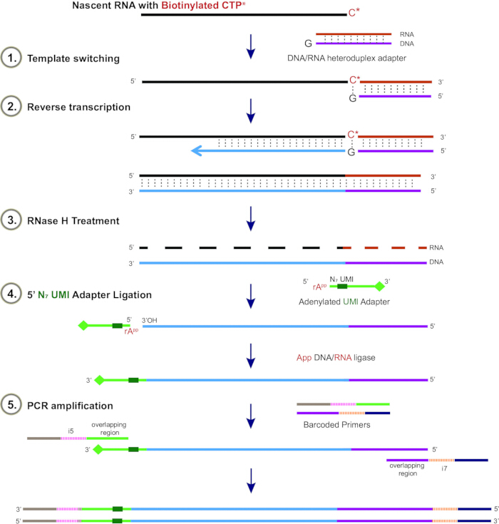
Figure 2: Detailed schematic representation of CoSTseq library preparation. Nascent RNA bearing a biotinylated 3'-end is incubated with a template switching adapter containing a G overhang that is complementary to biotin-CTP. For fragmented mature RNA (DMS-MaPseq), an adaptor with N-overhang is used (see Figure 1). cDNA synthesis is carried out using a highly processive reverse transcriptase, such as thermostable group II intron reverse transcriptase (TGIRT), followed by RNase H treatment to degrade the RNA strand. Subsequently, 5' App DNA/RNA ligase links the ribose-adenylated 5'-end (rApp) of the N7 UMI linker to the 3' OH of the single-stranded cDNA. The N7 UMI adapter includes a blocked 3′ end, preventing self-ligation among adapters. Finally, PCR amplification is carried out using primers that include overlapping regions and 5′ overhangs bearing unique Illumina i5 and i7 barcodes assigned to each sample. Please click here to view a larger version of this figure.
7. 5' Adapter ligation for preparation of final cDNA libraries for paired-end tag sequencing
- Preparation of 5'-end adapter
- Adenylate the 5'-end adapter using an RNA ligase-based reaction (e.g., Mth RNA Ligase, listed in Table of Materials) with the following reaction components: 8 µL of 10x 5'-DNA adenylation reaction buffer, 8 µL of 1 mM ATP, 100 µM R1R DNA (this is the TruSeq primer containing a unique molecular identifier or UMI), 8 µL of Mth RNA ligase, 52 µL of nuclease-free water.
NOTE: For details pertaining to the N7 UMI adapter design, see Table 2 and Figure 2. - Incubate this reaction at 65 °C for 1 h, then at 85 °C for 5 min. Proceed to purify the product using EtOH precipitation or by a silica-membrane spin column-based DNA cleanup method to isolate cDNA, according to the manufacturer's instructions.
- For EtOH precipitation, DNA can be precipitated at -20 °C (minimum 20 min) using sodium acetate (pH 5.2) at a final concentration of 0.3 M and 2x-2.5x of 95%-100% cold EtOH. Centrifuge the EtOH-precipitated reaction for 15 min at maximum speed in a pre-chilled (4 °C) centrifuge, wash the pellet using 70% EtOH, re-centrifuge, and air-dry the pellet. Resuspend (or elute, if using a spin-column) the pellet in 40 µL of nuclease-free water. Store at -20 °C long-term.
- Adenylate the 5'-end adapter using an RNA ligase-based reaction (e.g., Mth RNA Ligase, listed in Table of Materials) with the following reaction components: 8 µL of 10x 5'-DNA adenylation reaction buffer, 8 µL of 1 mM ATP, 100 µM R1R DNA (this is the TruSeq primer containing a unique molecular identifier or UMI), 8 µL of Mth RNA ligase, 52 µL of nuclease-free water.
- 5' adapter ligation
- For a single 20 µL reaction volume, add the following components to 10 µL of cDNA: 2 µL of 10x NE Buffer 1, 2 µL of 50 mM MnCl2, 2 µL of 5' AppDNA/RNA ligase, 4 µL of 10 µM (100 ng/µL) of the adenylated adapter from step 7.1.
- Incubate this reaction at 65 °C for 1 h, then 3 min at 90 °C. Clean the reaction using the minElute PCR purification kit and elute in a final volume of 20 µL nuclease-free water.
- Minimal PCR amplification of cDNA libraries with pilot test
- Before proceeding, prepare a 10 µM stock of index primers containing unique barcodes. Each unique CoSTseq or DMS-MaPseq sample should have a unique barcode associated with it for smooth downstream analysis. See primer sequences in Table 2. The forward and reverse individual barcode-containing primers can be combined in a 1:1 mixture and stored as a barcoding premix.
- Using 2 µL of the PCR product from step 7.2, add 2.5 µL of high-fidelity PCR buffer (e.g., a commercial HiFi buffer), 0.375 µL of dNTP (10 mM), 0.75 µL of barcoding premix (or 0.375 µL of individual i5 and i7 unique barcoding primers), 0.25 µL of high-fidelity hot-start DNA Polymerase (e.g., commercial HiFi hot-start enzyme), 6.625 µL of nuclease-free water.
- Incubate this reaction at 95 °C, and repeat [15 s 98 °C, 30 s 62 °C, 30 s 72 °C] for 23 cycles, end with 1 min incubation at 72 °C. Run on a 1% agarose gel to decide on the optimal cycling number.
NOTE: To test lower cycling conditions, a sample can be prepared at the end of a lower cycle (e.g., end of 15 or 16 cycles) and run on an agarose gel to compare the intensity of the cDNA library smear on an agarose gel across cycling lengths.
- Final PCR for library amplification
- Using 18 µL of the PCR product from step 7.2, add the following from the KAPA HiFi PCR Kit: 22.5 µL of high-fidelity PCR Buffer, 3.375 µL of dNTP (10 mM), 6.75 µL of barcoding premix (or 3.375 µL of individual i5 and i7 unique barcoding primers), 2.25 µL of high-fidelity hot-start DNA Polymerase, and 59.625 µL of nuclease-free water.
- Incubate this reaction at 95 °C, and repeat [15 s 98 °C, 30 s 62 °C, 30 s 72 °C] for the number of cycles determined in step 7.3. End with 1 min incubation at 72 °C.
- Size selection
- Perform size selection on the PCR product using paramagnetic bead-based purification (e.g., AMPure XP or equivalent). Use 1.3x the volume of beads (e.g., for a 50 µL PCR reaction, use 65 µL of bead slurry) and mix well until homogeneous. Let the beads bind the sample for 10 min at room temperature.
NOTE: This 1:1.3x sample to beads ratio enables the removal of primer adapter dimers and ensures DNA fragments retained are no smaller than 150 bp and not larger than 800 bp. - Remove the supernatant without disturbing the beads and proceed to wash the beads with 80% EtOH according to the manufacturer's protocol.
- Elute the final product in 11 µL. Run 1 µL on an agarose gel to verify libraries. Submit the final product for sequencing.
- Perform size selection on the PCR product using paramagnetic bead-based purification (e.g., AMPure XP or equivalent). Use 1.3x the volume of beads (e.g., for a 50 µL PCR reaction, use 65 µL of bead slurry) and mix well until homogeneous. Let the beads bind the sample for 10 min at room temperature.
8. Data analysis
NOTE: The complete CoSTseq package and analysis code are available on GitHub (https://github.com/NeugebauerLab/CoSTseq), and the workflow for analysis is shown in Figure 3. This pipeline is designed to handle both CoSTseq and DMS-MaPseq sequencing data. CoSTseq uses Snakemake, a bioinformatics workflow that readily parallelizes analysis by optimizing the number of available CPU cores19. The workflow is defined by a Snakefile, which consists of a set of rules that establish the analyses that will be run. There is no need to alter the Snakefile that is available upon installing the GitHub package.
- Adjust the configuration file (.yaml file) with correct file paths to where the raw data are stored. Included in these settings should be a path to the adapter sequences and the associated sample names. Alter the config file to run specific analyses defined in the Snakefile.
- To begin analysis, download raw paired end reads in fastq.gz format. Process reads using fastp20 to trim adapter sequences and quality filter for reads with Phred scores of at least 20. Align these reads to the reference genome using STAR21 to generate BAM files.
- As an additional confirmation, visualize the BAM files generated after running the CoSTseq analysis pipeline in the Integrated Genome Viewer (IGV) browser. Using IGV, the BAM files should be readily interpretable to show reads along all parts of genes of interest, indicating that reads represent nascent RNA. As expected, mature RNA reads should show minimal coverage at intronic regions, given that these reads are expected to be derived from mature and likely efficiently spliced transcripts.
- Use the fastp output (an .html file that can be opened in a web browser) for an estimated percentage of duplicated reads. Note that CoSTseq reads may be very highly duplicated, highlighting the criticality of the next step.
- Using UMICollapse22, deduplicate reads to collapse reads with identical UMIs and alignments, allowing for the preservation of unique reads only and mitigating PCR bias. The resulting files should be free of duplicated reads and in BAM format, where reads aligning to rRNA will be separated from those associated with non-rRNA genes.
- For the final major component of the modular CoSTseq pipeline, use a custom Python implementation to calculate the mutation counts and read coverage over each nucleotide. The functions describing this implementation can be found in the source code on GitHub, and the Snakefile details the input, output, parameters, and functions to generate these data that are stored in a .pkl format.
NOTE: There are a multitude of other analyses that can be performed using the CoSTseq analysis pipeline, although parameters may require optimization for personalized analysis needs. A valuable method adapted for CoSTseq is HDProbe, an R package that enables genome-wide comparisons of mutation rates10. In brief, HDProbe requires data from all replicates and can methodically categorize significant differences in mutation rates compared to the control. Additional types of analyses one may wish to perform include structure prediction based on the experimental DMS reactivities. This can be performed using the RNAstructure package23.
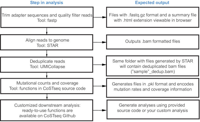
Figure 3: Steps of analysis and expected outputs of the modular CoSTseq analysis pipeline. Steps of analysis, indicating recommended use of existing tools in addition to the ready-to-use CoSTseq analysis pipeline with the expected output files and content. Please click here to view a larger version of this figure.
Access restricted. Please log in or start a trial to view this content.
Results
This section presents the actual results generated by implementing the CoSTseq workflow and analysis, as described in this protocol. Firstly, this section describes expected outcomes following quality assessment of successful library preparations before and after sequencing. Prior to sequencing, researchers can use a test PCR and TapeStation analysis to confirm the presence of RNA following biotin enrichment. This indicates successful isolation of nascent RNA during CoSTseq library preparation. Data quality assessment af...
Access restricted. Please log in or start a trial to view this content.
Discussion
RNA starts to fold co-transcriptionally due to faster kinetics of base-pairing compared to the rate of synthesis24,25,26,27. Our current knowledge of nascent RNA folding comes from single-molecule studies of prokaryotic RNAs, in vitro probing, or in silico approaches. In this protocol, a detailed workflow of CoSTseq is presented; this technique allows in vivo detectio...
Access restricted. Please log in or start a trial to view this content.
Disclosures
The authors have nothing to disclose.
Acknowledgements
The authors would like to thank Dr. SK Boopathy Jegathambal for coding and the members of the Neugebauer lab, especially P. Bech, for helpful discussions. This work was supported by the National Institutes of Health (R01GM112766 to KMN) and a predoctoral fellowship from the American Heart Association (908949 to LS). LPS was supported by an NIH training grant 5T32GM14943803. LRAB was supported by a postdoctoral fellowship from the American Heart Association (26POST1569544). Data acquisition at Yale Center for Genomic Analysis was supported by the National Institute of General Medical Sciences of the National Institutes of Health under Award Number 1S10OD03036301A1. This work is solely the responsibility of the authors and does not necessarily represent the official views of the NIH.
Access restricted. Please log in or start a trial to view this content.
Materials
| Name | Company | Catalog Number | Comments |
|---|---|---|---|
| 10mM ATP | Invitrogen | 18330019 | |
| 10mM Biotin-11-CTP | Jena biosciences | NU-831-BIOX | |
| 10mM GTP | Invitrogen | 18332015 | |
| 10mM UTP | Invitrogen | 18333013 | |
| 10X NEBuffer 1 | New England Biolabs | B7001S | Used in step 7.2.1 |
| 10X Phosphate Buffered Saline (PBS) | Gibco | 70011-044 | |
| 20% SDS | RPI | L23100-500 | |
| Acid phenol:chloroform | Ambion | AM9722 | |
| AMPure XP beads for size selection | Beckman Coulter | A63880 | Used for Size Selection in step 7.5.1 |
| Bacto Peptone | Gibco | 211677 | |
| Bacto Yeast Extract | Gibco | 212750 | |
| Bicine | Sigma Aldrich | B3876-100G | |
| Chloroform | Sigma Aldrich | 319988 | |
| D-(+)-GLUCOSE | Sigma Aldrich | G5767-500G | |
| DIFCO AGAR | BD DIFCO | 214010 | |
| Dimethyl Sulfate | Sigma Aldrich | D186309-5ML | |
| Dynabeads™ MyOne Streptavidin C1 beads | Invitrogen | 65001 | Used for Biotin Pulldown in Section 4.1 |
| Dynabeads™ mRNA DIRECT™ Purification Kit | Invitrogen | 61011 | Used for Poly A selection in Section 5.1 |
| EDTA, pH 8, 0.5M | Sigma Aldrich | 03690-100ML | |
| Ethanol | Sigma Aldrich | E7023-500ML | |
| GlycoBlue | Invitrogen | AM9516 | Co-precipitant used in step 4.2.7 and 5.2.4 |
| Induro® Reverse Transcriptase | New England Biolabs | M0681S | RT enzyme used in step 6.3.1 |
| Isoamyl alcohol | Sigma Aldrich | W205710-1KG-K | |
| Isopropanol | JT-BAKER | 9084-05-01 | |
| KAPA HiFi HotStart PCR Kit | Roche | 07958889001 | High-fidelity DNA Pol used in 7.3.2 and 7.4.1 for PCR reactions |
| Magnesium Chloride | Sigma Aldrich | SLCM2154 | |
| MinElute PCR Purification Kit | Qiagen | 28004 | Used for DNA clean up in step 6.3.3 and 7.2.2 |
| Mth RNA Ligase | New England Biolabs | M2611A | |
| Oligo Clean & Concentrator | Zymo Research | D4060 | Suggested for DNA Oligo clean up in step 7.1.2 |
| Potassium Acetate | Sigma Aldrich | 236497-500G | |
| Potassium Chloride | JT Baker | 3040-01 | |
| Potassium Hydroxide | Avantor | 6984-04-01 | |
| RNA Clean & Concentrator-5 | Zymo Research | R1014 | RNA clean up kit used in step 5.3.2 after PNK treatment |
| RNaseOUT™ Recombinant Ribonuclease Inhibitor | Thermo Fisher Scientific | 10777019 | RNase inhibitor used in step 5.3.1 during PNK treatment |
| Sarkosyl | IBI Scientific | IB07080 | |
| Sodium Acetate | Quality Biological | 351-035-721 | |
| Sodium Chloride | Sigma Aldrich | S5150-1L | |
| Sodium Hydroxide | Macron | 7708-10 | |
| SUPERase·In™ RNase Inhibitor (20 U/μL) | Thermo Fisher Scientific | AM2696 | RNase inhibitor used in step 6.3.1 |
| T4 Polynucleotide Kinase | New England Biolabs | M0201S | |
| Thermostable 5’ App DNA/RNA Ligase | New England Biolabs | M0319L | |
| Tris-HCl, pH 7.4, 1M | Thermo Scientific | J60202.K2 | |
| Triton X-100 | TEKNOVA | T1105 | |
| TRIzol™ Reagent | Invitrogen | 15596-026 | For nascent RNA elution in Section 4.2; also referred as "RNA reagent" in Section 4 |
| β-mercaptoethanol | Sigma Aldrich | M6250-1L |
References
- Zhang, J., Fei, Y., Sun, L. Advances and opportunities in RNA structure experimental determination and computational modeling. Nature Methods. 19 (10), 1193-1207 (2022).
- Bonilla, S. L., Jones, A. N., Incarnato, D. Structural and biophysical dissection of RNA conformational ensembles. Current Opinion in Structural Biology. 88, (2024).
- Bose, R., Saleem, I., Mustoe, A. M. Causes, functions, and therapeutic possibilities of RNA secondary structure ensembles and alternative states. Cell Chemical Biology. 31 (1), 17-35 (2024).
- Rapakko, J., Scaravilli, M., Änkö, M. L. Interplay between RNA-protein interactions and RNA structures in gene regulation. FEBS Open Bio. , (2025).
- Rodgers, M., Woodson, S. A. A roadmap for rRNA folding and assembly during transcription. Trends in biochemical sciences. 46 (11), 889-901 (2021).
- Tomezsko, P., Swaminathan, H., Rouskin, S. DMS-MaPseq for Genome-Wide or Targeted RNA Structure Probing In Vitro. Methods in Molecular Biology. 2254, 219-238 (2021).
- Xu, H., Douglas, C. W., Lambowitz, A. M. Improved TGIRT-seq methods for comprehensive transcriptome profiling with decreased adapter dimer formation and bias correction. Scientific Reports. 9 (1), (2019).
- Lopes, R., Agami, R., Korkmaz, G. GROseq, A Tool for Identification of Transcripts Regulating Gene Expression. Methods in molecular biology (Clifton, N.J). 1543, 45-55 (2017).
- Mahat, D. B., et al. Base-Pair Resolution Genome-Wide Mapping Of Active RNA polymerases using Precision Nuclear Run-On (PROseq). Nature protocols. 11 (8), 1455-1476 (2016).
- Schärfen, L., Vock, I. W., Simon, M. D., Neugebauer, K. M. Rapid folding of nascent RNA regulates eukaryotic RNA biogenesis. Molecular Cell. 85 (8), 1561-1574 (2025).
- Hawley, D. K., Roeder, R. G. Functional steps in transcription initiation and reinitiation from the major late promoter in a HeLa nuclear extract. Journal of Biological Chemistry. 262 (8), 3452-3461 (1987).
- Hawley, D. K., Roeder, R. G. Separation and partial characterization of three functional steps in transcription initiation by human RNA polymerase II. Journal of Biological Chemistry. 260 (13), 8163-8172 (1985).
- Zubradt, M., et al. DMS-MaPseq for genome-wide or targeted RNA structure probing in vivo. Nature Methods. 14 (1), 75-82 (2017).
- Tijerina, P., Mohr, S., Russell, R. DMS foot-printing of structured RNAs and RNA-protein complexes. Nature Protocols. 2 (10), 2608-2623 (2007).
- Liebig, A., Waldsich, C. W. Probing RNA structure within living cells. Methods in enzymology. 468, 219-238 (2009).
- Pelechano, V., Chávez, S., Pérez-Ortín, J. E. A Complete Set of Nascent Transcription Rates for Yeast Genes. PLOS ONE. 5 (11), (2010).
- Wery, M., Descrimes, M., Thermes, C., Gautheret, D., Morillon, A. Zinc-mediated RNA fragmentation allows robust transcript reassembly upon whole transcriptome RNA-Seq. Methods. 63 (1), 25-31 (2013).
- Wang, L. K., Lima, C. D., Shuman, S. Structure and mechanism of T4 polynucleotide kinase: an RNA repair enzyme. The EMBO Journal. 21 (14), 3873-3880 (2002).
- Köster, J., Rahmann, S. Snakemake a scalable bioinformatics workflow engine. Bioinformatics. 28 (19), 2520-2522 (2012).
- Chen, S., Zhou, Y., Chen, Y., Gu, J. fastp: an ultrafast all-in-one FASTQ preprocessor. Bioinformatics (Oxford, England). 34 (17), i884-i890 (2018).
- Dobin, A., et al. STAR: ultrafast universal RNA-seq aligner. Bioinformatics (Oxford, England). 29 (1), 15-21 (2013).
- Liu, D. Algorithms for efficiently collapsing reads with Unique Molecular Identifiers. PeerJ. 7, (2019).
- Spasic, A., Assmann, S. M., Bevilacqua, P. C., Mathews, D. H. Modeling RNA secondary structure folding ensembles using SHAPE mapping data. Nucleic Acids Research. 46 (1), 314-323 (2018).
- Saldi, T., Fong, N., Bentley, D. L. Transcription elongation rate affects nascent histone pre-mRNA folding and 3′ end processing. Genes & Development. 32 (3-4), 297-308 (2018).
- Pan, T., Artsimovitch, I., Fang, X., Landick, R., Sosnick, T. R. Folding of a large ribozyme during transcription and the effect of the elongation factor NusA. Proceedings of the National Academy of Sciences. 96 (17), 9545-9550 (1999).
- Heilman-Miller, S. L., Woodson, S. A. Effect of transcription on folding of the Tetrahymena ribozyme. RNA. 9 (6), 722-733 (2003).
- Yu, A. M., et al. Computationally reconstructing co-transcriptional RNA folding from experimental data reveals rearrangement of nonnative folding intermediates. Molecular Cell. 81 (4), 870-883 (2021).
- Yu, G., et al. Genome-wide probing of eukaryotic nascent RNA structure elucidates cotranscriptional folding and its antimutagenic effect. Nature communications. 14 (1), (2023).
Access restricted. Please log in or start a trial to view this content.
Reprints and Permissions
