Here we present a protocol to illustrate the isolation of rare cancer cells from a contrived sample containing whole blood and spiked in lung cancer cells. The protocol utilizes a semi-automated, label-free cell isolation platform. Enriched cells are subsequently analyzed for epidermal growth factor receptor mutations using droplet digital PCR.
Method Article
Profiling Epidermal Growth Factor Receptor DNA Mutations in Circulating Rare Cells Using a Cell Isolation System and Droplet Digital PCR
In This Article
Summary
Abstract
Mutations found within the epidermal growth factor gene (EGFR) are critical for determining the most effective treatments for patients with lung cancer. Routine testing for EGFR mutations can take the form of tumor biopsies or analysis of ctDNA from plasma of patients with lung cancer. Although testing blood samples from patients with lung cancer is performed routinely to provide insight into the presence or recurrence of lung cancer, via ctDNA detection or CTC detection, it typically does not involve the subsequent molecular analysis of the captured cells. Detection of individual mutations within the EGFR gene present within rare circulating tumor cells (CTCs) can offer additional insights into the potential efficacy of targeted therapies and beyond that, patient prognosis, especially in non–small cell lung cancer (NSCLC). Unfortunately, the rarity of such circulating tumor cells and lack of streamlined procedures to interrogate their genomic information present a significant detection challenge.
The goal of this protocol is to illustrate the realistic potential of capturing CTCs from whole blood and assessing their molecular content using ddPCR. A semi-automated, size-based cell isolation platform was used to capture circulating tumor cells, which were subsequently recovered for downstream molecular analysis. To determine the presence of specific EGFR mutations within the genomic DNA of captured cells, ddPCR was performed using assays for EGFR pT790M and EGFR pL858R.
Two separate lung cancer cell lines (A549 and H1975) were actively enriched from whole blood and accurate quantification of mutations present within the cell lines H1975 was performed using ddPCR.
Introduction
With continued translational research into the value of liquid biopsies, and recent publications specifically illustrating the clinical value of ctDNA monitoring in therapy selection1 liquid biopsies are coming of age. Liquid biopsies provide an alternative, less invasive method for cancer monitoring and prognosis when compared to conventional tissue biopsy methods. Extensive research has focused on fragmented DNA found in blood (cell-free DNA, cfDNA), and specifically the sub compartment that is tumour derived, known as circulating tumor DNA (ctDNA). ctDNA has been studied extensively over the last two decades and shown to contain tumor-specific gene mutations e.g. SNVs and Structural variants, as well as methylation markers and a distinct fragment pattern when compared to non-tumour cfDNA. These specific ctDNA characteristics are now utilized to monitor cancer patients’ molecular status across early detection, therapy response, minimal residual disease (MRD) and resistance to therapy status. Similarly, circulating tumor cells (CTCs), detached from the primary tumor site into blood vessels, have recently evolved as promising biomarkers whose existence and quantity in blood have shown a close correlation to disease progression and recurrence potential. Whilst the very early initial studies on CTCs mostly relied on their protein marker expression and enumeration2, there has been meaningful progress in studying CTCs at the molecular level including genomic mutation detection in Estrogen receptor 1 (ESR1) for breast cancer, and epidermal growth factor gene (EGFR) for various cancers.
EGFR mutations are critical for both determination of pathogenesis and prognosis but also in deciding the most effective treatments for patients with lung cancer3. Routine testing for EGFR mutations can take the form of solid tumor biopsies at diagnosis or progression or more recently, sampling of ctDNA from plasma of patients with relapsed/refractory lung cancer4. However, unlike ctDNA-based approaches, analysis of CTC-derived genomic material enables direct assessment of intact tumor cells and their mutational profiles. Along with ctDNA MRD studies, detection of mutations within the EGFR gene of rare CTCs can provide additional understanding into the potential efficacy of treatment regimens and beyond that, patient prognosis, especially in non-small cell lung cancer (NSCLC). The rarity of CTCs and the lack of streamlined protocols utilizing user-friendly instruments for clinics constitutes a serious impediment to broader adoption. This limitation highlights the need for a standardized and accessible workflow that enables both efficient CTC isolation and downstream molecular analysis. Therefore, the goal of this study is to present a reproducible workflow for the enrichment of circulating tumor cells and subsequent detection of EGFR mutations using a combined cell isolation and digital PCR approach.
The Genesis Cell Isolation System with microfluidic cell capture slides can capture CTCs in a semi-automated process with high viability and high capture efficiency, based on their larger size and reduced deformability compared to the primary white and red blood cell population5,6. Captured CTCs can then be recovered from the microfluidic cell capture slides by reverse flow off the slide for further downstream genomic analysis. To determine the presence of specific EGFR mutations within the genomic DNA of captured cells, ddPCR was performed on CTC derived genomic DNA using assays for EGFR T790M and L858R.
This protocol, using a model CTC sample and commercially available ddPCR assays, is designed to exemplify an entry point for targeted detection of cancer-associated mutations in CTCs. This protocol provides a practical approach for detecting cancer-associated mutations in circulating tumor cells and may support further research into liquid biopsy applications.
Protocol
Ethical statement:
Blood samples were obtained from authorized sources for research use, and written informed consent was obtained from all donors prior to sample collection. All experimental protocols involving human blood samples were reviewed and approved by the Institutional Review Board of a blood vendor, Discovery Life Sciences (Alabama, US). All procedures involving human blood samples were conducted in accordance with the ethical standards of the institutional research committee and with the Declaration of Helsinki.
1. Cancer cell enrichment using semi-automated cell isolation system
Note: This section describes enrichment of circulating tumour cells (CTCs) using a semi-automated cell isolation platform, followed by EGFR mutation detection by droplet digital PCR. A model CTC sample is used to demonstrate feasibility, and assay validation is recommended before clinical application. Two non–small cell lung cancer (NSCLC) cell lines were used: NCI-H1975 (EGFR L858R/T790M mutations) and A549 (EGFR wild-type), which served as a negative control for mutation detection.
- Blood Sample
- Ensure informed consent is obtained from blood donors/patients before blood collection. Obtain Institutional Review Board (IRB) approval, where required, for research involving human specimens, including blood.
- For feasibility testing, obtain a blood sample from authorized 3rd party vendor for research use only.
- Collect peripheral blood in Ethylenediaminetetraacetic acid (EDTA) tubes and process the sample through the Genesis Enrichment protocol within 48 h.
Note. For downstream analysis and Genesis Enrichment protocol, EDTA tubes are recommended. Streck and EDTA tube can be used for on slide immunostaining with Genesis Enumeration protocols (direct/indirect). - Store blood samples collected in EDTA tubes at 2–8 ˚ C until use. For Genesis cell isolation, no pre-treatment is required and whole blood up to 10 mL can be used for the Enrichment protocol.
- Model CTC Sample/Clinical Sample Preparation
- Prepare a cell stock solution at a concentration of 100–1,000 cells in 10 µL appropriate cell culture media or phosphate-buffered saline (PBS) buffer. Skip this step if clinical samples are in use.
NOTE: The spiking concentrations used in this study are higher than typically observed clinical CTC levels and are intended for method demonstration purposes only. - Add the 10 µL of the cell stock solution into up to 10 mL of whole blood from a healthy donor to prepare a model CTC sample. For clinical samples, do not include any cancer cells into the whole blood samples from patients.
- Mix the blood sample with an equal volume of PBS (1x) and gently agitate on a blood rocker at room temperature until use. If storage exceeds one hour, store at 4 °C.
- Prepare a cell stock solution at a concentration of 100–1,000 cells in 10 µL appropriate cell culture media or phosphate-buffered saline (PBS) buffer. Skip this step if clinical samples are in use.
- CTC Enrichment
- Power on the Genesis Cell Isolation System and wait for the system to be initialized. This can take up to 5 min.
- Place the waste jar in position at the base of the station bays and attach the waste jar tubing to the waste jar.
- Press the tubing into the pinch valve until you feel it click into place, and attach Genesis System tubing to the waste jar outlet.
- Run a Self-Test by pressing “start” from the home screen and touching the ‘Self-Test’ on the screen.
- When the Self-Test is complete, select ‘2.0 Kit Enrichment’ from the select protocol options.
- Follow the instructions on the screen, populate the required data entry fields and finish the Priming and Assembly procedure. Successful priming using 70% ethanol and 1x PBS yields best cell capture performance and recovery for the DNA extraction. Priming takes about 20 min to complete.
- Ensure that the correct enrichment program (2.0 Kit Enrichment) is selected and that all tubing connections and slide placements are properly secured before starting the run. Avoid introducing air bubbles during sample loading, as this may affect flow consistency and capture efficiency.
- Gently dispense the model sample prepared from Step 1.2.3 (Model CTC Sample/Clinical Sample Preparation) into the inlet funnel and continue the Enrichment protocol by pressing ‘Resume’ on the system screen.
- When the CTC capture is completed, the semi-automated cell isolation system will prompt the user to remove the waste jar and reattach the priming reservoir.
- Detach the Genesis System tubing from the waste jar and pipet 4 mL of dilution buffer into the priming reservoir.
- Attach the backflow adaptor to the priming reservoir and reattach to the Genesis System tubing. Press Resume on the screen to continue the Enrichment protocol. This releases the captured cells in the microfluidic slide to the inlet funnel by applying backflow.
- Collect recovered cells by pipetting out from the inlet funnel to a 5.0 mL Eppendorf tube. Ensure that you collect all of the aqueous samples in the inlet funnel, which can often exceed 4 mL.
- Centrifuge the recovered sample at 600 g for 5 min to pellet the captured cells before cell lysis.
- Remove the supernatant carefully and proceed to DNA Extraction protocol (Section 1.4).
- DNA Extraction
Caution: DNAzol BD is harmful if it comes into contact with skin or is ingested. Avoid contact with skin and eyes. Use only within a chemical fume hood environment and do not breathe any mist/vapor/spray. In case of contact with skin wash with water immediately.- Add 1 mL of DNAzol BD directly into the tube from Step 1.3.14.
- Transfer the entire volume of the sample to a new Eppendorf DNA LoBind 1.5 mL Microcentrifuge tube and shake vigorously.
- Incubate the tube at room temperature for 5 min. Vortex the tube vigorously and incubate at room temperature for at least 4 min.
- Ensure complete mixing during lysis and precipitation steps to maximize DNA recovery. Optional RNase treatment may be included if RNA contamination is a concern.
- Add 400 µL of isopropanol to the DNAzol BD-cell lysate sample mixture directly.
- Centrifuge the tube at 10,000 g for 6 minutes to precipitate DNA.
- After centrifugation, remove the supernatant and gently add 0.5 mL of DNAzol BD to the pelleted DNA.
- Vortex the tube until the DNA pellet is completely dispersed.
- Centrifuge the mixture at 10,000 g for 5 min.
- Remove the supernatant and wash the DNA pellet by pipetting 1 mL of 75% ethanol.
- Repeat steps 9–10 for a total of two washes.
- Remove any residual ethanol in the bottom of the tube using a micropipette.
NOTE: Ensure that the pellet does not become completely dry. - Add 20 µL of DNase-/RNase-free deionized (DI) water to solubilize the DNA from the CTC samples.
- Quantify double-stranded DNA (dsDNA) in the sample using dsDNA quantification assay kits/platforms, such as Qubit 1x dsDNA High Sensitivity Assay Kit or Bioanalyzer High Sensitivity DNA Analysis.
- Store extracted DNA samples at 4 °C or -20 °C to prevent degradation.
Pause Point: The extracted DNA samples can be stored at -20 °C for short-term storage or at -80 °C for long-term storage before proceeding to downstream analyses.
2. EGFR Mutation Analysis using ddPCR
Digital PCR is a suitable method for detecting rare mutations with high sensitivity and precision. ddPCR mutation detection assays provided in a single tube typically include a FAM labelled probe targeting a mutant allele and a second HEX labelled probe targeting a wild-type allele.
- Reaction setup and droplet generation
- Thaw ddPCR Supermix, ddPCR Mutation Detection Assays on ice.
- Vortex each tube to ensure the contents are homogeneous, since a concentration gradient may form during storage at -20 °C.
- Centrifuge briefly to sediment contents at the bottom of the tube and place the tube on ice.
- Prepare extracted DNA samples at the desired concentration before preparing the reaction mix according to the recipe in Table 1.
- Make a premix containing super-mix, assays and restriction enzymes, plus water for all required samples for testing and add sample DNA individually as the last reaction component. Make sure final concentration of DNA samples is 50 fg–100 ng.
Note: DNA fragmentation by restriction digestion prior to droplet generation is recommended to improve accuracy, reduce viscosity, and enhance template accessibility. Direct digestion using 2–5 U of restriction enzyme during ddPCR setup is recommended. For EGFR point mutation assays (T790M/L858R), MseI is recommended; refer to assay data sheets for compatible enzymes before customization. Data sheets are available for download for individual assays from the Bio-Rad digital assays portal (Bio-Rad Droplet Digital PCR Assays | Home) - Prepare a negative control for at least one well, containing only wild-type template at a concentration similar to the concentration of unknown samples.
- Vortex the reaction tubes to mix thoroughly and centrifuge briefly to sediment all samples to the bottom of each reaction tube. Allow the reaction tubes to equilibrate at room temperature for 2 min.
- Load 20 µL of each reaction mix into a sample well of a microfluidic cartridge (DG8 Cartridge) followed by 75 µL of Droplet Generation Oil for Probes into the oil wells.
- Cover the cartridge with a gasket and place it in the QX200 Droplet Generator.
- Press the button on the top of the Droplet Generator to close the lid. When the lid is closed, droplet generation automatically begins. This procedure combines the samples and oil within the microchannels of the microfluidic cartridge to create an emulsion of ~20,000 nanoliter-sized droplets for each sample.
- PCR amplification (Thermal cycling conditions)
- Program thermal cycling conditions on a thermal cycler as recommended in Table 2
- When droplet generation is complete, all lights on the instrument are solid green. Open the instrument by pressing the button on the Droplet Generator again and remove the cartridge holder and cartridge.
- Remove the disposable gasket and aspirate 45 µL of droplets from the cartridge, taking care to ensure a slight angle on the pipette tip to prevent droplet shredding.
- Dispense droplets into the ddPCR 96-Well Plates and seal the plate with PCR Plate Heat Seal using the PX1 PCR Plate Sealer.
- After sealing the 96-well plate containing droplets, carefully transfer the 96-well plate to the thermal cycler that was prepared in Step 2.2.1 and start the thermal cycling.
- Droplet readout and data analysis
- Initiate the ddPCR Droplet Reader instrument (QX200 Droplet Reader or QX600 Droplet Reader) and a connected computer on which the QX Manager Software is installed and configured.
- Once the thermal cycling step is complete, move the sealed 96-well plate to a Droplet Reader instrument.
- Open QX Manager Software to configure a new plate layout based on experimental design. Click the ‘Add Plate’ tab and the software will enable the Configure Plate button. Click the ‘Configure Plate’ to assign sample details to the plate.
- Fill in the plate and well information details, select the wells, designate the sample condition, experiment type, target name(s), target type(s), and reference: ch1 for FAM and ch2 for HEX color.
- Click ‘Apply’ to load the well and once finished, select ‘OK’.
- When the plate layout is complete for all samples, select ‘Run’ to start the droplet reading process.
- Upon data acquisition, select samples in the well selector under ‘Analyze’ and set appropriate threshold for the ddPCR Mutation Detection Assay to distinguish mutant from wild-type signals.
- For duplex mutation assays such as those used in this protocol, thresholds may be automatically set using the QX manager software default settings (ref to fig in results).
- Alternatively, thresholds may be manually set using the cross hairs or lasso methods, ensuring that thresholds or lasso boundaries separate positive and negative clusters and are positioned based on clusters locations defined by both positive and negative controls included in each run.
NOTE: Thresholds should be set consistently across samples within a batch using positive and negative controls to ensure reproducible mutation calling. - The concentration reported is copy numbers per µL of the final 1x ddPCR reaction.
Results
The Genesis cell isolation system with microfluidic cell capture slides isolated an average of 84% of spiked cancer cells in blood samples at a concentration of 25–250 cells·mL-1 (Figure 3A). This high capture efficiency demonstrates the effectiveness of the enrichment step for isolating rare circulating tumor cells from whole blood samples. Optional immunostaining of cells captured in microfluidic cell capture slides using the system Enumeration protocol demonstrated rare cell identification using two positive antibodies against cytokeratin, DAPI to identify nucleated cells and one negative antibody against CD45 that is expressed on most white blood cells but typically not expressed on CTCs (Figure 3B). This staining pattern confirms the successful identification of circulating tumor cells while excluding background white blood cells.
ddPCR mutation detection assays enabled identification and profiling of EGFR mutations in enriched cancer cells. This study demonstrated the detection of gDNA mutations in cells, initially enriched by the Genesis platform, using ddPCR and Mutation Detection Assays. ddPCR plots (Figure 4) of cancer cells lines A549 (EGFR wild type) and H1975 (L858R/T790M) for the two tested EGFR mutations showed ddPCR can sensitively detect mutations present in the cell line H1975 after spiking into whole blood and subsequent enrichment via the Genesis platform. Positive mutation signals were observed in H1975 cells, whereas no mutation signals were detected in A549 cells, confirming assay specificity. The samples spiked with control cell line A549 showed no mutation present droplets confirming that the detection of mutations at L858R and T790M are associated specifically with the H1975 cell line. This demonstration of CTC isolation on the Genesis platform followed by molecular analysis utilizing ddPCR can offer a powerful molecular diagnostic tool for liquid biopsy studies and preclinical studies using clinical samples to monitor disease progression and personalized treatment. Overall, these results demonstrate that the combined enrichment and ddPCR workflow can reliably detect and quantify EGFR mutations in circulating tumor cells.
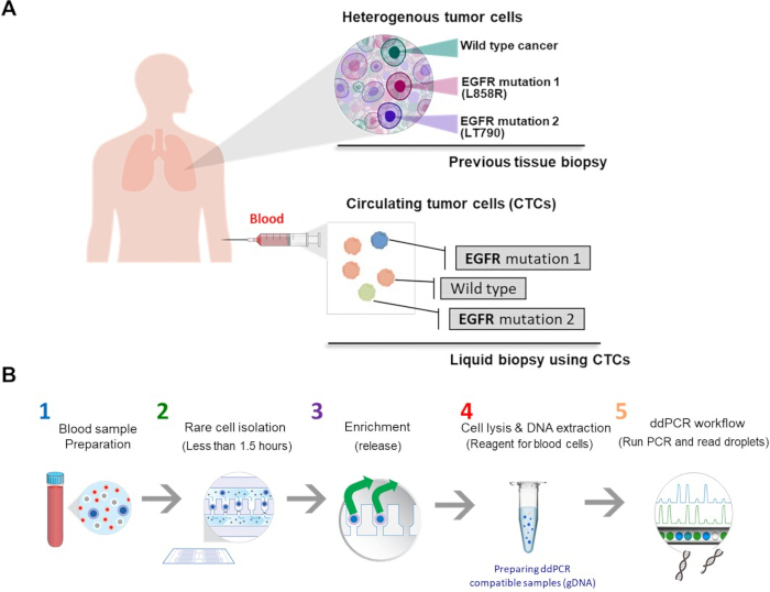
Figure 1: Liquid biopsy workflow for profiling EGFR DNA mutations in lung cancer. (A) Illustration of tumor heterogeneity showing different EGFR mutation profiles detected using conventional tissue biopsy and circulating tumor cells (CTCs) from liquid biopsy. (B) Schematic workflow for EGFR DNA mutation profiling in circulating tumor cells using liquid biopsy. Please click here to view a larger version of this figure.
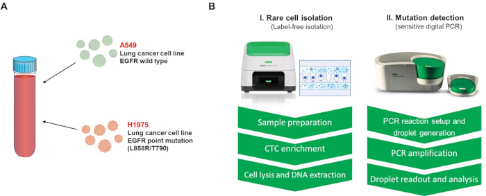
Figure 2: Demonstration of EGFR DNA mutation profiling using contrived samples. (A) Preparation of a model CTC sample by spiking lung cancer cell lines with defined EGFR mutation status into whole blood from a healthy donor. (B) Workflow illustrating EGFR DNA mutation detection in circulating tumor cells using commercially available platforms. Please click here to view a larger version of this figure.
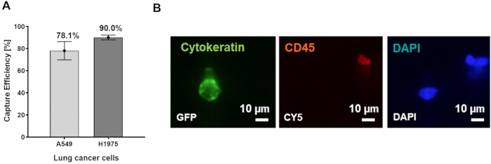
Figure 3: Label-free isolation and identification of circulating tumor cells. (A) Capture efficiency of lung cancer cell lines (A549 and H1975) spiked into whole blood samples from healthy donors using a semi-automated rare cell isolation platform. (B) Immunostaining of captured cells showing cytokeratin-positive tumor cells. Scale bar = 10 µm. Please click here to view a larger version of this figure.
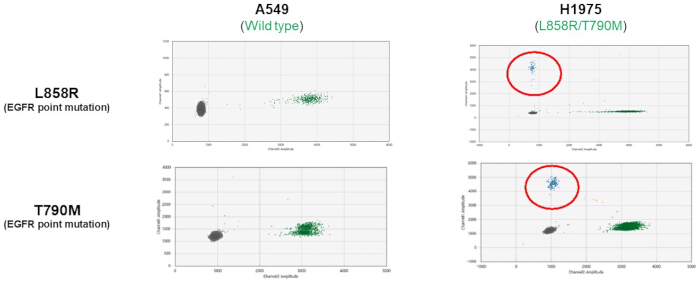
Figure 4. EGFR mutation profiling using droplet-based digital PCR. Two-dimensional amplitude plots showing EGFR mutation detection in enriched cancer cells following cell lysis and DNA extraction. A549 cells (wild type) show absence of mutation-specific signals, whereas H1975 cells exhibit positive signals for EGFR L858R and T790M mutations. The assay enables sensitive detection and quantification of mutant and wild-type DNA populations. Please click here to view a larger version of this figure.
| Component | Volume per reaction (µl) | Final concentration |
| 2x ddPCR Supermix for Probes (no dUTP) | 10 | 1x |
| 20x target (FAM) and wild-type (HEX) primers/probe | 1 | 1x |
| Restriction enzyme, diluted (Msel) | 1 | 2-5 U/reaction |
| Target sample and/or DNase-free water (NTA) | Variable | Variable |
| Total volume | 20 | - |
Table 1: Reaction setup for droplet-based digital PCR mutation detection. Composition and final concentrations of reagents used for detecting rare EGFR mutations in cancer cell-derived DNA samples.
| Thermal cycling* step | Temperature | Time | Ramp Rate | Number of cycles |
| (ºC) | (ºC/sec) | |||
| Enzyme activation | 95 | 10 min | 2 | 1 |
| Denaturation | 94 | 30 sec | 2 | 40 |
| Annealing/extension | 55 | 1 min | 2 | 40 |
| Enzyme deactivation | 98 | 10 min | 2 | 1 |
| Cooling | 4 | 30 min | 2 | 1 |
| Hold (optional) | 4 | ∞ (infinite) | 1 | 1 |
| Use a heated lid set to 105ºC and set the sample volume to 40μl for the C1000 Touch Thermal Cycler with 96-Deep Well Reaction Module. | ||||
Table 2: Thermal cycling conditions for digital PCR amplification. Thermal cycling parameters used for amplification of EGFR mutation targets using a thermal cycler with a 96-well reaction module.
Discussion
The presence of Circulating tumor cells in blood of patients with solid tumors has been a topic of investigation for over 60 years7 and has been recognized as a primary liquid biopsy from before their first inclusion into the collective term in 20108. However, CTCs are rare, and they are difficult to selectively isolate from whole blood9. There are however numerous methods for capturing and enriching CTCs, as illustrated by the following reviews10,11, covering physical/microfluidic approaches as well as cell surface marker selection methods. Many of these methods are time consuming, labor intensive and often involve multiple steps/systems to achieve the required selectivity.
Capture of CTCs from whole blood in this protocol is reliant on their size (> 8 µm) and lack of deformity in the background of phenotypically normal red and white blood cells that pass through the microfluidics pores within the microfluidic cell capture slides. Therefore, correct sample loading, proper slide priming, and prevention of clogging are critical steps for maintaining capture efficiency and sample recovery.
For the workflow to be successful, blood sample management is the key factor; ensuring that fresh blood is utilized for sample processing and maintaining the correct transport and storage conditions to prevent hemolysis. Degradation of red blood cells or white blood cells will impact quality of CTC capture and sensitivity of detection of any rare genomic variants, respectively. If hemolysis, clotting, or visible sample degradation is observed, the sample should not be processed, as these conditions may reduce CTC recovery and downstream mutation detection sensitivity.
Since CTCs can be present at levels below 1 CTC per 10 million white blood cells in whole blood, it is essential to capture as large a volume as is permissible for enrichment. The microfluidic cell capture slides can accommodate 10 mL of blood in line with standard blood capture tubes such as Streck and EDTA, which typically hold ~7.5–10 mL blood. A reduction in blood volume collected will directly impact on CTC capture sensitivity and subsequent genomic variant detection. Obtaining volumes beyond this routine from patients undergoing treatment can be challenging both clinically and ethically. Beyond increasing volume more than 10ml or utilizing diagnostic leukapheresis samples to increase WBC/CTC populations12, increasing sensitivity with larger volumes is beyond the scope of the Genesis platform currently.
The protocol was run as outlined in the standard SOP but with care taken to ensure fresh blood collection, comprehensive mixing of EDTA with blood on collection and careful storage of collected blood prior to use, illustrating the potential for this protocol to be utilized in a clinical setting where routine standard operating procedures are valued. However, because this protocol was demonstrated using contrived samples, validation with clinical patient specimens is required before routine clinical implementation.
The sensitivity of detection of mutations within a CTC population is dependent on both the presence of CTCs within a standard whole blood sample and subsequent detection of somatic mutations within that CTC population, which is dependent upon the frequency of the mutation in the CTC subpopulation. These two confounding factors will ultimately limit overall sensitivity of detection of mutations in a captured CTC population; having a greater impact in early detection samples, compared to late-stage samples that are likely to have higher CTC counts and higher mutation bearing CTC populations.
Current blood based liquid biopsy studies are heavily biased towards circulating tumor DNA present in plasma as an indication of tumor presence. Typically, detection of ctDNA is based on the presence of tumor specific genomic markers e.g. SNVs, structural variants, MSI-H, TMB and CGP. CTCs as a lesser utilized and yet equally viable biomarker in liquid biopsy are known to shed into the blood stream from growing and potentially metastasizing tumors8. Analysis of CTCs is primarily focused on cell surface markers used for their capture or for post capture validation utilizing established or custom antibody panels e.g. pan-Cytokeratin, Vimentin, PD-L1, EpCAM etc. In contrast, molecular analysis of CTCs is limited in adoption due to the challenges of selectively enriching and capturing CTCs from whole blood followed by precise analysis of the CTC subpopulation. The current method utilizes a standardized workflow incorporating established protocols for CTC capture followed by molecular analysis using ddPCR with off-the-shelf assays for known pathologically relevant genomic mutations in contrived whole blood samples. This approach is significant because it enables mutation profiling directly from enriched CTCs, complementing plasma ctDNA-based liquid biopsy approaches.
Utilizing the current approach, captured rare CTCs can be investigated for both their presence in whole blood and the presence of pathologically relevant genomic markers within the CTC population. Studies have shown the value of assessing both the ctDNA profile of patients with cancer and the CTC reservoir circulating in parallel to ctDNA13, with the assessment of both sources of mutations expanding the overall molecular landscape of individual patients. This has the potential for the development of pathology testing paradigms within the scope of translational research initially, but ultimately in a clinical setting. The new paradigm would incorporate both CTC presence as an indication of cancer presence/progression and pathological mutations present within the CTC captured cells potentially indicating clonal expansion or therapeutic resistance14. The implication for this is that the development of metastasis and clonal expansion, followed by subsequent drug resistance could be monitored at both the ctDNA level and the CTC level in concert, directly impacting on therapy selection in later stage cancer.
Future applications may include adapting this workflow to additional cancer-associated mutations and testing its performance in longitudinal clinical samples.
Acknowledgements
The authors thank Abiodun Bodunrin,Cynthia Shu, Ajitha Cristie-David, Andrew Prantner and Errile Pusod for their assistance with ddPCR experiments, cancer cell handling and cancer cell isolation experiments for this study.
Materials
| Name | Company | Catalog Number | Comments |
|---|---|---|---|
| Genesis Cell Isolation System | Bio-Rad Laboratories | 12019822 | Instrument (Refered as "Semi-automated cell isolation system") |
| QX200 Droplet Generator | Bio-Rad Laboratories | 1864002 | Instrument |
| QX200 Droplet Reader or QX600 Droplet Reader | Bio-Rad Laboratories | 1864003 or 12013328 | Instrument (Refered as "Droplet Reader instrument ") |
| PX1 PCR Plate Sealer | Bio-Rad Laboratories | 1814000 | Instrument |
| C1000 Touch Thermal Cycler with 96–Deep Well Reaction Module | Bio-Rad Laboratories | 1851197 | Instrument |
| Qubit 4 Fluorometer | Thermo Fisher Scientific Inc. | Q33226 | Instrument |
| Celselect Slides | Consumable (Refered as Microfluidic Cell Capture Slides) | ||
| Celselect Slides Enrichment Kit 2.0 | Bio-Rad Laboratories | 17009455 | Consumable |
| DNAzol BD | Molecular Research Center, Inc. | DN129 | Reagent |
| MseI restriction enzyme | New England Biolabs | R0525S | Reagent |
| ddPCR Mutation Detection Assay: EGFR T790M | Bio-Rad Laboratories | Assay ID dHsaMDV2010019 | Assay |
| ddPCR Mutation Detection Assay: EGFR L858R | Bio-Rad Laboratories | Assay ID dHsaMDV2010021 | Assay |
| Qubit 1X dsDNA High Sensitivity Assay Kit | Invitrogen | Q33230 | Assay kit |
| DG8 Cartridges for QX200 Droplet Generator | Bio-Rad Laboratories | 1864008 | Consumable |
| Droplet Generation Oil for Probes | Bio-Rad Laboratories | 1863005 | Consumable |
| Automated Droplet Generation Oil for Probes | Bio-Rad Laboratories | 1864110 | Consumable (optional) |
| ddPCR Supermix for Probes (No dUTP) | Bio-Rad Laboratories | 1863023 | Reagent |
| PCR Plate Heat Seal | Bio-Rad Laboratories | 1814040 | Pierceable foil seal |
| DNA LoBind Microcentrifuge Tube, 1.5 ml | Eppendorf | 022431021 | Consumable |
| ddPCR 96-Well Plates | Bio-Rad Laboratories | 12001925 | Consumable |
References
- Bidard, F. C., et al. First-line camizestrant for emerging ESR1-mutated advanced breast cancer. N Engl J Med. 393 (6), 569-580 (2025).
- Pawlikowska, P., et al. Circulating tumor cells (CTCs) for the noninvasive monitoring and personalization of non-small cell lung cancer (NSCLC) therapies. J Thorac Dis. 11, Suppl 1. S45-S56 (2019).
- Tian, Z., et al. EGFR mutations in non-small cell lung cancer: Classification, characteristics and resistance to third-generation EGFR-tyrosine kinase inhibitors (Review). Oncol Lett. 30 (2), 375(2025).
- Riely, G. J., et al. NCCN Clinical Practice Guidelines in Oncology: Non-Small Cell Lung Cancer. , Version 5.2026, National Comprehensive Cancer Network. Available from: https://www.nccn.org (2026).
- Gogoi, P., et al. Development of an automated and sensitive microfluidic device for capturing and characterizing circulating tumor cells (CTCs) from clinical blood samples. PLoS One. 11 (1), e0147400(2016).
- Kang, Y. T., et al. Label-free rapid viable enrichment of circulating tumor cell by photosensitive polymer-based microfilter device. Theranostics. 7 (13), 3179-3191 (2017).
- Alexander, R. F., Spriggs, A. I. The differential diagnosis of tumour cells in circulating blood. J Clin Pathol. 13 (5), 414-424 (1960).
- Pantel, K., Alix-Panabières, C. Circulating tumour cells in cancer patients: Challenges and perspectives. Trends Mol Med. 16 (9), 398-406 (2010).
- Allard, W. J., et al. Tumor cells circulate in the peripheral blood of all major carcinomas but not in healthy subjects or patients with nonmalignant diseases. Clin Cancer Res. 10 (20), 6897-6904 (2004).
- Descamps, L., Le Roy, D., Deman, A. L. Microfluidic-based technologies for CTC isolation: A review of 10 years of intense efforts towards liquid biopsy. Int J Mol Sci. 23 (4), 1981(2022).
- Rushton, A. J., Nteliopoulos, G., Shaw, J. A., Coombes, R. C. A review of circulating tumour cell enrichment technologies. Cancers (Basel). 13 (5), 970(2021).
- Fehm, T. N., et al. Diagnostic leukapheresis for CTC analysis in breast cancer patients: CTC frequency, clinical experiences and recommendations for standardized reporting. Cytometry A. 93 (12), 1213-1219 (2018).
- Markou, A. N., et al. Preoperative mutational analysis of circulating tumor cells (CTCs) and plasma-cfDNA provides complementary information for early prediction of relapse: A pilot study in early-stage non-small cell lung cancer. Cancers (Basel). 15 (6), 1877(2023).
- Smilkou, S., et al. Detection rate for ESR1 mutations is higher in circulating-tumor-cell-derived genomic DNA than in paired plasma cell-free DNA samples as revealed by ddPCR. Mol Oncol. 19 (7), 2109-2119 (2025).
- Schmid, S., Li, J. J. N., Leighl, N. B. Mechanisms of osimertinib resistance and emerging treatment options. Lung Cancer. 147, 123-129 (2020).
Reprints and Permissions
