This protocol provides a standardized method for isolating and enriching primary myoblasts from adult and neonatal mouse skeletal muscle through tissue dissociation, sequential filtration, preplating, and defined culture conditions, enabling the establishment and maintenance of satellite cell-derived myoblast cultures for downstream experimental applications.
Method Article
Isolation of Skeletal Muscle Satellite Cells for In Vitro Myogenesis Studies
* These authors contributed equally
In This Article
Summary
Abstract
Skeletal muscle regeneration and growth depend on satellite cells, the muscle tissue's resident stem cells. Currently, these cells serve as the in vitro model that most accurately represents natural myogenic processes. Nonetheless, the isolation of satellite cells is technically challenging as they exist in only small numbers within skeletal muscle and the typically heterogeneous nature of the isolated cell populations. In this study, we describe a standardized method for isolating and enriching primary myoblasts from skeletal muscles of both adult and neonatal mice. The procedure outlines muscle dissection, enzymatic dissociation, sequential filtration, preplating steps to limit non-myogenic cells, and defined culture conditions for establishing satellite cell-derived myoblast cultures. The protocol provides practical guidance on timing, handling, and critical steps for establishing a successful culture. Primary myoblasts generated using this protocol can be used for a variety of downstream applications, including studies of myogenesis, functional analysis, genetic manipulation, live-cell imaging, or drug testing. The cells reliably fuse and mature, making them well-suited for studying myoblast fusion, muscle development, regenerative mechanisms to restore skeletal muscle function, as well as their metabolism.
Introduction
The capacity of skeletal muscle to regenerate following any insult to its integrity is fundamentally dependent on a specialized population of myogenic precursor cells, known as satellite cells (SCs), named for their satellite position along the myofiber. They localize beneath the muscle fiber lamina, attached to the multinucleated muscle fibers1,2,3. SCs are characterized by the expression of the paired box protein Pax7, which is conserved across multiple species, including human, mouse, monkey, and pig. Pax7, which was identified as the first detectable marker for SCs in both quiescent and activated states, is essential for the development and survival of SCs4. Under normal conditions, SCs are retained in a quiescent state (G0 phase of the cell cycle), characterized by low metabolic activity and being transcriptionally inactive5,6. Upon muscle injury or mechanical stress, they become activated, resume the cell cycle, expand in number, and differentiate to contribute to new myofibers or repair damaged ones7,8,9. During this process, a subset of SCs must undergo self-renewal, returning to a quiescent (Pax7⁺) state to maintain the long-term integrity of the stem cell pool. Self-renewal occurs through either asymmetric division, where one daughter cell retains stem-like characteristics while the other becomes a committed myogenic progenitor, or symmetric division, where both daughter cells either maintain a stem-like identity or differentiate, depending on the physiological and niche context10,11,12.
SCs play a vital role in muscle regeneration, making them a key focus in skeletal muscle biology, regenerative medicine, and muscular dystrophy research. Understanding how these cells behave and function not only sheds light on the fundamental processes of muscle repair but also paves the way for developing therapies to treat muscle-wasting conditions and enhance regeneration.
Isolation of SCs from muscle tissue is a crucial step in in vitro studies, enabling investigation of their molecular characteristics, proliferation potential, differentiation pathways, and responses to various stimuli. However, this process possesses several technical challenges due to the muscle's complex structure, the relatively low abundance of satellite cells compared to other cell types, and the need to preserve their viability and stemness during isolation.
Currently, three primary techniques are employed for the isolation of SCs: the preplating method, fluorescence-activated cell sorting (FACS), and magnetic-activated cell sorting (MACS). Each method offers distinct advantages and limitations in terms of purity, yield, cell viability, and SCs preservation.
The preplating method exploits the differential adhesive properties of muscle-derived cells, with SCs being among the least adherent. After digesting skeletal muscle enzymatically, the resulting heterogeneous cell suspension is plated onto culture plates typically coated with collagen type I. After 1-24 h (depending on the protocol) incubation at 37°C, non-adherent cells are collected and transferred to a Matrigel-coated plate for further culture13,14,15,16. The resulting population usually includes both SCs and fibroblasts as well as a small number of adipocytes and endothelial cells17,18. Repeating the preplating process every 24-48 h for up to a week for SCs isolated from adult mice and up to 2 weeks for neonates can improve the purity of the cell population. While this method is cost-effective and technically straightforward, it is labor-intensive and often yields cultures with variable composition. The preplating method is suitable for laboratories without FACS or MACS, offering a simple way to enrich satellite cells while preserving viability and stem-like properties. Therefore, it is generally recommended to combine this approach with additional purification methods to enhance the overall purity of the cell population. In the current protocol, we optimized the preplating step by using collagen-coated plates and adjusting the timing depending on the model. This differential adhesion accelerates fibroblast removal, shortening the preplating period, using growth factors, and reducing both handling time and cellular stress during isolation.
FACS is a widely used method for isolating satellite cells based on the expression of specific surface markers (α7-integrin, CD34, CD29, β1-integrin, CXCR4), enabling high purity and specificity19,20. However, it is often challenging due to technical complexity, high cost of antibodies, and the need for specialized equipment. Moreover, the mechanical and biochemical stress imposed during sorting can compromise cell viability. The requirement for generating a single-cell suspension may result in significant cell loss, while reliance on specific fluorescent markers can lead to an incomplete representation of satellite cell heterogeneity. Additionally, enzymatic digestion, which is employed to dissociate cells, has the potential to degrade critical surface antigens, thereby affecting accurate identification and isolation as well, and may also impair myoblast function21,22. FACS is suitable for isolation highly pure populations within a short period, particularly when precise phenotypic resolution is required. However, this method is less appropriate when maintaining intact, viable cells is critical, due to the mechanical and biochemical stresses imposed during sorting, or for labs lacking specialized equipment.
MACS, on the other hand, is a technique used to isolate satellite cells by binding magnetic beads conjugated to antibodies against specific surface markers of fibroblasts and general immune cells to their surfaces and then separating them from unwanted cell populations using a magnetic field. The elution of the negative selection from the stationary phase is the satellite cell population of interest. MACS enables efficient enrichment of satellite cells with relatively high purity and scalability, while requiring less specialized equipment and technical expertise compared to FACS. Furthermore, its gentle magnetic separation process preserves cell integrity, making it suitable for downstream applications such as cell culture, gene expression analysis, and transplantation studies. However, MACS is limited by lower purity when multiple markers are needed, possible non-specific binding or bead carryover, and a lack of quantitative data on marker expression23,24,25.
Despite the availability of protocols for skeletal muscle cell isolation, obtaining reproducible and compositionally uniform primary cultures remains challenging, particularly due to developmental and tissue-specific variability. SCs isolation from neonatal muscles has been less extensively characterized than from adult muscles, with relatively fewer published protocols, many of which require further optimization depending on technical conditions and experimental purpose26,27,28. Adjustment of parameters, including enzyme concentration, digestion duration, and culture conditions, is often needed for different developmental stages-especially in neonates-to achieve maximal yield, viability, and purity of isolated satellite cells. In this article, we demonstrate an optimized protocol for the isolation of satellite cells from skeletal muscles of adult and neonatal mice (postnatal days 6-14, P6-P14), combining preplating and MACS sorting with adjustments to preplating timing and medium composition. This protocol offers a reproducible cell population across developmental stages, with isolated cells showing high purity and viability. Immunostaining indicates that the proportion of Pax7+ cells typically exceeds 97%. Moreover, these myoblasts exhibit robust differentiation potential, efficiently forming multinucleated myotubes upon replacement of the growth medium with differentiation medium. Differentiated myotubes can be observed using phase-contrast microscopy or verified through immunostaining for myosin heavy chain (MHC), a marker indicative of mature muscle fibers.
Protocol
Adult male mice (3 months old) of the C57BL/6J × STOCK Tyrcch Bmp5se+/+ Myo6sv/J genotype, as well as both male and female newborn mice (postnatal days 6-14, P6-P14) of the C57BL/6J x flox/MK5/flox genotype, were used in this study. Mice were kept and bred under pathogen-free conditions within the animal facility of the Nencki Institute of Experimental Biology, PAS. Throughout the experiments, mice were provided with a conventional rodent diet. Animal care and euthanasia were conducted in accordance with the European Communities Council directives adopted in Poland (Act of January 15, 2015, regarding the use of animals in research). As all studies used tissues from animals without causing suffering, approval from an ethics committee was not mandated. Procedures were performed under supervision and authorization of the Director of the Nencki Institute of Experimental Biology. The following internal approvals were granted: 410P/2021/IBD and 487W/2022/IBD. For the experiments, the following hindlimb muscles were analyzed in adult animals: gastrocnemius, soleus, extensor digitorum longus, and tibialis anterior. In the newborn, the whole hindlimb and forelimb triceps muscles were examined.
A schematic overview of the procedure for isolating and purifying primary myoblasts from the hind limb muscles of mice is shown in Figure 1.
Information on the reagents and equipment used in this study is provided in the Table of Materials.
1. Muscle dissection
- Euthanize the mouse in accordance with the IACUC-approved guidelines established in the institution.
- Dissect the hindlimb muscles outside the biosafety cabinet. Spray the hindlimbs with 70% ethanol and position the mouse in a supine orientation.
NOTE: Sterile technique is not required for this step, aside from the use of autoclaved dissection instruments and 70% ethanol for surface decontamination. - Muscle collection
- Carefully remove the skin from both hind limbs using autoclaved scissors to expose the underlying musculature. Using sterile scissors and forceps, make an incision in the dorsal skin and gently peel it back to fully expose the hindlimb muscles. Remove any visible adipose tissue and tendons to ensure clean preparation.
- Carefully peel off the fascia surrounding the belly of the muscle to ensure a more homogeneous isolation and less contamination. Continue isolating the tissues and collect muscles from one hind limb. Immediately transfer muscles to a culture plate containing 1x Phosphate-Buffered Saline with 1% Penicillin and Streptomycin (PBS + P/S). Repeat the dissection for the opposite hind limb and place the muscles into a second culture plate containing PBS + P/S.
NOTE: The optimal age range for newborn mice is postnatal day 6 (P6) to postnatal day 14 (P14). Below P6, muscle isolation is less precise and may lead to contamination from surrounding tissues, thereby reducing culture homogeneity. Starting from P6, isolation of muscle bellies-from origin to insertion tendons-becomes cleaner and thus improves the overall quality of the explants. For newborn mice, muscles from several animals may be pooled into a single plate. From this point onward, all procedures should be carried out under sterile conditions in a biosafety cabinet to avoid contamination, and at room temperature (RT) if not specified otherwise.
- Spray the exterior of the plate with 70% ethanol before transferring it into a sterile biosafety cabinet. Using a second pair of autoclaved forceps, transfer the muscles from the culture plate to the first well of a 6-well plate containing PBS + P/S, then add 2-3 drops of betadine (100 mg/mL) to decontaminate the tissue. After gentle shaking for less than 10 s, rapidly transfer the muscles through the second and third wells containing fresh PBS + P/S to thoroughly rinse off betadine and eliminate any remaining hair, connective tissue, or other debris.
NOTE: Muscles should not be exposed to betadine for an extended period, as it can be toxic to cells.
2. Muscle digestion
- Using a second pair of autoclaved forceps, transfer the washed muscles into two 1.5 mL tubes (one tube per hind limb muscle group). Finely mince the tissue with autoclaved scissors until the muscle is reduced to small, uniform fragments approximately 1-2 mm in size, ensuring even exposure during subsequent enzymatic digestion.
- Collagenase digestion
- Transfer the minced muscles into tissue dissociation tubes, containing 0.2% collagenase in Dulbecco's Modified Eagle Medium (DMEM) filtered with a 0.2 µm pore filter. Use one dissociation tube for each hind limb muscle group, maintaining a ratio of 500 mg of tissue per 3 mL of collagenase solution.
NOTE: Collagenase is a proteolytic enzyme that can cause skin and eye irritation and respiratory sensitization if inhaled. Handle the powder in a chemical fume hood and wear appropriate personal protective equipment.
Thorough mincing of the muscle tissue at this stage is critical for achieving optimal myoblast yield. However, if the total tissue mass exceeds 500 mg, increase the collagenase amount proportionally, as excessive tissue may compromise enzymatic digestion efficiency at the enzyme concentration described below. - Incubate the samples for 30 min at 37 °C to initiate enzymatic digestion.
- Transfer the minced muscles into tissue dissociation tubes, containing 0.2% collagenase in Dulbecco's Modified Eagle Medium (DMEM) filtered with a 0.2 µm pore filter. Use one dissociation tube for each hind limb muscle group, maintaining a ratio of 500 mg of tissue per 3 mL of collagenase solution.
- Perform tissue dissociation using a mechanical tissue dissociator with the m_muscle_01 program (refers to a specific, predefined, and optimized program on the dissociator consisting of controlled mechanical agitation) designed to facilitate enzymatic digestion while preserving cell viability. Apply the program twice to enhance the mechanical disruption of tissue.
- If a tissue dissociator is not available, pass the minced muscles through a 20 G syringe needle several times until a homogeneous suspension without large tissue fragments is obtained.
Alternatively, transfer the minced muscle into a 15 mL conical tube with 0.2% collagenase instead.
- If a tissue dissociator is not available, pass the minced muscles through a 20 G syringe needle several times until a homogeneous suspension without large tissue fragments is obtained.
- Incubate the samples again for an additional 30 min at 37 °C to complete the digestion process.
- Repeat step 2.3 once more to ensure thorough mechanical dissociation of the remaining tissue.
NOTE: Carefully observe the muscle digestion at this stage. The tissue should be almost completely dissociated, with minimal visible fragments remaining, and the digestion medium should appear cloudy. Excessive incubation damages the cells.
3. Cell filtration
- Place a 100 µm cell strainer on top of a 50 mL conical tube. Prewet the strainer with 1 mL of prewarmed (to 37 °C) neutralizing medium (NM) containing high glucose DMEM with 10% fetal bovine serum (FBS) and 1% P/S.
- Pour the digested muscle suspension over the prewetted 100 µm strainer. Rinse the strainer with an additional 1 mL of NM to remove residual tissue debris.
- In a new 50 mL conical tube, place a 70 µm cell strainer and prewet it with 1 mL of NM medium. Pass the filtrate from the 100 µm strainer through a prewetted 70 µm strainer to further remove larger infiltrating cells, including macrophages. Rinse the 70 µm strainer with 1 mL of NM to recover any remaining cells.
- In a new 50 mL conical tube, place a 40 µm cell strainer and pass the cell mixture through the prewetted 40 µm strainer. Rinse the 40 µm strainer with 1 mL of NM to recover any remaining cells.
- Prepare a 30 µm cell strainer on a 15 mL conical tube and prewet it with 1 mL of NM. Pass the filtrate from the 40 µm strainer through the prewetted 30 µm strainer to further refine the cell suspension. Wash the 30 µm strainer with 1 mL of NM to maximize cell recovery.
- Centrifuge the collected cell suspension at 300 × g for 15 min at RT. Confirm that a white cell pellet is visible at the bottom of the tube. Working under a sterile biosafety cabinet, carefully remove and discard the supernatant (digestion medium).
NOTE: Following centrifugation, the cell pellet can be resuspended in myoblast proliferation medium (MPM) and either proceed directly to section 5 for myoblast enrichment or, optionally, be subjected to magnetic-activated cell sorting (MACS, described below) to further increase the purity of the myoblast population.
4. (Optional) Magnetic-activated cell sorting
- Resuspend the cell pellet obtained in step 3.6 in freshly prepared MACS sorting buffer composed of 1x PBS (pH 7.2), 0.5% FBS, and 2 mM EDTA. Use 80 µL of buffer per gram of original tissue.
NOTE: Maintain the MACS sorting buffer at 2-8 °C throughout the procedure. - Add 20 µL of magnetic labeling reagent for satellite cell isolation per up to 1 g of tissue.
NOTE: For samples smaller than 1 g, use the same reagent volumes as indicated for 1 g. For samples larger than 1 g, scale all reagent volumes proportionally (e.g., for 2 g of tissue, use 2x the reagent volume). - Gently mix the sample to ensure homogeneous labeling and incubate for 15 min at 2-8 °C on ice.
- Following incubation, adjust the total volume to 500 µL with MACS sorting buffer for samples derived from up to 5 g of tissue. For larger tissue amounts, divide the suspension equally into multiple tubes to maintain optimal separation efficiency.
- Magnetic separation and satellite cells collection
NOTE: Perform magnetic separation using a magnetic cell separation system according to the manufacturer's instructions.- Place a separation column in the magnetic field of the magnetic separator. Wash the separation column with 3 mL of sorting buffer to equilibrate it before sample application.
- Apply the labeled cell suspension onto the column and collect the eluent fraction, which contains the satellite cell-enriched population.
- Wash the column with two sequential 1 mL portions of sorting buffer. Add each aliquot only after the column reservoir has emptied. Collect the cells that pass through and combine them with the eluent from step 4.5.3.
- Centrifuge the combined cell suspension at 300 × g at RT for 10 min to pellet the isolated satellite cells in MPM.
5. Myoblast purification (preplating step)
- Prepare collagen-coated plates.
- Dilute type I rat collagen in 20 mM acetic acid (dissolved in sterile distilled water) to a working concentration of 50 µg/mL and add to the culture vessel. Incubate at RT for 1 h.
NOTE: Approximately 5 mL of 50 µg/mL collagen is required to coat a 100 mm plate with a surface area of ≈80 cm2. - Carefully aspirate solution from the plates.
- Thoroughly rinse the plates 3x with sterile 1x PBS in equal volumes to clear any remaining acid.
NOTE: Plates are suitable for immediate use or can be air dried and stored at 2-8 °C until needed.
- Dilute type I rat collagen in 20 mM acetic acid (dissolved in sterile distilled water) to a working concentration of 50 µg/mL and add to the culture vessel. Incubate at RT for 1 h.
- Resuspend cell pellet obtained in step 3.6 or step 4.5.4 in 10 mL of myoblast proliferating medium (MPM) prewarmed to 37 °C.
- Prepare MPM for adult mouse myoblasts containing high glucose DMEM, 20% FBS, 10% horse serum (HS), 0.5% of chicken embryo extract (CEE), 20 ng/mL basic fibroblast growth factor (bFGF) dissolved in sterile distilled H2O, and 1% of P/S or MPM for neonatal mouse myoblasts containing F10 medium (supports more controlled and selective growth for neonatal culture, which contains a higher proportion of fibroblasts), 20% FBS, 10% HS, 0.5% CEE, 20 ng/mL bFGF, and 1% of P/S.
NOTE: Add bFGF directly to the culture medium to a final concentration of 20 ng/mL. bFGF is essential for preserving the undifferentiated state of myoblasts and promoting their proliferation29.
CEE also promotes myoblast outgrowth and proliferation, as well as plays an important role in sustaining healthy myoblast cultures by preventing spontaneous myoblast fusion14,30,31. - Once the culture reaches greater myogenic purity (typically after three passages), the medium in neonatal myoblasts can be transitioned to MPM consisting of a 1:1 mixture of high-glucose DMEM and F10, and subsequently to the standard MPM formulation used for adult mouse myoblasts.
NOTE: It is important to verify the initial purity beforehand, as changes in medium will eliminate the selective growth advantage of myoblasts over fibroblasts. - Preplate cells.
- First preplating
- Adult myoblasts: Seed cells on collagen-coated plates. Keep the plate in a 37 °C, CO2 incubator for 24 h.
- Neonatal myoblasts: Plate the cells on collagen-coated dishes and incubate for 24 h. Maintain cultures in MPM supplemented with F10 medium (see section 5.4), unless otherwise specified.
NOTE: At this stage, microscopic examination reveals a heterogeneous mixture of muscle fragments and cells (Figure 2A).
Neonatal fibroblasts display high proliferative capacity, increased cytoskeletal flexibility (characterized by a less rigid actin-myosin network and more dynamic focal adhesion turnover), and display distinctive integrin expression profiles, compared to adult fibroblasts32,33. These characteristics enable efficient adhesion to culture surfaces even in the absence of exogenous extracellular matrix coatings, such as collagen. Therefore, for neonatal myoblasts, collagen-coated plates could be substituted for standard uncoated tissue culture plastic, although this may increase the number of required pre-plates.
- Second preplating
- Adult myoblasts: Using a 10 mL serological pipette, gently transfer the cells still suspended in MPM to a collagen-coated plate prewarmed to 37 °C. Rinse the original plate with 1-2 mL of warm MPM to recover any remaining unattached cells. Keep the plate in a 37 °C, CO2 incubator for 6-8 h.
NOTE: The process must be carefully monitored by phase-contrast microscopy to ensure that all non-adherent cells, representing the full spectrum of myogenic cells, are collected and transferred to new culture plates. - Neonatal myoblasts: Repeat a second preplating on collagen-coated plates for another 16 h, rinsing the original plate with 1-2 mL of warm MPM F10 to recover any remaining unattached cells.
NOTE: Large cells, predominantly fibroblasts, will remain attached to the bottom of the collagen-coated plate (Figure 2B and Figure 3).
- Adult myoblasts: Using a 10 mL serological pipette, gently transfer the cells still suspended in MPM to a collagen-coated plate prewarmed to 37 °C. Rinse the original plate with 1-2 mL of warm MPM to recover any remaining unattached cells. Keep the plate in a 37 °C, CO2 incubator for 6-8 h.
- First preplating
- Plating cells on Matrigel-coated plates
- Prepare a 10% Matrigel solution by diluting Matrigel with cold DMEM. Using a dispenser tip, evenly coat the plate's surface until it is completely covered. Allow Matrigel to polymerize at 37 °C for 15 min.
NOTE: Matrigel coating should be performed on ice, as it rapidly polymerizes at RT. - Transfer non-adherent cells from a collagen-coated plate to a Matrigel-coated plate. Rinse the collagen-coated plate with 1-2 mL of warm MPM to recover any remaining cells. Keep the plate in the CO2 incubator at 37 °C for 72 h.
- For adult myoblasts, confirm that the culture predominantly contains adherent, small, rounded myoblasts after 72 h (Figure 2C). A few large, triangular-shaped fibroblasts may also be present, but at very low abundance. If a large number of fibroblasts is still present after seeding on Matrigel, repeat the preplating steps of 2-6 h, depending on fibroblast abundance.
- For neonatal myoblasts, carry out additional passages combined with a preplating step to remove fibroblasts and progressively enrich the myoblast population from the initial heterogeneous culture.
- Prepare a 10% Matrigel solution by diluting Matrigel with cold DMEM. Using a dispenser tip, evenly coat the plate's surface until it is completely covered. Allow Matrigel to polymerize at 37 °C for 15 min.
6. Maintenance and expansion of myoblasts
- Maintain purified myoblasts in culture by incubating them in MPM, replacing the medium every other day.
NOTE: Avoid allowing cells to exceed 60-70% confluency, as higher density can trigger spontaneous myoblast fusion and initiate differentiation. - If needed, expand the culture by splitting cells onto multiple plates coated with 10% Matrigel.
NOTE: Adult myoblasts: 2-3 passages are generally recommended, as additional passages increase cell elongation and spontaneous fusion. Neonatal myoblasts can typically undergo 5-6 passages without significant spontaneous fusion.- To expand the myoblast population, use a 1:1 mixture of high-glucose DMEM and F10 in MPM beginning at passage 3, once fibroblast contamination has substantially diminished. By passage 4, the culture typically consists of a nearly pure myoblast population; switch to MPM for neonatal mouse myoblasts.
7. Myoblast differentiation
- Visualize the cells under a microscope. Confirm that cells are relatively uniform, small, and round or spindle-shaped, actively proliferating, and not overcrowded. This ensures the culture is ready for differentiation.
- Coat the tissue culture surface appropriate for the experimental design with 10% Matrigel, as described in section 5.6.1.
NOTE: Although imaging quality is superior on glass surfaces, myogenic cells exhibited reduced attachment and viability in glass chambers, likely due to lower Matrigel protein adhesion compared to tissue culture plastic34. - Detach the cells by adding 2 mL of 0.25% trypsin-EDTA and incubate at 37 °C in a CO₂ incubator for 2-5 min.
- Examine the culture under a microscope to confirm that all cells have detached. Once detachment is complete, add 2 mL of neutralizing medium (see section 3.1).
- Centrifuge the cell suspension at 300 × g at RT for 10 min to pellet the cells.
- Resuspend and seed primary myoblasts onto Matrigel-coated culture plates and incubate at 37 °C in a CO₂ incubator for 24 h.
- Next day, replace MPM with myoblast differentiation medium (MDM) containing high-glucose DMEM, 5% HS, and 1% P/S.
NOTE: To achieve high-quality myotube formation, ensure that myoblasts reach 80-90% confluency before switching to differentiation medium. If the desired confluency is not reached the following day, continue incubation until optimal density is achieved.
8. Immunostaining of myoblasts and myotubes
- Wash the cells 2x with 1x PBS to remove residual medium.
- Fix the cells in 4% paraformaldehyde (PFA) in 1x PBS for 15 min at RT. Further steps will be carried out at RT if not specified otherwise.
NOTE: PFA is toxic. Always handle PFA solutions in a chemical fume hood while wearing gloves, lab coat, and eye protection. Avoid direct contact with skin or eyes. Dispose of PFA-containing waste according to institutional chemical and biohazard regulations. - Incubate the fixed cells with 50 mM ammonium chloride (NH₄Cl) for 30 min at RT (preferably on a shaker, 50-100 rpm) to quench free aldehyde groups and reduce nonspecific binding during subsequent staining steps.
- Wash the cells for 3 x 5 min at RT with 1x PBS (on a shaker, 50-100 rpm).
- Perform permeabilization of the cells with 0.2% Triton X-100 in 1x PBS for 10 min (on a shaker).
- Wash the cells for 2-3 x 5 min with 1x PBS (on a shaker).
- Block nonspecific antibody binding by incubating the cells in blocking solution (2% horse serum in 1x PBS containing 0.02% Triton X-100) for 1.5 h at RT (on a shaker, 50-100 rpm).
- Incubate the cells overnight at 4 °C with primary antibodies diluted (Pax7 - 1:100; MHC - 1:50) in blocking solution (on a shaker, 50-100 rpm).
- Wash the cells 3 x 10-15 min with 1x PBS containing 0.02 % Triton X-100 (on a shaker, 50-100 rpm).
- Incubate the cells with appropriate secondary antibodies diluted 1:250 in blocking solution for 1-1.5 h at RT (on a shaker and protected from light).
- Wash the cells for 3 x 10-15 min with 1x PBS containing 0.02 % Triton X-100 (on a shaker, 50-100 rpm).
- Mount the samples using an appropriate mounting medium and a coverslip, as specified by the experimental design.
- For long-term storage, keep the mounted samples at 4 °C, protected from light.
Results
Successful execution of this protocol yields highly viable, morphologically distinct primary myoblast cultures derived from hindlimb muscles of both adult and neonatal mice. By combining sequential filtration through cell strainers of decreasing pore sizes with optional magnetic-activated cell sorting (MACS), this protocol enables the isolation of a highly purified myoblast population. To further improve the purity of the isolated Pax7+ cells, an optimized preplating step was incorporated. After this step (protocol section 5), most fibroblasts and other non-myogenic cells remain adherent (Figure 3) to the initial collagen-coated surface, whereas myoblasts stay in suspension and subsequently attach to the Matrigel-coated plates (Figure 2B). Building on the previously demonstrated effectiveness of specific components of the myoblast proliferation medium (MPM), including high serum concentration, supplementation with bFGF and chicken embryo extract (CEE), and the use of Matrigel coating-this formulation, optimized by us, provides a distinct proliferative advantage for myoblasts over non-myogenic cells14,30,31,35. Thus, within 72 h of culture in MPM, small, round to spindle-shaped myoblasts with a high nuclear-to-cytoplasmic ratio should predominate (Figure 2C). A minor population of large, flattened fibroblast-like cells may remain, typically representing less than 3% of the culture.
Cell yield after 72 h post isolation using this protocol depends on the age of the donor animals. For myoblasts isolated from one adult mouse using four hindlimb muscles (gastrocnemius, soleus, tibialis anterior, and extensor digitorum longus) from both legs, the typical yield is approximately 2-4 × 10⁶ cells per animal. More than 97% of the cells isolated from adult (**p < 0.01) and neonatal mice (***p < 0.0001) were myogenic progenitors, as indicated by Pax7+ expression (Figure 4A,B), confirming the high enrichment of myogenic progenitors. This Pax7-based purity assessment was performed after completion of the full enrichment workflow, including preplating followed by MACS and/or expansion and not on the early adherent preplate fractions.
Neonatal myoblasts achieved high purity with or without MACS (Figure 4A,B), making this protocol suitable for laboratories with varying levels of technical support. However, when MACS is omitted, additional preplating steps are required to reach comparable purity.
For adult myoblasts, direct statistical comparison of MACS-sorted versus unsorted populations was not performed. However, given lower proliferative capacity and greater commitment to differentiation, MACS is expected to enhance enrichment relative to preplating alone.
Upon switching to differentiation medium (MDM) after reaching 80-90% confluency, myoblasts efficiently fuse to form elongated, multinucleated myotubes within 48-72 h. These myotubes are readily visualized by phase-contrast microscopy (Figure 2D) and can be further confirmed by immunostaining for myosin heavy chain (MHC) (Figure 5A,B). The presence of organized sarcomeric structures in differentiated myotubes (Figure 5B) indicates structural maturation consistent with myogenic differentiation under these culture conditions.
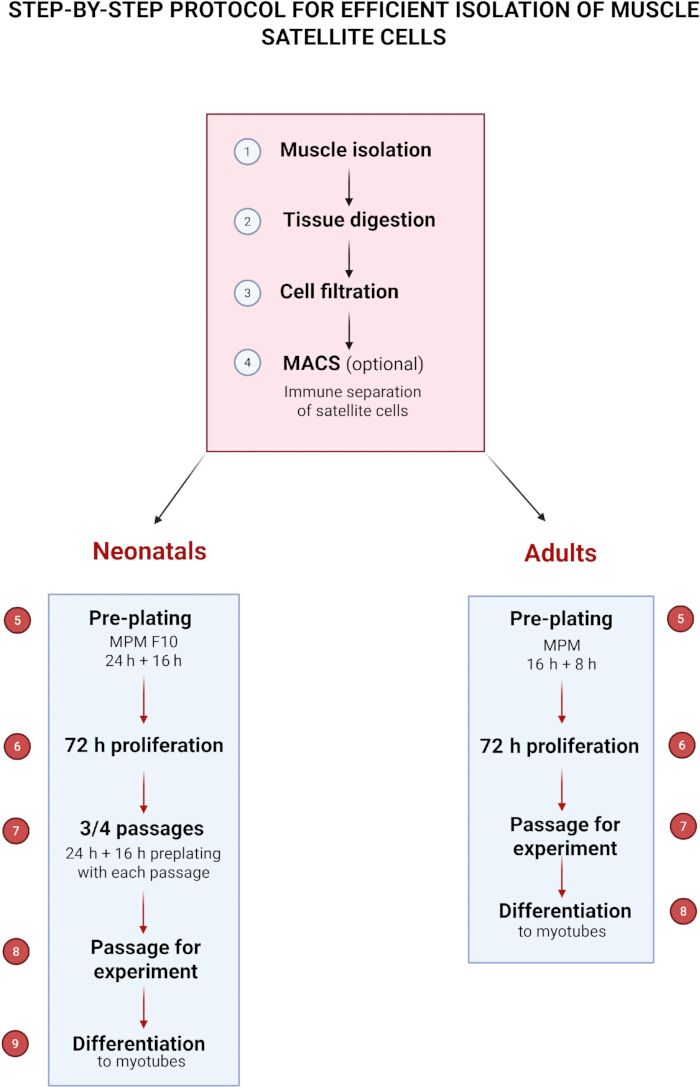
Figure 1: Step-by-step protocol for the efficient isolation of muscle satellite cells. Please click here to view a larger version of this figure.
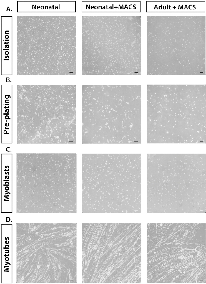
Figure 2: Representative phase-contrast microscopy images of the different methodologies discussed in the text. (A) Phase-contrast (20x) images of the isolation of the cells after digestion. (B) Phase-contrast (20x) of the preplating of the cultures with attached fibroblast, together with unattached myoblasts. (C) Phase-contrast (20x) of proliferating myoblasts once attached. (D) Phase-contrast (20x) of differentiated myotubes. Scale bar = 50 µm. Please click here to view a larger version of this figure.
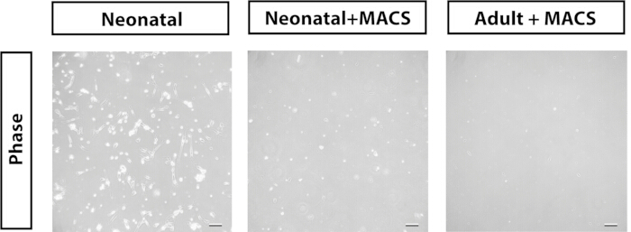
Figure 3: Representative phase-contrast microscopy images (20×) of cells remaining attached following the first preplating step. Scale bar = 50 µm. Please click here to view a larger version of this figure.
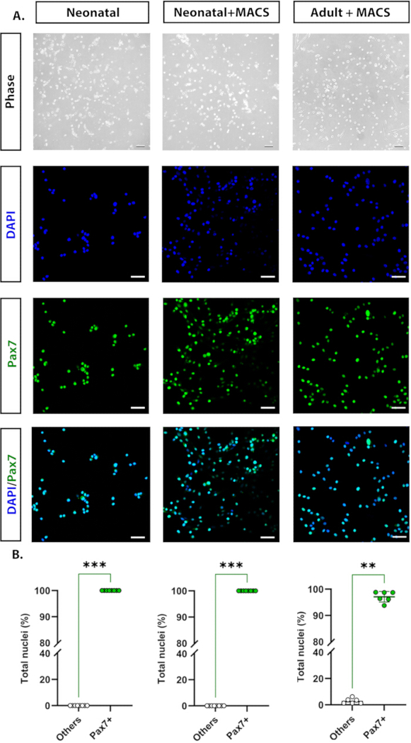
Figure 4: Representative images taken under phase-contrast microscope (20x) and immunocytochemistry (40x). (A) Pax7+ (green) and nuclei stained with DAPI (light blue), and their colocalization in the merged channels. (B) Analysis of the purity of myoblast culture, quantifying the percentage of Pax7+ cells normalized to nuclei per field acquired (one field per well, in at least six wells). Immunostaining was repeated at least three times, with two to three technical replicates per experiment. **p < 0.01, ***p < 0.0001 (Statistical significance was analyzed using Mann Whitney U test). Scale bar = 50 µm. Please click here to view a larger version of this figure.
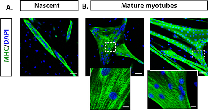
Figure 5: Representative immunofluorescence images showing myotubes differentiated from adult mouse myoblasts. (A) Nascent myotubes (3 days in culture) imaged at 20x of MHC (green) and nuclei stained with DAPI (light blue). (B) Mature myotubes (6 days in culture) imaged at 20x and details of the structure of the sarcomeric pattern defined by the myosin staining. Immunostaining was repeated at least three times, with two to three technical replicates per experiment. Scale bar = 50 µm; zoomed-in scale bar = 10 µm. Please click here to view a larger version of this figure.
Discussion
Satellite cells (SCs) constitute the principal myogenic stem and progenitor population responsible for skeletal muscle growth, repair, and regeneration36,37. Their ability to re-enter the cell cycle and differentiate into mature myofibers makes them indispensable for studying skeletal muscle biology. When isolated and cultured under defined in vitro conditions, SCs proliferate as myoblasts that can be induced to differentiate and fuse into multinucleated myotubes, recapitulating key aspects of myogenesis observed in vivo.
Our method enables the recovery of highly viable, homogeneous cell populations by carefully adjusting key parameters, including enzyme concentration, digestion duration, and culture conditions, particularly tailored for neonatal tissues. The combination of enzymatic and mechanical dissociation, followed by multi-step filtration and optional magnetic-activated cell sorting (MACS) together with preplating, ensures high cell yield and purity. Importantly, this protocol enables the generation of cultures that typically exeeds 97% Pax7+ myogenic progenitors, making them suitable for downstream differentiation assays and molecular studies.
The preplating technique relies on the differing adhesion properties of fibroblasts and SCs16,38,39. This step is particularly critical in determining culture purity, and minor deviations in timing or washing can lead to fibroblast contamination or reduced myoblast recovery. The inclusion of a precisely timed preplating step is particularly advantageous, as it efficiently separates myogenic cells from fibroblasts and other non-myogenic populations. This approach can be employed in the absence of magnetic separation equipment or as an additional measure to further increase the purity of myoblast cultures. In our protocol, we optimized this step by using collagen-coated plates and a precisely timed preplating period, which efficiently separates myogenic cells from fibroblasts and other non-myogenic populations.
Another methodological advancement of the current protocol is the use of a defined myoblast proliferation medium (MPM) optimized for both adult and neonatal cells. The combination of high serum concentration, chicken embryo extract (CEE), and basic fibroblast growth factor (bFGF) supports sustained proliferation and maintenance of an undifferentiated phenotype. CEE, in particular, contributes growth-promoting factors that enhance myoblast viability and delay spontaneous fusion, while bFGF maintains SCs identity through activation of canonical signaling pathways29,31. The use of Matrigel-coated surfaces further improves cell adhesion and proliferation efficiency, contributing to the high viability and morphological homogeneity observed within 72 hours post isolation.
Upon transition to differentiation medium, the purified myoblasts readily fuse into multinucleated, contractile myotubes expressing myosin heavy chain (MHC), demonstrating preserved myogenic potential and structural maturation. The resulting cultures are highly suitable for studies of skeletal muscle regeneration, cell signaling, transcriptional regulation, and disease modeling. Confluence is a critical factor, as cultures with very low confluence will fail to progress, while excessively high confluence will induce myoblast fusion and differentiation. Myoblasts should not be maintained in the same culture dish for more than 5 days, regardless of confluence, as prolonged incubation can also promote differentiation.
Despite these advantages, certain steps remain failure-sensitive. Enzymatic digestion must be carefully calibrated to balance tissue dissociation with cell viability. Preplating timing, cell density prior to differentiation, and gentle handling during dissociation all strongly influence yield, purity, and fusion potential. Common issues such as persistent fibroblast contamination, low yield, premature differentiation, poor attachment, or weak myotube formation can often be addressed by minor adjustments in preplating, medium supplementation, or plating technique.
Troubleshooting and practical considerations:
Isolation purity and enzymatic digestion require careful attention, as variations in enzyme type, concentration, incubation time, or tissue mincing can strongly influence both cell yield and culture purity. Under-digestion may leave tissue partially intact, reducing the release of SCs and clogging the strainer in the next step, whereas over-digestion may appear visually similar to a biofilm and can compromise cell viability. Careful optimization is therefore essential for reproducible results.
Filtration and the use of strainers are also critical, since incomplete or inefficient filtration can leave tissue debris or aggregates in the cell suspension, which may interfere with cell attachment and reduce culture purity. Using appropriately sized strainers, gently passing the suspension through them, and pre-wetting the filters can improve debris removal while minimizing mechanical stress on the cells. If larger tissue fragments clog the filter, it should be replaced with a new strainer rather than forcing the suspension through, as cells can become trapped on the filter surface, leading to decreased yield. Repeating filtration with a fresh filter when necessary can further enhance the recovery of viable myoblasts.
Premature or failed differentiation can occur when cultures become overly dense, as this may trigger spontaneous differentiation or premature fusion, thereby compromising experimental timing. Monitoring cell density and adjusting seeding density or medium volume prior to differentiation is therefore critical. In cases of excessive confluence, partial splitting or gentle reseeding can help maintain proliferation without inducing unintended myotube formation. Conversely, if the desired confluency is not reached before the start of differentiation, incubation should be continued until optimal density is achieved to ensure effective differentiation. Maintaining myoblasts beyond the recommended timeframe (>5 days) increases the risk of spontaneous differentiation and reduces proliferative capacity, regardless of initial confluence. Regular assessment of cell morphology, adherence to culture timelines, and timely medium changes are essential to preserve both viability and differentiation potential.
Persistent fibroblast contamination can be minimized through meticulous tissue processing, including careful removal of connective and adipose tissue during muscle dissection. Optimization of the preplating step is particularly important, as rapidly adhering fibroblasts attach to uncoated surfaces while SCs remain in suspension. The use of collagen-coated culture plates in combination with a defined myoblast proliferation medium supplemented with bFGF promotes selective myoblast expansion while limiting fibroblast growth. Additional purification can be achieved through negative selection strategies such as MACS to deplete fibroblast populations. Regular microscopic monitoring is essential to detect early fibroblast overgrowth and enable timely intervention.
In summary, this protocol advances the reproducibility and efficiency of primary myoblast isolation by integrating sequential purification, optimized culture conditions, and adaptable purification options. Importantly, it reliably yields highly enriched Pax7+ cells, which can be further used for controlled studies of SCs biology, myogenic commitment, and differentiation dynamics.
In contrast to protocols optimized for prolonged serial expansion, which can alter cell behavior and reduce myogenic potential and fusion ability, our method is intentionally designed to preserve cells with a robust myogenic identity and high fusion competence. This focus is advantageous for applications that require physiological relevance, efficient myogenic differentiation, and retention of defining SCs characteristics, particularly in experiments centered on studying myogenic differentiation and regeneration.
Disclosures
The authors have no conflicts of interest to declare.
Acknowledgements
Confocal imaging was performed at the Laboratory of Imaging Tissue Structure and Function, which serves as an imaging core facility at the Nencki Institute of Experimental Biology and is a part of the infrastructure of the Polish Euro-BioImaging Node.
This work was supported by National Science Center, Poland (Sonata grant 2022/47/D/NZ3/02737 to Lilya Lehka and Sonata bis grant 2020/38/E/NZ4/00314 to Grzegorz Sumara), and statutory funds from the Ministry of Science and Higher Education to the Nencki Institute. This work was supported by the project financed by the Minister of Education and Science based on contract No 2022/WK/05 (Polish Euro-BioImaging Node "Advanced Light Microscopy Node Poland").
Materials
| Name | Company | Catalog Number | Comments |
|---|---|---|---|
| 0.25% trypsin-EDTA | Gibco | 25200-056 | |
| 100 µm cell strainer | VWR | 732-2759 | |
| 30 µm strainer | Miltenyi Biotec | 130-041-407 | |
| 40 µm cell strainer | VWR | 732-2757 | |
| 70 µm cell strainer | VWR | 130-095-823 | |
| Acetic acid | POCH | 568760114 | Dissolved in dH2O to a final concentration of 20 mM and sterilized through a 0.2 µm filter |
| Alexa Fluor 488, donkey anti-Rabbit | Invitrogen | A21206 | Dissolved in blocking solution (see Section 8.7) at a 1:250 dilution |
| Alexa Fluor 488, goat anti-mouse | Abcam | ab150113 | Dissolved in blocking solution (see Section 8.7) at a 1:250 dilution |
| Ammonium chloride (NH4Cl) | BioShop | AMC303.500 | Dissolve in water to a final concentration 50 mM |
| Antifade Mounting Media | Vectashield | H-2000-10 | |
| Anti-PAX7 antibody | Abcam | ab187339 | Dissolved in blocking solution (see Section 8.7) at a 1:100 dilution |
| Basic fibroblast growth factor | Corning | 354060 | Dissolved in sterile dH2O to a stock concentration of 10 µg/mL |
| Betadine, 100 mg/mL | EGIS Pharmaceuticals | ||
| Cell culture plate, 6 well, Cell+ | SARSTEDT | 83.3902.300 | |
| Chicken embryo extract | MP Biomedicals | 2850145 | |
| Collagen I, rat tail | Gibco | A10483-01 | Dissolved in 20 mM acetic acid |
| Collagenase, type I | Sigma | C0130-500MG | Dissolved in DMEM and sterilized through a 0.2 µm filter |
| Dissociation tube (gentleMACS C Tubes) | Miltenyi Biotec | 130-093-237 | |
| Dulbecco's Modified Eagle Medium, high glucose | Gibco | 31966-021 | |
| EDTA | Invitrogen | AM9260G | |
| Falcon Conical Centrifuge Tubes, 15 mL | Corning | 352096 | |
| Falcon Conical Centrifuge Tubes, 50 mL | Corning | 352070 | |
| Fetal Bovine Serum | Gibco | 10082147 | |
| Horse serum | Gibco | 26050088 | |
| Magnetic labeling reagent (Satellite Cell isolation Kit) | Miltenyi Biotec | 130-104-268 | |
| Magnetic separator (MidiMACS Separator) | Miltenyi Biotec | 130-042-302 | |
| Matrigel | Corning | CLS356234 | Dissolved in DMEM to 10 % concentration |
| Paraformaldehyde, 4% in PBS | Thermo Fisher Scientific | J61899.AK | |
| Penicillin-Streptomycin | Gibco | 15140122 | |
| Phosphate-Buffered Saline | VWR | 392-0334 | |
| Separation column (LS Columns) | Miltenyi Biotec | 130-042-401 | |
| Skeletal Muscle Myosin Antibody, MHC | Santa Cruz | sc-32732 | Dissolved in blocking solution (see Section 8.7) at a 1:50 dilution |
| TC-dish, 100 x 20 mm | SARSTEDT | 83.3902 | |
| Tissue dissociator (GentleMACS Dissociator) | Miltenyi Biotec | 130-093-235 | |
| Triton X-100 | Sigma-Aldrich | X100-1L |
References
- Yeh, C. -J., Sattler, K. M., Lepper, C. Molecular regulation of satellite cells via intercellular signaling. Gene. 858, 147172(2023).
- Gonzalez, M. L., Busse, N. I., Waits, C. M., Johnson, S. E. Satellite cells and their regulation in livestock. J Anim Sci. 98 (5), skaa081(2020).
- Mauro, A. Satellite cell of skeletal muscle fibers. J Biophys Biochem Cytol. 9 (2), 493-495 (1961).
- Yin, H., Price, F., Rudnicki, M. A. Satellite cells and the muscle stem cell niche. Physiol Rev. 93, 23-67 (2013).
- Kahn, R. E., Dayanidhi, S., Lacham-Kaplan, O., Hawley, J. A. Molecular clocks, satellite cells, and skeletal muscle regeneration. Am J Physiol Cell Physiol. 324 (1), C1332-C1340 (2023).
- Abreu, P., Mendes, S. V. D., Ceccatto, V. M., Hirabara, S. M. Satellite cell activation induced by aerobic muscle adaptation in response to endurance exercise. Life Sci. 170, 33-40 (2017).
- Dueweke, J. J., Awan, T. M., Mendias, C. L. Regeneration of skeletal muscle after eccentric injury. J Sport Rehabil. 26 (2), 171-179 (2017).
- Michele, D. E. Mechanisms of skeletal muscle repair and regeneration in health and disease. FEBS J. 289 (21), 6460-6462 (2022).
- Domingues-Faria, C., Vasson, M. -P., Goncalves-Mendes, N., Boirie, Y., Walrand, S. Skeletal muscle regeneration and impact of aging and nutrition. Ageing Res Rev. 26, 22-36 (2016).
- Tierney, M. T., Sacco, A. Satellite cell heterogeneity in skeletal muscle homeostasis. Trends Cell Biol. 26 (6), 434-444 (2016).
- Troy, A., et al. Coordination of satellite cell activation and self-renewal by Par-complex-dependent asymmetric activation of p38α/β MAPK. Cell Stem Cell. 11 (4), 541-553 (2012).
- Dumont, N. A., Wang, Y. X., Rudnicki, M. A. Intrinsic and extrinsic mechanisms regulating satellite cell function. Development. 142 (9), 1572-1581 (2015).
- Danoviz, M. E., Yablonka-Reuveni, Z. Skeletal muscle satellite cells: background and methods for isolation and analysis. Myogenesis. DiMario, J. X. 798, Humana Press. 21-52 (2012).
- Gharaibeh, B., et al. Isolation of a slowly adhering cell fraction containing stem cells from murine skeletal muscle by the preplate technique. Nat Protoc. 3 (9), 1501-1509 (2008).
- Benedetti, A., et al. A novel approach for the isolation and long-term expansion of pure satellite cells based on ice-cold treatment. Skelet Muscle. 11 (1), 7(2021).
- Yoshioka, K., et al. A modified preplating method for high-yield and high-purity muscle stem cell isolation. Front Cell Dev Biol. 8, 793(2020).
- Uezumi, A., Fukada, S., Yamamoto, N., Takeda, S., Tsuchida, K. Mesenchymal progenitors distinct from satellite cells contribute to ectopic fat formation. Nat Cell Biol. 12 (2), 143-152 (2010).
- Christov, C., et al. Muscle satellite cells and endothelial cells: close neighbors and privileged partners. Mol Biol Cell. 18 (4), 1397-1409 (2007).
- Elizalde, G., Munoz, A. Z., Almada, A. E. Protocol for the isolation of mouse muscle stem cells using fluorescence-activated cell sorting. STAR Protoc. 4 (4), 102656(2023).
- Pasut, A., Oleynik, P., Rudnicki, M. A. Isolation of muscle stem cells by fluorescence-activated cell sorting. Myogenesis. DiMario, X. 798, Humana Press. 53-64 (2012).
- Lee, J. D., Sinha, B. C., Larkin, L. M., Vandenburgh, H. H. Isolation and purification of satellite cells for skeletal muscle tissue engineering. J Regen Med. 3 (2), (2015).
- Llufrio, E. M., Wang, L., Naser, F. J., Patti, G. J. Sorting cells alters their redox state and cellular metabolome. Redox Biol. 16, 381-387 (2018).
- Cusmano, A. A., et al. Efficient isolation and ex vivo differentiation of murine satellite cells. Stem Cell. Turksen, K. 2939, Springer. 37-54 (2025).
- Park, Y. G., Moon, J. H., Kim, J. A. Comparative study of magnetic-activated cell sorting and preplating for myoblast purification. Yonsei Med J. 47 (2), 179-186 (2006).
- Park, Y. G., Baek, A., Kim, S. Myoblast purifying by magnetic-activated cell sorting. Methods Cell Biol. 112, Elsevier. 439-447 (2012).
- Lee, H., Han, N. R., Kim, S. J., Yun, J. I., Lee, S. T. Development of a high-yield isolation protocol for active muscle satellite cells. Int J Stem Cells. 15 (3), 283-290 (2022).
- Cui, Y., et al. An easy and practical method for isolation, culture and identification of mouse skeletal muscle satellite cells. Int J Clin Exp Med. 9, 19424-19431 (2016).
- Ding, S., et al. Characterization and isolation of highly purified porcine satellite cells. Cell Death Discov. 3, 17003(2017).
- Pawlikowski, B., Vogler, T. O., Gadek, K., Olwin, B. B. Regulation of skeletal muscle stem cells by fibroblast growth factors. Dev Dyn. 246 (5), 359-367 (2017).
- Shahini, A., et al. Efficient and high-yield isolation of myoblasts from skeletal muscle. Stem Cell Res. 30, 122-129 (2018).
- Lee, D. H., Kwak, K. B., Park, Y. C., Chung, C. H., Ha, D. B. Purification and characterization of myoblast fusion inhibitor. Mol Cells. 1, 177-182 (1991).
- Moulin, V., et al. Fetal and adult human skin fibroblasts display intrinsic differences in contractile capacity. J Cell Physiol. 188 (2), 211-222 (2001).
- Wilson, C. G., et al. Age-dependent expression of collagen receptors in rat cardiac fibroblasts. Microsc Microanal. 17 (4), 555-562 (2011).
- Kohen, N. T., Little, L. E., Healy, K. E. Characterization of Matrigel interfaces during defined hESC culture. Biointerphases. 4 (4), 69-79 (2009).
- Wang, Z., et al. In vitro culture system supporting robust expansion of myogenic progenitors. BioRes Open Access. 3 (3), 79-87 (2014).
- Péault, B., et al. Stem and progenitor cells in skeletal muscle development and therapy. Mol Ther. 15 (5), 867-877 (2007).
- Le Grand, F., Rudnicki, M. A. Skeletal muscle satellite cells and adult myogenesis. Curr Opin Cell Biol. 19 (6), 628-633 (2007).
- Rando, T. A., Blau, H. M. Primary mouse myoblast purification and transplantation. J Cell Biol. 125 (6), 1275-1287 (1994).
- Hindi, L., McMillan, J., Afroze, D., Hindi, S., Kumar, A. Isolation, culturing, and differentiation of primary myoblasts. Bio Protoc. 7 (9), (2017).
Reprints and Permissions
