Here, we present a protocol for combining iterative protein staining (IBEX) with RNAscope in situ hybridization technology for the deep spatial phenotyping of intact fixed frozen tissues for high-resolution spatial profiling applications.
A subscription to JoVE is required to view this content. Sign in or start your free trial.
Method Article
Bex-Plex Enables Simultaneous Visualization of Protein Markers and Ribonucleic Acid Transcripts for Deep Spatial Profiling of Intact Tissue
In This Article
Summary
Abstract
Here, we present a protocol to enable simultaneous spatial profiling of protein and ribonucleic acid (RNA) markers at single-cell resolution in intact tissues. One of the core issues surrounding solid tumor cancers is the inability to predict patient responsiveness to therapeutic intervention. Deciphering these complexities requires quantitative measurement of cellular behaviors, interactions, and phenotypes within intact tissues. Specifically, the proteome and transcriptome provide overlapping but nonredundant information that can be critical to designing rational immunotherapeutics. However, there is not currently a straightforward and cost-effective way to deeply profile both the transcriptional and functional state of all cells in the tumor microenvironment while retaining their critical spatial localization information. To this end, we have successfully combined the IBEX (iterative bleaching extends multiplexity) platform with HiPlex RNAscope to simultaneously visualize high-plex protein markers and high-plex ribonucleic acid (RNA) transcripts with single-cell resolution. This approach incorporates a modified RNA target retrieval step to preserve nuclear morphology and enable accurate image alignment across cycles. “Bex-Plex” enables us to perform an unmodified IBEX protocol on fixed frozen tissues followed by the HiPlex RNAscope protocol, in which we use a modified RNA target retrieval to ensure nuclear-level alignment with IBEX cycles. This protocol is optimized for fixed frozen tissues and enables integrated spatial analysis of transcriptional and functional cellular states. The power to profile at this single-cell level not only provides a deep well of information about the tissue itself but can also empower predictions about the nuanced cellular interactions that drive therapeutic responsiveness, providing a framework for studying spatial biology in tumor microenvironments and related systems.
Introduction
Since their introduction in 2011, cancer immunotherapies that decrease the suppression of cytotoxic T cells by blocking inhibitory immune checkpoint molecules such as PDL-1 and CTLA-4 have emerged as the gold standard for solid tumor treatment1. However, despite large-scale use in patients, the clinical efficacies of these drugs vary widely because many solid tumor cancers either intrinsically present with or eventually generate resistance to these therapies2. In many cases this is due to the extremely heterogeneous nature of solid tumors, even among the same cancer type3. A more in depth understanding of the ....
Access restricted. Please log in or start a trial to view this content.
Protocol
The procedures outlined in this protocol were approved by the Institutional Animal Care and Use Committee of the University of California, San Diego (protocol number S24049). Male and female mice 7-12 weeks of age on a C57BL/6 genetic background and housed under specific pathogen-free conditions were used for these experiments.
1. Tissue preparation
- Euthanize experimental mice by approved primary and secondary measures (e.g. CO2 euthanasia and cervical dislocation) and harvest tissues of interest into supplemented media.
- Prepare a fresh stock of tissue fixative by diluting a 4% stock ....
Access restricted. Please log in or start a trial to view this content.
Results
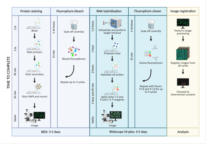
Figure 1 : Overview of Bex-Plex workflow.
Fixed frozen tissues sectioned onto chrome alum-coated slides undergo up to 3 cycles of standard IBEX followed by up to three cycles of RNAScope HiPlex wherein the initial target retrieval step is p.......
Access restricted. Please log in or start a trial to view this content.
Discussion
The Bex-Plex workflow integrates the iterative protein staining platform, IBEX, with RNAscope in situ hybridization to enable simultaneous detection of proteins and RNA transcripts at single-cell resolution. The primary goal of this protocol is to provide a reproducible method for integrated spatial analysis of protein and RNA expression within intact tissues. The ability to measure both protein and RNA expression in the same tissue has previously presented technical challenges: high costs, complex antigen retri.......
Access restricted. Please log in or start a trial to view this content.
Disclosures
The authors declare no competing financial interests. No AI-based tools were used in the preparation of this manuscript.
Author contributions:
SK contributed to data curation, data analysis, investigation, and writing. CM contributed to data curation. PH contributed to data analysis. AV contributed to data curation. VM contributed to conceptualization, data curation, data analysis, supervision, and writing.
Acknowledgements
This work was supported by the University of California San Diego Department of Pediatrics, Division of Allergy, Immunology, and Rheumatology and NIH NIGMS grant R00GM147841 (MOSAIC K99/R00) and NCI U54CA272220 (UC San Diego FIRST grant). The authors would additionally like to thank Andrea Radtke for establishing the original IBEX method, without which we would have been unable to create Bex-Plex.
....Access restricted. Please log in or start a trial to view this content.
Materials
| Name | Company | Catalog Number | Comments |
|---|---|---|---|
| 1.5 ml microcentrifuge tubes | Biopioneer | MCNT-1.5F | |
| 200 proof EtOH | Fisher Scientific | BP28184 | |
| 20X SSC buffer | Invitrogen | AM9765 | |
| 24 well plates | |||
| Alexa Fluor 488 anti-mouse CD11b Antibody | Biolegend | 101217 | |
| Alexa Fluor 523 anti-mouse CD3e Antibody | Invitrogen | 58-0032-82 | |
| Alexa Fluor 647 anti-mouse Ly6G Antibody | Biolegend | 127610 | |
| Alexa Fluor 700 anti-mouse CD68 Antibody | Biolegend | 137025 | |
| BD Cytofix/Cytoperm fixation and permeablization solution | BD | 554722 | |
| Chrome alum gelatin | Fisher | NC1692262 | |
| Confocal microscope (e.g Leica Stellaris) | |||
| Cryomolds | VWR | 25608-922 | |
| Cryostat (e.g CryoStar NX50 Cryostat) | Thermofisher Scientific | 957100 | |
| Cryostat blades (e.g Leica Low Profile Disposable Blades DB80LX) | Leica Biosystems | 14035843496 | |
| DAPI | Thermofisher Scientific | 62248 | |
| Dissection microscope (e.g Zeiss SteREO Discovery.V8) | |||
| Dissection tools | |||
| Dry Ice | |||
| Dunk tank for slide staining (e.g Simport Scientific EasyDip Slide Staining Jars) | Fisher Scientific | 22-038-489 | |
| Fluoromount-G | Fisher | OB10001 | |
| Heated water bath | |||
| Histology brushes (e.g. Ted Pella Camel Hair Brushes) | Fisher Scientific | NC2023341 | |
| Hybridization oven (e.g. HybEZ II Hybridization System) | ACD | 321710 | |
| Image analysis software of choice | |||
| ImmEdge Hydrophobic Barrier PAP Pen | Vector Laboratories | H-4000 | |
| Kimwipes | VWR | 21905-026 | |
| Lithium borohydride | STREM Chemicals | 93-0397 | |
| Micropipettes (P-10, P-20, P-200, P-1000) | |||
| Mini Microcentrifuge (e.g. Corning LSE Mini Microcentrifuge) | Corning | 6770 | |
| Mouse BD Fc Block | BD | 553142 | |
| No 1.5 Coverglass | VWR | 48393-241 | |
| Pipette tips (P-10, P-20, P-200, P-1000) | |||
| RNAscope HiPlex Probe, mm-CD274-T7 | ACD | 420501-T7 | |
| RNAscope HiPlex Probe, mm-IL-10-T6 | ACD | 317261-T6 | |
| RNAscope HiPlex12 Reagents Kit v2 (includes detection kit, cleaving stock solution, protease reagents, target retreival reagents, and wash buffer) | ACD | 324409 | |
| RNAscope Protease III | ACD | 322337 | |
| RNase ZAP | Millipore Sigma | R2020-250 | |
| Simple ITK software: https://github.com/niaid/sitk-ibex | |||
| Slide staining tray/humidity chamber | |||
| Sucrose Bioxtra | Sigma | S7903-1KG | |
| Superfrost Plus Microscope slides | VWR | 48311-703 | |
| Tissue-Tek OCT | VWR | 25608-930 | |
| Tween-20, Molecular Biology grade | Promega | H5152 | |
| UltraPure DNase/RNase free water | Fisher Scientific | 10977023 |
References
- Ribas, A., Wolchok, J. D. Cancer immunotherapy using checkpoint blockade. Science. 359 (6382), 1350-1355 (2018).
- Zhao, X., Subramanian, S. Intrinsic resistance of solid tumors to immune checkpoint blockade therapy. Cancer Res. 77 (4), 817-822 (2017).
- Bailey, P.,
Access restricted. Please log in or start a trial to view this content.
Reprints and Permissions
This article has been published
Video Coming Soon