This protocol describes a standardized method for surgical harvesting, enzymatic digestion, and density-gradient purification of Lewis rat pancreatic islets, enabling high-yield, high-purity preparations for direct use in transplantation or in vitro studies.
Method Article
Pancreatic Islet Isolation and Purification from Lewis Rats Using Enzymatic Digestion and Density-Gradient Separation
* These authors contributed equally
In This Article
Summary
Abstract
Pancreatic islets are essential for both clinical islet transplantation and preclinical studies aimed at understanding the mechanisms and progression of diabetes. However, progress in these fields is often limited by the availability of high-quality islets, as emerging therapeutic strategies require larger, cleaner, and more standardized preparations. This article presents a high-throughput, standardized protocol for surgical harvesting, enzymatic digestion, and density-gradient purification of pancreatic islets from Lewis rats. The method combines intraductal pancreas perfusion with a defined enzyme blend, pooled digestion of multiple pancreases, and density-based separation to maximize yield while minimizing fragmentation and eliminating the need for routine hand-picking. Islet yield is quantified using two complementary approaches: a traditional aliquot-based counting method commonly used in clinical settings, and a semi-automated whole-preparation imaging-based method. In a representative isolation from 12 pancreases, manual counting yielded approximately 3,300 islet equivalents (IEQ) and 2,700 absolute islets per pancreas (~39,600 total IEQ), with a size distribution of 62% (50-100 µm), 17% (100-150 µm), 10% (150-200 µm), 5% (200-250 µm), and 6% (>250 µm). In comparison, whole-prep automated analysis reported 16,350 total IEQ (1,362 IEQ/pancreas), highlighting inherent variability in aliquot-based quantification. Preparations had a purity of 98% by dithizone staining, and islet viability was maintained at 98% in vitro on days 3 and 7 after isolation. Functional assessment by static glucose-stimulated insulin secretion demonstrated a 10-fold increase in insulin release following a 1-h stimulation with 16.7 mM glucose. Overall, this protocol provides a reproducible and efficient approach for generating high-purity, functionally intact islet preparations that can be used immediately without additional cleanup. Islets retain robust function in culture for up to seven days, enabling flexibility for large-scale in vitro assays as well as in vivo transplantation studies.
Introduction
Pancreatic islet transplantation represents a viable strategy for restoring endogenous insulin production in individuals with type 1 diabetes (T1D)1. Beyond T1D, islet biology is increasingly relevant to type 2 diabetes (T2D), a condition characterized by progressive β-cell dysfunction and loss, as well as to patients undergoing partial or total pancreatectomy for chronic pancreatitis, pancreatic tumors, or traumatic injury. In these contexts, access to high-quality islets is essential for advancing cellular therapies, modeling β-cell failure, studying islet-immune and islet-stromal interactions, and developing emerging regenerative approaches2. Consequently, robust and reproducible islet isolation procedures are critical to support both mechanistic research and translational development across a wide range of pancreatic endocrine disorders.
Although islets constitute less than 2% of the pancreatic mass, they exert a disproportionately significant role in glucose homeostasis. Their intrinsic vulnerability to hypoxia, enzymatic over-digestion, oxidative stress, and mechanical shear makes them particularly sensitive to isolation-related injury3,4,5. Even subtle deviations in temperature, oxygenation, or tissue handling can impair β-cell stimulus-secretion coupling or trigger apoptosis, owing to limited antioxidant defenses and high metabolic demand6,7. As a result, most conventional protocols emphasize rapid downstream use, typically within 24-48 h of harvesting in both clinical and experimental settings8,9,10,11,12. While this "immediate-use" framework minimizes deterioration, it restricts experimental flexibility, hampers longitudinal studies, and complicates efforts to accumulate sufficient islet numbers for large in vitro assays or in vivo transplantation experiments.
Among rodent models, Lewis rats are widely utilized for islet research due to their inbred genetic background, stable immune profile, and reproducible physiological characteristics14. Strain-dependent differences in islet yield, responsiveness, and resilience are well documented. De Groot et al. demonstrated that Lewis rat islets exhibit superior glucose-stimulated insulin secretion compared with other strains, underscoring their suitability for functional, metabolic, and transplantation studies15. Additional work has confirmed their reliability for immunological and allo-/xenotransplantation research14,15,16. However, across laboratories, variability in islet yield, purity, and function remains a significant barrier. Much of this variability arises from inconsistencies in enzyme blend, intraductal perfusion techniques, pancreas handling, digestion timing, and purification strategy17.
To address these limitations, we developed a reproducible, step-by-step protocol for surgical harvesting, enzymatic digestion, and density-gradient purification of pancreatic islets from Lewis rats. This workflow is optimized to maximize yield, minimize fragmentation, and preserve functional viability. Importantly, it establishes clear benchmarks, including islet equivalents (IEQ), viability over time, purity, and glucose-stimulated insulin secretion, that enable investigators to reliably distinguish successful from suboptimal isolations18,19,20. By improving both yield and longevity, the protocol enhances experimental flexibility and may reduce the number of donor animals required, supporting more ethical and scalable experimental design.
Distinct from many traditional approaches, the present method demonstrates that Lewis rat islets can be maintained in culture for up to 7 days with preserved viability and function, expanding their utility for extended in vitro studies. The protocol further incorporates a streamlined, pooled-processing strategy capable of handling up to 30 pancreases in a single batch, enabling uniform digestion and reducing inter-sample variability. With endocrine purity reaching 98%, the procedure eliminates the need for labor-intensive hand-picking and reduces opportunities for microbial contamination. Compared with single-step or bulk digestion methods that frequently produce impure or mechanically compromised preparations13, this high-throughput workflow reliably yields large numbers of clean, transplantation-quality islets suitable for in vitro assays, metabolic profiling, and in vivo transplantation studies.
Protocol
All procedures were approved by the Houston Methodist Institutional Animal Care and Use Committee (IACUC, protocol #IS00007362) and conducted in accordance with the National Institutes of Health Guide for the Care and Use of Laboratory Animals and the Animal Welfare Act.
NOTE: The materials and instruments used throughout the protocol can be found in the Table of Materials.
1.Preparation prior to harvesting and isolation day
- Prepare the following solutions under aseptic conditions inside a biosafety cabinet (see Table 1) and store all solutions at 4 °C until use.
Penicillin-streptomycin rinse solution (Pen-Strep).
Washing media.
Quenching buffer.
Islet culture media.
Krebs buffer. - Sterilize tools and equipment via autoclaving.
- Autoclave pancreas harvesting surgical pack: surgical drapes, gauze, rat-tooth tissue forceps, flat forceps, fine curved forceps, one skin scissor, micro-dissecting scissors, two small hemostats, and one curved hemostat.
NOTE: Autoclave two pancreas harvesting surgical packs if there will be two surgical operators. - Autoclave pancreas cleaning surgical pack: fine curved forceps and micro-dissecting scissors.
- Autoclave one 30 cm x 30 cm sheet of aluminum foil.
- Autoclave 1-6 600 mL and 1 L beakers, according to the number of pancreases to be processed (see Table 2).
- Autoclave one 500 µm sieve.
- Autoclave two 14 G spinal needles, in case one clogs with undigested pancreas tissue.
- Autoclave one syringe tube for each 50 mL syringe required (see Table 3).
- Autoclave pancreas harvesting surgical pack: surgical drapes, gauze, rat-tooth tissue forceps, flat forceps, fine curved forceps, one skin scissor, micro-dissecting scissors, two small hemostats, and one curved hemostat.
- Prepare the water bath for digestion.
- Fill the water bath equipped with an orbital shaker with autoclaved ultrapure water until it is 3/4 inch above the level of the platform.
- Add 3 mL antimicrobial solution and keep the lid closed to minimize contamination.
2. Preparations on the harvesting and isolation day
- Prepare gradient syringes.
- Remove islet gradient solutions 1.108 g/cm³, 1.096 g/cm³, and 1.069 g/cm³ prepared and used in the next steps and "Islet Purfication" step 5.2, from 4 °C storage, and allow them to equilibrate to room temperature (about 1 h).
NOTE: Required gradient volume will differ by the number of pancreases to be processed; refer to Table 3 to ensure adequate thawed volumes of select gradients. - Preload and label the 50 mL syringes with 1.096 g/cm³ and 1.069 g/cm³ gradients.
- Vigorously mix the 1.069 and 1.096 g/cm³ density gradient solutions.
- Load the appropriate volumes, depending on Table 3, into 50 mL syringes fitted with surgical tubing and a 16 G needle.
- After filling, cap the needles to maintain sterility and set the syringes aside for subsequent use in "Islet Purification" step 5.5.
- Keep 1.108 g/cm3 gradient at room temperature until later use.
- Remove islet gradient solutions 1.108 g/cm³, 1.096 g/cm³, and 1.069 g/cm³ prepared and used in the next steps and "Islet Purfication" step 5.2, from 4 °C storage, and allow them to equilibrate to room temperature (about 1 h).
- Make Working dissociation media (WDM) enzyme solution, load into syringes for perfusion and autoclaved beakers.
- Work under sterile conditions in a biosafety cabinet and keep all solutions on ice.
- Thaw aliquots of collagenase, thermolysin, and DNase I on ice.
- Dilute enzyme reagents in a sterile 1 L beaker using WDM to achieve the following final concentrations: collagenase 0.15 mg/mL, thermolysin 5 µg/mL, and DNase I 0.1 mg/mL (Table 2).
- Mix WDM gently using a swirling motion 10 times. Do not mix with a pipette; preserve enzyme activity by gently swirling.
- One by one, load 10 mL of the WDM enzyme solution into each 10 mL syringe using a 16 G needle for rapid filling, then replace with a capped 25 G X ½" inch needle for later pancreas perfusion.
- Wrap loaded syringes with autoclaved aluminum foil and place on ice until use.
- Dispense excess WDM equally into sterile 600 mL beakers to hold harvested pancreases according to Table 2. Cover each beaker with sterile aluminum foil and keep it on ice (Table 2).
- Prepare cell culture workspace.
- Turn on the orbital water bath shaker, set to 130 rpm and 37 °C. Leave as such during pancreas harvesting.
- Place the following items under UV light in the biosafety cabinet for 1 h for further sterilization.
Sterile 500 µm sieve.
Sterile 1 L beaker.
Syringe pump. - Ensure the cell culture incubator is set to 25 °C 5% CO2.
- Set the centrifuge to 4 °C, with acceleration and deceleration at 9.
- Gather materials for harvesting and proceed to the surgical room.
- Designated pancreas cleaner:
- Gather one large ice bucket to hold the required number of 600 mL WDM beakers and the pancreas rinse solution (see Table 1 and Table 2).
- Gather 1 sterile Petri dish for every 6 pancreases to be processed.
- Gather 1 sterile micro-dissecting scissors and 2 small, curved delicate forceps.
- Designated pancreas harvester:
- Gather a sterile harvesting surgical pack.
- Gather sterile surgical gloves.
- Gather an ice tray with preloaded 10 mL WDM syringes sealed in autoclaved aluminum foil.
- Designated pancreas cleaner:
- In the surgical room, prepare the pancreas harvesting station.
- Turn on the glass bead sterilizer to sterilize surgical tools by submerging them for 1 min in the sterilizer between each animal. Instruments may be cooled rapidly by immersion in sterile 1x PBS.
- Place an absorbent pad under the microscope's field of view for any blood or leaked WDM enzyme solution.
- Set the microscope to a total magnification of 6.3x between the ocular and objective lenses.
- Open the surgical pack and don sterile surgical gloves using aseptic technique.
- Proceed with surgical harvesting.
- In the surgical room, prepare the pancreas cleaning station.
- Pour enough Pen-Strep solution to fill half of a 100 mm Petri-dish (about 6 mL) to submerge the isolated pancreases and place on ice.
- Open the surgical pack and don sterile surgical gloves using aseptic technique.
- In the surgical room, prepare the euthanasia chamber.
- Place reservoir trays into an empty, clean, and re-sealable rat cage.
- Fill trays halfway full of isoflurane.
- Set the perforated tray on top of the reservoirs to prevent spilling while rats are placed inside.
- Seal the chamber top with the cage lid.
- Proceed to pancreas harvesting.
3. Pancreas harvesting
- Place rats one at a time into the euthanasia chamber on top of the tray for rapid isoflurane overdose.
NOTE: Euthanasia time required for the rat will vary, especially as the isoflurane evaporates. - Confirm absence of pulse and respiration for each rat.
- Confirm euthanasia with a secondary method that does not disturb the abdominal cavity (e.g., cardiac puncture).
NOTE: Other primary euthanasia methods are acceptable; however, isoflurane overdose is preferred, as rapid euthanasia minimizes islet hypoxia. - Spray each rat's abdominal fur with 70% ethanol to mat and prevent loose hair in the surgical field.
- Place the euthanized rat supine under the microscope with the head distal to the operator. Ensure the upper abdominal cavity is under the microscope field of view.
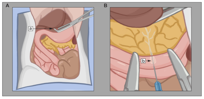
Figure 1: Surgical exposure and intraductal perfusion of the rat pancreas. Illustration of the key steps in pancreas exposure and perfusion through the Sphincter of Oddi. (A) Euthanized Lewis rat with the abdominal cavity opened. (Aa) Clamp placement of the common bile duct and hepatic vein. (B) Magnified view of the duodenum showing hemostats marking the region of the Sphincter of Oddi. (Bb) Perfusion needle positioned within the main pancreatic duct. Please click here to view a larger version of this figure.
- Harvest the pancreas (pancreas harvester role):
- Make a "v-shaped" incision, using skin scissors, starting from the pubic symphysis toward each lateral ribcage to access the abdominal cavity (see Figure 1A).
- Retract the small and large intestines to the left side of the animal using flat forceps to clear the abdominal cavity "workspace".
- Unfurl the duodenum using the flat forceps and curved hemostats. Ensure the common bile duct and hepatic portal vein are in the microscope field of view.
- Retract the liver cephalad and clamp the common bile duct and hepatic vein using the curved hemostats. Clamp 1-2 mm from the liver (see Figure 1Aa).
NOTE: Steps 3.6.1-3.6.4 and 3.6.8 may be done through direct visualization. Use the microscope for all other steps to ensure delicate tissue manipulation. - Under the view of the microscope, using the flat forceps and flat hemostats, gently mobilize the contents in the duodenum to clear the Sphincter of Oddi.
NOTE: Ensure duodenum contents are displaced from the intended needle insertion site. - Isolate the region surrounding the Sphincter of Oddi by gently clamping the duodenum with the flat hemostats approximately 5 mm from each side of the sphincter. Ensure minimal compression to preserve tissue integrity (see Figure 1B).
- Using a sterile WDM syringe, insert the needle carefully through the duodenal wall, pass it through the Sphincter of Oddi, and advance into the main pancreatic duct for perfusion (see Figure 1Bb).
- As soon as the needle goes through the Sphincter of Oddi, maintaining constant pressure, perfuse 7-10 mL of WDM into the pancreas at a rate of about 500 μL/s until the pancreas is fully distended.
NOTE: Ensure the bevel is up and the needle is parallel to the pancreatic duct to minimize trauma. Begin injecting immediately upon insertion. This will help dilate the sphincter and duct, facilitating advancement of the needle. The pancreas is considered fully distended when the tail of the pancreas, located near the spleen, becomes visibly perfused.
- As soon as the needle goes through the Sphincter of Oddi, maintaining constant pressure, perfuse 7-10 mL of WDM into the pancreas at a rate of about 500 μL/s until the pancreas is fully distended.
- Excise the pancreas by gently dissecting it free from surrounding structures in the following order: spleen, stomach, large intestine, duodenum, and mesentery.
NOTE: Use blunt dissection to minimize tissue damage. Proceed sequentially to maintain anatomical orientation and reduce tension on vascular and ductal connections.- Remove both flat hemostats from the duodenum.
- Use the flat forceps to clamp the splenopancreatic ligament (connects the pancreas and spleen) and apply gentle traction.
- Using the fine curved dissecting forceps, carefully mobilize and separate the pancreas from the adjacent splenic attachments.
- Detach the pancreas from the stomach by lifting the pancreatic tissue with fine curved forceps and applying gentle counter-traction with flat forceps.
- Then, using the flat forceps, apply firm traction to avulse the large intestine from the pancreas.
- Lift the small intestine using the flat forceps and dissect the mesentery from the pancreas using the fine curved forceps.
- Gently lift the perfused pancreas with the flat forceps and use the fine dissecting scissors to sever any remaining attachments to connective tissue.
- Immediately transfer the excised pancreas into a Petri dish containing ice-cold rinse solution and immediately proceed to harvest the next pancreas, as the pancreas cleaner begins pancreas cleaning.
- Clean the pancreas (pancreas cleaner role):
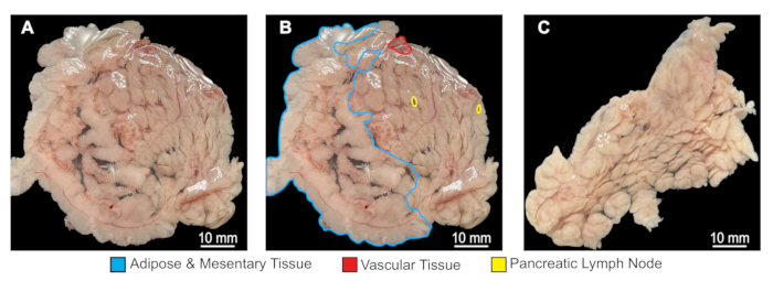
Figure 2: Cleaning of the perfused rat pancreas. Sequential visualization of the pancreas before and after removal of non-essential tissue. (A) Perfused pancreas immediately after dissection, with adjacent adipose tissue, lymph nodes, and connective structures visible. (B) Perfused pancreas with major anatomical landmarks labeled. (C) Cleaned pancreas following removal of excess tissue, showing the final appearance prior to digestion. Please click here to view a larger version of this figure.
- Use the fine curved forceps and fine dissecting scissors to carefully cut away lymph nodes, adipose tissue, mesentery, and large vasculature from the perfused pancreas, if present (see Figure 2).
- Transfer the cleaned pancreas to a sterile 600 mL beaker containing 25 mL of cold WDM. Cover the beaker with sterile aluminum foil to retain cleanliness between the transfer from the surgical workspace to the cell culture workspace.
- Replace the rinse solution in the Petri dish every 6 pancreases to prevent contamination.
- Record the time when the first pancreas is placed on ice. This will be used to document the cold ischemia time.
- Once all pancreases are procured and cleaned, quickly proceed to the digestion phase in the cell culture workspace.
4. Pancreas digestion
- Pause orbital shaker revolutions and secure the covered beaker(s), containing pancreases, into the designated holders of the water bath.
- Quickly, restart the orbital shaker set at 130 rpm and start a 10-min timer for the digestion time (see Figure 3A).
NOTE: Record this point as the end of the cold ischemia period. Log each islet harvest with its corresponding total cold ischemia time in a laboratory record. - At the 5-min mark, manually swirl the beaker clockwise, then counterclockwise to aid tissue dissociation.
- At the 8-min mark, retrieve the quenching solution from cold storage and transfer it into the biosafety cabinet for later immediate use (see Table 1).
- At the 10-min mark, rapidly transfer and submerge beakers into ice to slow enzyme activity (see Figure 3B).
NOTE: From this point onward, handle all tissues, cells, and solutions on ice to preserve them until step 4.20 when using room temperature density gradients. - Transfer the beaker exterior and tray into a biosafety cabinet.
- Add quenching buffer at a 1:1 ratio with the solution in the beaker to stop digestion (approximately 75-100 mL per beaker).
- Aspirate and expel the tissue suspension gently and repeatedly using a 50 mL syringe fitted with a 14 G spinal needle to dissociate large tissue fragments (see Figure 3C).
- Transfer the suspension to 200 mL conical tubes.
- Rinse the beaker with 5 mL of washing media and decant into the 200 mL conical tube containing the islet digest to collect any remaining digest.
- Centrifuge digests at 200 × g for 5 min at 4 °C. Ensure use of appropriate centrifuge bottle adapters and cushions for safety.
- Decant the supernatant carefully and resuspend the pellet in 100 mL of washing media using the same 14 G spinal needle as before.
- Filter the suspension through a sterile 500 µm sieve placed over a sterile 600 mL beaker to remove any large, undigested tissue.
NOTE: If pancreas perfusion and cleaning were performed optimally, minimal adipose tissue and no unperfused pancreas will remain atop the sieve. - Rinse the sieve with 10 mL of washing media to recover entrapped tissue.
- Transfer the filtrate into new 200 mL conical tubes.
- Centrifuge at 200 × g for 5 min at 4 °C.
- Decant the supernatant and resuspend the pellet in 100 mL washing media using a 10 mL serological pipette.
NOTE: Ensure minimal cell loss by pre-wetting pipette tips with FBS-containing buffer before exposure to islets. - Centrifuge at 600 × g for 5 min at 4 °C.
- Carefully aspirate the supernatant to obtain a dry pellet.
- Set the centrifuge temperature to 22 °C in preparation for gradient separation.
NOTE: This can be expedited by running the centrifuge at 2000 x g for 15 min at 22 °C, without samples.
5. Islet purification
- Refer to Table 3 to determine the number of 50 mL conical tubes required.
NOTE: Refer to Table 3 to distribute the total pellet volume evenly, ensuring that approximately 2-3 mL of pellet digest is divided into each gradient tube. If weak islet band separation is later seen, use additional gradient tubes to enhance separation resolution. - Pre-wet a 10 mL serological pipette tip with the 1.108 g/cm³ density gradient solution to prevent islets from adhering to the inner surface of the tip.
- Resuspend each tissue pellet in 15 mL of 1.108 g/cm³ gradient solution per 2 mL of pellet volume using the pre-wetted 10 mL serological pipette. Despite the total volume exceeding 10 mL, the smaller pipette width facilitates more controlled mixing and efficient pellet dissociation. Mix gently until uniform.
- Dispense 15 mL of the cell suspension into the bottom of each 50 mL conical tube carefully, avoiding contact with the tube walls to ensure clean gradient levels.
- Using the preloaded gradient syringes, sequentially overlay 10 mL of 1.096 g/cm³ gradient, followed by 10 mL of 1.069 g/cm³ gradient.
- Dispense each gradient at a rate of 5 mL/min using a syringe pump, positioning the 16 G needle tip against the inner wall of the tube to prevent gradient mixing.
- Centrifuge the tubes at 700 × g for 15 min at 22 °C, with the centrifuge acceleration set to 3 and deceleration set to 0 to prevent gradient mixing.
NOTE: Given the extended deceleration time, the entire centrifugation cycle takes approximately 45 min. Monitor the time closely, as islets must be collected promptly to prevent gradient mixing and potential cell damage. If needed, prepare counterbalance tube(s) by weight, not volume, to account for density differences. After removing the tubes, reset the centrifuge to 4 °C, and acceleration and deceleration to 9 for future steps. - Following centrifugation, identify the islet layer located at the interfaces between the 1.069-1.096 g/cm³ and 1.096-1.108 g/cm³ gradient layers (see Figure 3D).
- Aspirate and discard any tissue collected at the top layer carefully using a Pasteur pipette pre-wetted with washing media.
- Wet another Pasteur pipette, collect the islet layer, and transfer it to a pre-chilled 200 mL conical tube containing 5 mL of washing media.
- Bring up the pre-chilled 200 mL conical tube volume to 200 mL with washing media.
- Centrifuge at 600 × g for 5 min and decant the supernatant.
- Resuspend the pellet in 30 mL of washing media using a pre-wetted 10 mL serological tip.
- Transfer resuspension to a new 50 mL conical tube using a pre-wetted 10 mL serological tip for better islet pelleting.
- Rinse the same 200 mL conical tube with 20 mL of washing media and transfer to the 50 mL conical tube containing the remaining islets.
- Centrifuge the 50 mL conical with islets and washing media at 300 × g for 5 min at 4 °C.
- Decant the supernatant and resuspend the islet pellet in 50 mL of islet culture media (see Table 1).
NOTE: Remove 100 μL aliquots during this step if counting islets with the manual quantification method. - Evenly plate islets into non-tissue-culture-treated 100 mm Petri dishes, using one plate for every five Lewis rat pancreases processed.
- Calculate the total islet media volume required using the formula (number of pancreases ÷ 5 × 10 mL).
- Resuspend the islet pellet in half of the calculated media volume, then evenly distribute the suspension across the determined number of plates.
- Rinse the 50 mL conical tube containing residual islets with the remaining half of the calculated media and pipette this wash across the same plates to reach a final volume of 10 mL per dish (see Figure 3E).
- Incubate overnight at 25 °C in 5% CO₂. Add 3 mL of islet culture media every 24 h to the side of each petri dish and then gently swirl to replenish the media.
- Proceed to islet counting (step 6).
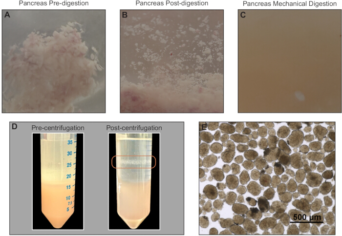
Figure 3: Pancreas digestion and purification of isolated islets. Representative images of the sequential steps from pooled pancreas digestion to islet purification. (A) Pooled perfused pancreases prior to enzymatic digestion. (B) Fragmented pancreases post-enzymatic digestion. (C) Uniform tissue suspension, post-mechanical. (D) Density-gradient tubes before centrifugation (left) and after centrifugation (right), with visible islet band at the gradient interface circled. (E) Brightfield image of Day 0 islets immediately after collection. Please click here to view a larger version of this figure.
6. Islet counting
- Collect manual islet quantification.
- During step 5.16, withdraw three 100 µL aliquots of the islet suspension using a 200 μL pipette and normal 200 μL tips.
NOTE: Ensure the islets are equally suspended in media via pipette mixing, when aliquots are taken for measurement. - Transfer each aliquot into its own 2 mm × 2 mm gridded Petri dish containing 1 mL of 1× PBS.
- Count islets under an inverted microscope, cataloging them by diameter with an eyepiece reticle lens insert.
NOTE: Not all islets will be circular; adhere to either horizontal or vertical measurements throughout counting. - Calculate the total number of 150 µm islet equivalents using the standardized conversion table (see Table 4).
NOTE: Compare each sample's IEQ. If the IEQ of the aliquots differs by more than 30%, proceed with the average of the most similar aliquots.
- During step 5.16, withdraw three 100 µL aliquots of the islet suspension using a 200 μL pipette and normal 200 μL tips.
7. Safety precautions
- Perform all surgical and tissue handling procedures under aseptic conditions inside a biosafety cabinet when appropriate. Use personal protective equipment (PPE), including safety goggles, lab coats, gloves, and surgical masks, throughout handling of reagents and tissues.
- Perform pancreas harvesting under sterile conditions with adequate surgical tools and PPE. Handle isoflurane and other anesthetics in accordance with institution guidelines to prevent inhalation exposure.
- Dispose of animal tissues, sharps, and biohazard waste in compliance with institutional biosafety protocols. Maintain enzymatic reagents and biological materials on ice unless otherwise specified to preserve activity and viability.
Results
The following results represent a typical islet isolation performed using the described method, with twelve Lewis rat pancreases processed in parallel.
Yield and size distribution
Manual aliquot counting and IEQ conversion
Two independent 100 µL aliquots were obtained from the mixed islet suspension, as described in section "Islet Counting," Step 6.1, and counted by diameter bins (50-100 µm, 100-150 µm, 150-200 µm, 200-250 µm, 250-300 µm, 250-300 µm). Counts were converted to islet equivalents (IEQ; 150 µm reference) using standard equivalency tables and extrapolated to the total suspension volume (see Figure 4A). Across the test isolation (n = 12), yield was 3330 IEQ/pancreas with a median islet diameter in the 50-100 µm bin. Replicate aliquots agreed by 18.8% per bin (coefficient of variation), indicating moderate variation yet expected with the use of manual counting methods. Over the previous 14 islet isolations, this method produced an average of 1353 (SD 607) IEQ/pancreas.
Automated whole-plate quantification
To benchmark manual IEQ, brightfield images were acquired at D1 using a microscope and stitched to generate whole-dish composites (4× objective; 1/100 s exposure). Automated sizing and object quantification were performed using Fiji and coding with a predefined size threshold of 50-600 µm. Images were first preprocessed to reduce background variation and enhance contrast uniformity. Islet structures were identified based on intensity differences relative to surrounding tissue, generating binary object masks. Adjacent or partially touching islets were separated to avoid underestimation of object number. Objects outside the specified size range were excluded to remove debris and non-islet tissue. For each retained object, morphometric parameters including area and x- and y-axis diameters were extracted. Islets were then binned by diameter, and IEQ was calculated by multiplying the number of islets in each size bin by the corresponding conversion factor. Automated sizing yielded 98,832 total objects, with mean x- and y-axis diameters of 117 µm and 116 µm, respectively, and an estimated total of 16,350 IEQ, which strongly correlated with manual IEQ measured at D0 (Spearman r = 0.937, p = 0.0048; see Figure 4A-C).
Islet cell shedding
Whole-plate images were acquired 2 h post-purification and again at Day 1 (D1), using the same methodology in "Automated whole-plate quantification" to evaluate early islet stabilization ("shedding"). Object counts changed by 0.9% and IEQ by -6.11% from 2 h to D1 (see Figure 4D). Paired comparison of islet diameters between D0 and D1 showed a statistically significant median decrease of 2 µm (Wilcoxon signed-rank test, p = 0.0136, n = 2820). However, the small magnitude of this change is unlikely to be biologically or clinically meaningful.
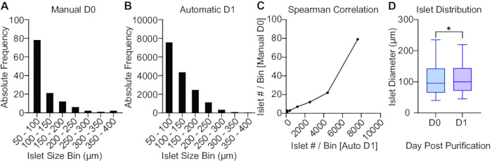
Figure 4: Quantitative assessment of islet size distribution, correlation, and early shedding. Graphical summary of islet measurements obtained through manual and automated analyses. (A) Day 0 (D0) islet size distribution based on manual quantification across standardized islet-equivalent IEQ size bins. (B) Day 1 (D1) islet size distribution determined by automated image-based measurements across the same standardized IEQ size bins. (C) Correlation of counts between manual D0 and automated D1 quantification across IEQ size bins. (D) Box plot of the distribution of islet diameters between D0 and D1 for automated measurements of the same plate. Please click here to view a larger version of this figure.
Purity (endocrine vs. exocrine content)
Dithizone (DTZ) staining
On D1, an aliquot of islets (n = 451) was stained with DTZ for 30 s at room temperature and immediately imaged using brightfield microscopy (4× objective; 1/20 s exposure). In Fiji, background pixels were removed such that the remaining pixel area corresponded to the total islet area. DTZ-negative exocrine tissue regions, identified by darker color contrast, were manually selected using a lasso tool, and the area of each region of interest was automatically recorded in pixels. Endocrine purity was calculated as the percentage of DTZ-positive pixels relative to total islet pixels, yielding a purity of 98.56 ± 0.38% (Figure 5A).
Viability and functional performance
Glucose-stimulated insulin secretion (GSIS)
At each time point (D1, D3, D7), 15 size-matched islets were hand-picked and placed into individual wells of a 24-well plate, prepared in quadruplicate. The islets were incubated at 37 °C 5% CO2 in Krebs buffer under a low-high-low glucose stimulation protocol: first in low glucose (2.8 mM, 60 min), followed by high glucose (16.7 mM, 60 min, see Table 1). Effluent fractions were collected at the end of each incubation period, and insulin concentrations were quantified using a rat insulin ELISA assay. The stimulation indices were calculated as the ratio of insulin secretion during high to low glucose conditions. Stimulation indices were 9.37, 8.34, and 6.7, at days 1, 3, and 7, respectively, with no significant differences (Figure 5B).
Live/Dead staining
At D1, D3, and D7, 15 islets were hand-picked and stained with the Live/Dead viability kit according to the manufacturer's instructions. Briefly, islets were incubated in the Live/Dead staining solution for 30 min at 25 °C, then imaged under consistent microscope settings to ensure comparability across time points (4X objective; 1/30 s exposure, Texas Red and GFP filters). Viability was quantified using a Python script employing scikit-image for islet segmentation and fluorescence measurement. Islets were identified by combining the green and red fluorescence channels, applying Li's thresholding method to generate a binary mask, and using watershed segmentation to separate touching islets. Objects were filtered by area to exclude debris and artifacts. For each islet, viability was calculated as the percentage of live integrated density (green pixels) relative to total integrated density (green plus red pixels).The results showed viability of 68 ± 9% at D1, 98.2 ± 0.6% at D3, and 100% at D7. A positive control sample treated with 70% ethanol for 10 min exhibited 2.7 ± 7.2% viability (see Figure 5C-F).
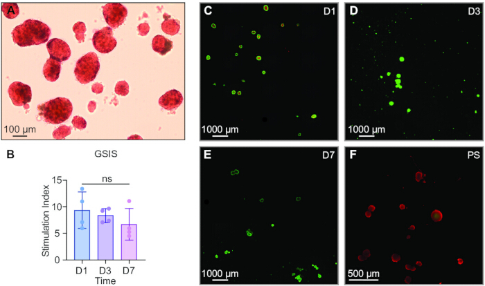
Figure 5: Assessment of islet purity, viability, and functional responsiveness. Representative images and quantitative summaries of isolated islet characteristics over time. (A) Representative 10× brightfield image of islets stained with dithizone (DTZ), showing red-stained endocrine islets and unstained exocrine tissue. (B) Glucose-stimulated insulin secretion (GSIS) values of high-glucose/low-glucose (HG/LG) stimulation index for Days 1, 3, and 7. (C-E) Live/dead fluorescence images of islets at Day 1 (C), Day 3 (D), and Day 7 (E). (F) Live/dead fluorescence image of positive-control islets treated with ethanol. Please click here to view a larger version of this figure.
| (A) Washing Media | ||
| Component | Final concentration | Amount (mL) |
| RPMI 1640 | - | 500 mL |
| Fetal Bovine Serum | 10% | 50 mL |
| Pen-Strep (10,000 U/mL) | 1% | 5.5 mL |
| HEPES | 25 mM | 12.5 mL from 1M stock |
| L-glutamine | 2 mM | 5 mL from 200 mM stock |
| (B) Islet Culture Media | ||
| Component | Final concentration | Amount (mL or g) |
| RPMI 1640, No glucose | - | 500 mL |
| Fetal Bovine Serum | 10% | 50 mL |
| Pen-Strep (10,000 U/mL) | 1% | 5.5 mL |
| HEPES | 20 mM | 11 mL from 1M stock |
| Sodium Pyruvate | 1 mM | 5.5 mL from 100 mM stock |
| Glucose | 5.5 mM | 2.75 mL from 200 g/L stock |
| (C) Penicillin-Streptomycin Rinse Solution | ||
| Component | Final concentration | Amount (mL or g) |
| Saline Solution, 0.9% | - | 500 mL |
| Penicillin‚ Streptomycin (10,000 U/mL) | 2% | 10 mL |
| (D) Quenching Buffer | ||
| Component | Final concentration | Amount (mL or g) |
| HBSS | - | 500 mL |
| Fetal Bovine Serum | 20% | 100 mL |
| HEPES (1 M) | 2.50% | 2.5 mL |
| (E) Krebs Buffer | ||
| Component | Final concentration | Amount (mL or g) |
| dH2O | - | 975 mL |
| NaCl | 115 mM | 6.72 g |
| NaHCO3 | 24 mM | 2.02 g |
| KCl | 5 mM | 0.3728 g |
| HEPES (1 M) | 25 mM | 25 mL |
| MgCl2 | 1 mM | 0.0952 g |
| BSA | 0.10% | 1 g |
| CaCl2 | 2.5 mM | 0.2775 g |
Table 1: Solution list. Composition and preparation details for all solutions used throughout the islet isolation and purification protocol. Each subsection lists components, final concentrations, preparation volumes, and catalog numbers. (A) Washing Media, used for rinsing and washing tissue during digestion and purification. (B) Islet Culture Media, used for post-isolation islet maintenance and viability. (C) Penicillin-Streptomycin Rinse Solution, used to sterilize and rinse harvested pancreases. (D) Quenching Buffer, used to terminate enzymatic digestion and preserve cell viability. (E) Krebs Buffer, used during glucose-stimulated insulin secretion (GSIS) assays.
| Number of Pancreases | Collagenase (15.38 mg/mL) | Thermolysin (1 mg/mL) | DNAse (100 mg/mL) | Working buffer | 600 mL Beakers to use | Volume/Beaker |
| 30 | 4.5 mL | 2.25 mL | 450 µL | 450 mL | 5 | 25 mL |
| 24 | 3.6 mL | 1.8 mL | 360 µL | 360 mL | 4 | 25 mL |
| 20 | 3 mL | 1.5 mL | 300 µL | 300 mL | 3 | 25 mL |
| 16 | 2.4 mL | 1.2 mL | 240 µL | 240 mL | 2 | 25 mL |
| 12 | 1.8 mL | 0.9 mL | 180 µL | 180 mL | 2 | 25 mL |
| 10 | 1.5 mL | 0.75 mL | 150 µL | 150 mL | 2 | 25 mL |
| Final conc. | 0.15 mg/mL | 5 mg/mL | 0.1 mg/mL | |||
Table 2: Digestive enzyme solution by pancreas quantity. Volumes of collagenase, thermolysin, and DNase I required to prepare the Working Dissociation Medium (WDM) for varying numbers of rat pancreases. Final concentrations of each enzyme are listed at the bottom of the table.
| Number of Pancreases | Number 50 mL tubes | Volume/ syringe | Number of 1.096 syringes | Number of 1.068 syringes |
| 30 | 10 tubes | 50 mL | 2 | 2 |
| 24 | 8 tubes | 40 mL | 2 | 2 |
| 20 | 6 tubes | 30 mL | 2 | 2 |
| 16 | 4 tubes | 40 mL | 1 | 1 |
| 12 | 4 tubes | 40 mL | 1 | 1 |
| 10 | 2 tubes | 20 mL | 1 | 1 |
Table 3: Gradient loading by pancreas quantity. Number of 50 mL conical tubes and corresponding gradient volumes required for islet purification based on the number of pancreases processed.
| Islet Diameter Range (µm) | Islet Particle Number (AI) | IEQ Conversion Factor | IEQ per Range |
| 50-100 | a | * 0.167 | a * 0.167 |
| 101-150 | b | * 0.648 | b * 0.648 |
| 151-200 | c | * 1.685 | c * 1.685 |
| 201-250 | d | * 3.500 | d * 3.500 |
| 251-300 | e | * 6.315 | e * 6.315 |
| 301-350 | f | * 10.352 | f * 10.352 |
| > 350 | g | * 15.833 | g * 15.833 |
Table 4: Islet equivalence conversion. Conversion of islet diameter ranges to standardized islet equivalents (IEQ). The table lists the observed islet diameter bins, the number of islet particles per range, the corresponding IEQ conversion factor for each size range, and the resulting IEQ contribution per range.
Discussion
This protocol outlines a high-throughput approach for isolating and purifying pancreatic islets from Lewis rats. The success of the method depends on several critical steps that collectively preserve islet integrity and function. Among these are the precise and complete intraductal perfusion of the pancreas, the careful cleaning of the perfused pancreas, and the exact enzymatic digestion time. Consistent whole organ perfusion is crucial to achieve uniform distension of the pancreas and ensure even distribution of the collagenase-thermolysin-DNase blend, which acts respectively to digest the extracellular matrix, enhance enzyme penetration, and reduce DNA-mediated viscosity21,22. Under-perfusion can result in incomplete tissue digestion and loss of a significant portion of islets, as the under-digested tissue is filtered out with the sieve. Similarly, inadequate removal of adipose or vascular tissue before digestion can introduce non-endocrine impurities that lower purity. Finally, prolonged digestion results in islet desegregation from excessive extracellular matrix breakage, while suboptimal digestion yields undigested tissue resistant to mechanical digestion via spinal needle. These steps represent the most frequent technical pitfalls and should be carefully monitored during training and optimization.
While this method was designed specifically for Lewis rats, it likely can be adapted to other strains or species through minor re-optimization of digestion time and enzyme ratio23,24,25,26. Some laboratories may choose to adjust the ratio of collagenase, thermolysin, and DNase I in the Working Dissociation Medium (WDM) to optimize yield for their specific enzyme lot or tissue characteristics. To do so, laboratories may carefully follow the manufacturer's step-by-step guide for enzyme and tissue optimization and, in all instances, avoid adding components such as 0.1 M EDTA, cysteine, mercaptoethanol, protease inhibitors, serum, or albumin, as these may inhibit collagenase, thermolysin, and DNase 1 enzyme activity27. Although no definitive working time for the enzyme blend before dissociation has been published, prior data show that extended cold preservation of pancreases adversely affects islet yield and function28. Empirically, we have found that, in our protocol, successful isolations were consistently achieved when the total cold ischemia time, defined as the interval between placing the first pancreas on ice for cleaning and transferring it to the 37 °C water bath, was kept under 1.5 h. Further optimization of this parameter may be warranted depending on the end-user application.
Despite its robustness, this protocol has several practical and technical limitations. First, the enzyme blend (collagenase, thermolysin, and DNase I) and density gradients are essential for efficient tissue dissociation and islet isolation, yet impose significant cost. Our selection of enzyme combination was informed by the structural characteristics of the pancreatic extracellular matrix and the need to balance effective tissue dissociation with the preservation of islet integrity. Each enzymatic component facilitates a distinct role: collagenase targets fibrillar and basement-membrane collagens to liberate islets; thermolysin, a neutral protease, cleaves non-collagenous matrix proteins and cell-cell adhesion molecules, reducing the required collagenase exposure; and DNase I minimizes viscosity caused by DNA released from lysed cells29,30,31. Although lower-cost formulations of digestive enzymes are commercially available, their purity is variable and may compromise enzymatic performance13,23. Given that collagenase is isolated from bacterial cultures, additional purification steps reduce endotoxin contamination but substantially increase cost13,23.
For collagenase, we recommend preparations with an activity of approximately 2,000 to 4,000 Wünsch units per bottle, which supports efficient dissociation at moderate dosing while avoiding prolonged digestion. Notably, the blend should include a balanced ratio of collagenase class I to class II, corresponding to a collagenase II/total collagenase fraction of approximately 0.3-0.5. This ratio provides strong collagenolytic activity while limiting excessive degradation of non-collagenous extracellular matrix. Additionally, the collagenase mixture should be complemented by a moderate level of neutral protease activity (roughly 150,000-250,000 units) and DNase, for reasons already commented.
From a quality control standpoint, each lot should include documentation of low endotoxin burden (≤50 EU/mg protein), high enzyme purity (≥85% by HPLC), and verified lot-to-lot activity, as enzyme purity and composition can vary considerably by manufacturer. Finally, variations in lot composition may affect digestion consistency and islet yield, necessitating the performance of lot-specific internal validation. Such validation can be achieved by following the standardized digestion conditions described in this protocol and subsequently assessing digestion efficiency and islet morphology. Indicators of suboptimal enzyme performance include incomplete tissue dissociation, islets with irregular or rough borders, or the need for excessive mechanical shear to achieve separation, in which case titration of the digestion time may be necessary.
Similar considerations apply to density gradients. We selected a polysucrose-based gradient, a non-ionic, highly hydrophilic sucrose-epichlorohydrin copolymer that forms stable gradients up to 1.2 g/mL and has a long track record in islet isolation32,33. Iodixanol is a well-validated alternative, and studies in human islets show that iodixanol-based gradients can improve islet recovery while maintaining viability and function34,35,36,37. However, rodent data are inconclusive, with some studies reporting superior outcomes using iodixanol and others demonstrating better viability and function with polysucrose-based gradients32,35. Critically, the use of commercially manufactured, quality-controlled gradient solutions is recommended to enhance reproducibility across laboratories. Polysucrose-based gradients are widely available at optimal densities for islet isolation, whereas iodixanol gradients are typically prepared by in-house dilution, introducing additional variability. A second limitation is that surgical harvesting requires a high level of technical proficiency, particularly for intraductal perfusion through the Sphincter of Oddi and rapid pancreas excision. Even minor delays or misalignment can result in tissue damage, leakage, uneven perfusion, or prolonged warm ischemia, which may compromise islet yield and viability. To mitigate this limitation, the protocol includes detailed procedural descriptions and original graphical aids with color-coded anatomical landmarks to facilitate understanding. New operators are encouraged to become comfortable with the perfusion technique prior to performing full isolations, and practice on carcasses is advised before attempting live harvests.
Third, for maximum efficiency, this protocol requires coordinated teamwork. During pancreas harvesting and enzymatic digestion, three trained operators are typically required: one performing sterile surgery, one cleaning the pancreas under sterile conditions, and one circulating and euthanizing animals under non-sterile conditions. Following digestion, downstream purification steps can generally be performed by fewer operators once proficiency is achieved. While this coordination represents an upfront logistical demand, it enables the isolation of many islets from multiple animals within a relatively short timeframe, improving overall throughput compared to less coordinated workflows. While this protocol is technically demanding and resource-intensive, these limitations are common to all islet isolation methods. By emphasizing standardized digestion conditions, structured training, and coordinated execution, this high-throughput approach can reduce time and labor per isolation and yield consistent, reproducible results when performed by a trained team.
Compared with conventional rodent islet isolation protocols, this method offers substantial advantages in throughput, reproducibility, and consistency. Traditional single-organ digestions rely heavily on manual hand-picking and often yield variable purity13. In contrast, our protocol utilizes pooled digestion and density-gradient purification, substantially reducing preparation time and inter-sample variability while maintaining high islet viability and functional responsiveness. The isolated islets were comprehensively characterized for morphology (dithizone staining), viability (live/dead assay), and function (glucose-stimulated insulin secretion), confirming the robustness of the method. Collectively, this standardized workflow enables the generation of plentiful, clean, and functional islet preparations suitable for downstream applications, including encapsulation, transplantation, and microphysiological modeling38,39,40. Importantly, Lewis rat islets isolated with this technique have reversed diabetes in preclinical models, underscoring the translational relevance of this reproducible and scalable approach41.
Disclosures
A.G. is a co-founder and chief scientific advisor of Continuity Biosciences, LLC. A.G., J.P-M., and C.Y.X.C. are inventors of intellectual properties licensed by continuity Biosciences, LLC. All other authors declare no competing interests.
Acknowledgements
This study was supported by the National Institutes of Health (NIH) R01DK133610-01 (A.G., N.K.) and NIH NIDDK R01DK132104 (A.G.), Breakthrough T1D SRA-2021-1078-S-B (AG) and SRA-2022-1224-S-B (A.G., N.K.), and the Vivian L. Smith Foundation.
Materials
| Name | Company | Catalog Number | Comments |
|---|---|---|---|
| 21 G x 2 in. BD PrecisionGlide Needle | BD | 305129 | |
| 500 µm Brass Frame and 304 Stainless Steel Screen, 3" Diameter x 1" Deep (Sieve number 35) | McMaster-Carr | 34735K525 | |
| Air-Tite Sterile Syringes, Luer lock | Fisher Scientific | 14-817-178 | |
| Biogel PI UltraTouch M | Mölnlycke | 42665 | |
| Bonn Scissors; Straight; Sharp Points; 15 mm Blade Length; 3 1/2" Overall Length | Roboz | RS-5840 | |
| Braintree Scientific GERMINATOR 500 DRY STERIL | Fisher Scientific | NC9956482 | |
| Cell-IQ Series 5.8 cu.ft. CO2 Laboratory Incubator | PHCBI | MCO-170AICUVHL-PA | |
| Corning 100 mm Not TC-treated Culture Dish | Corning | 430591 | |
| Corning Fetal Bovine Serum, 500 mL, Regular, USDA Safety Tested (Heat Inactivated) | Corning | 35011CV | |
| Corning Islet Gradient 1.069 (density 1.069 g/cm3) | Corning | 99-815-CIS | Requires special request from company |
| Corning Islet Gradient 1.096 (density 1.096 g/cm3) | Corning | 99-691-CIS | Requires special request from company |
| Corning Islet Gradient 1.108 (density 1.108 g/cm3) | Corning | 99-692-CIS | Requires special request from company |
| Covidien Tendersorb Underpads | Fisher Scientific | 22-225-102 | |
| DNase I Grade II | Roche | 10104159001 | |
| Falcon 50 mL High Clarity Conical Centrifuge Tubes | Fisher Scientific | 14-432-22 | |
| Fisherbrand Aluminum Foil, Standard-Gauge Roll | Fisher Scientific | 01-213-100 | |
| Fisherbrand Nonwoven Gauze Sponges | Fisher Scientific | 22-028-559 | |
| Fisherbrand Reusable Glass Heavy-Duty Low-Form Beakers | Fisher Scientific | FB101600 | |
| Fluorescence Mircoscope BZ-X810 | Keyence | BZ-X810 | |
| Glucose Solution | Gibco | A2494001 | |
| Halsey Micro Needle Holder | Fine Science Tools | 12500-12 | |
| HBSS, calcium, magnesium, no phenol red | Gibco | 14025092 | |
| HEPES (1 M) | Gibco | 15630080 | |
| Hemostatic Forceps Classic Crile 5-1/2 Inch Length | McKesson | 1231217 | |
| Hudson (Ewald) Forceps | Roboz | RS-5238 | |
| Hypodermic Needle Monoject 1-1/2 Inch Length 16 Gauge Regular Wall Without Safety | McKesson | 43481 | |
| L-Glutamine (200 mM) | Gibco | 25030081 | |
| Liberase T-Flex, Research Grade | Roche | 5989132001 | |
| LIVE/DEAD Viability/Cytotoxicity Kit, for mammalian cells | Invitrogen | L3224 | |
| MATLAB software package | MathWorks | https://www.mathworks.com/pricing-licensing.html?prodcode=ML | |
| NE-4000 Two Channel Programmable Syringe Pump | SyringePump | NE-4000-US | |
| NEEDLE, HYPO STR DISP 27GX1/2"(100/BX) EXLINT | McKesson | 408877 | |
| Nunc Cell Culture/Petri Dishes | Thermo-Scientific | 174926 | |
| Olympus CKX41 Inverted Phase Contrast Microscope | Olympus | CKX41-B | |
| PBS, pH 7.4 | Gibco | 10010023 | |
| Penicillin‚ Streptomycin (10,000 U/mL) | Gibco | 15140122 | |
| RPMI 1640 | Gibco | 11875093 | |
| RPMI 1640, no glucose | Gibco | 11879020 | |
| Sodium Pyruvate (100 mM) | Gibco | 11360070 | |
| Sodium Pyruvate (100 mM stock) | Gibco | 11360070 | |
| Sorval ST 16R | Thermo-Scientific | 75004381 | |
| Stainless steel 304 syringe needle | Milipore Sigma | Z117013-1EA | |
| SURGICAL DRAPE, DISPOSABLE, 36 IN X 100 YD | Covetrus | 10408 | |
| Surgical Scissors - ToughCut | Fine Science Tools | 14054-13 | |
| Thumb Dressing Forceps 4.5" Serrated Delicate 1.3 mm Tip Width | Roboz | RS-8120 | |
| Thermo Scientific Nunc 200mL Centrifuge Tube | Thermo-Scientific | 14-962-17 | |
| Thermo Scientific Nunc Centrifuge Bottle Adapters and Cushions | Thermo-Scientific | 12-565-283B | |
| TSDUB15 15L Dubnoff Shaking Water Bath | Fisher Scientific | 300610298 | |
| TX-400 4 x 400 mL Swinging Bucket Rotor | Thermo-Scientific | 75003181 | |
| Tygon S-50-HL | Tygon | AAXoooo3 | |
| Ultra Sensitive Rat Insulin ELISA kit | Crystal Chem | 90060 |
References
- Grattoni, A., et al. Harnessing cellular therapeutics for type 1 diabetes mellitus: progress, challenges, and the road ahead. Nat Rev Endocrinol. 21 (1), 14-30 (2025).
- Páez-Mayorga, J., et al. Emerging strategies for β cell transplantation to treat type 1 diabetes mellitus. Cell Transplant. 31, 963689721004(2022).
- Cabrera, O., et al. The unique cytoarchitecture of human pancreatic islets and its relevance to β-cell function. Proc Natl Acad Sci U S A. 103 (7), 2334-2339 (2006).
- Brissova, M., et al. Pancreatic islet microvasculature and blood flow. J Clin Invest. 124 (4), 1871-1883 (2014).
- Jiang, F. X., Morahan, G. Pancreatic islet architecture and function. Cell Death Discov. 10, 170(2024).
- Sato, Y., et al. Hypoxia induces β-cell apoptosis and functional suppression in isolated islets. PLoS One. 9 (12), e114868(2014).
- Tiedge, M., Lortz, S., Drinkgern, J., Lenzen, S. Relation between antioxidant enzyme gene expression and antioxidative defense status of insulin-producing cells. Diabetes. 46 (11), 1733-1742 (1997).
- Ichii, H., et al. Shipment of human islets for transplantation. Am J Transplant. 7 (4), 1010-1020 (2007).
- Carobbio, S., et al. Rapid GSIS kinetics in freshly isolated mouse islets. Diabetologia. 47 (9), 1652-1662 (2004).
- Zawalich, W. S., Zawalich, K. C., Kelley, G. G. Immediate perifusion of fresh rat and mouse islets. Am J Physiol Endocrinol Metab. 294 (2), E444-E450 (2008).
- Wassmer, C. H., et al. Clinical use of freshly isolated islets transplanted within 48 h. Transplant Proc. 53 (6), 1907-1912 (2021).
- Tai, D. S., et al. Immediate autotransplantation of islets following total pancreatectomy. JAMA Surg. 150 (5), 472-479 (2015).
- Carter, J. D., Dula, S. B., Corbin, K. L., Wu, R., Nunemaker, C. S. A practical guide to rodent islet isolation and assessment. Biol Proced Online. 11, 3-31 (2009).
- Emamaullee, J. A., et al. Lewis rats in transplantation studies. Diabetes. 65 (5), 1350-1359 (2016).
- de Groot, M., et al. Rat islet isolation yield and function are donor strain dependent. Lab Anim. 38 (2), 200-206 (2004).
- Moazenchi, A. M., et al. Strain variability in islet yield and isolation. Islets. 2 (5), 283-289 (2010).
- Dolenšek, J., Slak Rupnik, M., Stožer, A. Isolation variability limits reproducibility in rat islet studies. PLoS One. 14 (5), e0216136(2019).
- Stehno-Bittel, L. The flaws and future of islet volume measurements. Cell Transplant. 27 (4), 571-578 (2018).
- Kissler, H. J., et al. Validation of methodologies for quantifying isolated human islets. Cell Transplant. 18 (6), 611-620 (2009).
- Papas, K. K., et al. Islet assessment for transplantation. Cell Transplant. 18 (6), 519-528 (2009).
- Dono, K., et al. Low temperature collagenase digestion for islet isolation from 48-hour cold-preserved rat pancreas. Transplantation. 57 (1), 22-26 (1994).
- Shapiro, A. M. J., et al. High yield of rodent islets with intraductal collagenase distension and warm incubation. Cell Transplant. 5 (6), 631-638 (1996).
- Corbin, K. L., Hughes, J. W., Nunemaker, C. S. A practical guide to rodent islet isolation and assessment revisited. Biol Proced Online. 23, 10(2021).
- de Haan, B. J., et al. Factors influencing isolation of functional pancreatic rat islets. Pancreas. 29 (1), e15-e22 (2004).
- Kenyon, N. S., et al. Long-term survival and function of intrahepatic islet allografts in baboons treated with humanized anti-CD154. Diabetes. 48 (7), 1473-1481 (1999).
- Kenyon, N. S., et al. Long-term survival and function of intrahepatic islet allografts in rhesus monkeys treated with humanized anti-CD154. Proc Natl Acad Sci U S A. 96 (14), 8132-8137 (1999).
- Liberase T-Flex Research Grade - Method Sheet. eLabDoc. , Roche Diagnostics GmbH. (2025).
- Miyazaki, Y., et al. Strategy towards tailored donor tissue-specific pancreatic islet isolation. PLoS One. 14 (5), e0216136(2019).
- Cross, S. E., et al. Key matrix proteins within the pancreatic islet basement membrane are differentially digested during human islet isolation. Am J Transplant. 17 (2), 451-461 (2017).
- O'Gorman, D., Kin, T., Pawlick, R., Imes, S., Senior, P. A., Shapiro, A. J. Clinical islet isolation outcomes with a highly purified neutral protease for pancreas dissociation. Islets. 5 (3), 111-115 (2013).
- Wang, W., et al. Adjustment of digestion enzyme composition improves islet isolation outcome from marginal grade human donor pancreata. Cell Tissue Bank. 8, 187-194 (2007).
- McCall, M. D., Maciver, A. H., Pawlick, R., Edgar, R., Shapiro, A. M. J. Histopaque provides optimal mouse islet purification kinetics: comparison study with Ficoll, iodixanol and dextran. Islets. 3 (4), 144-149 (2011).
- Ricordi, C., Lacy, P. E., Finke, E. H., Olack, B. J., Scharp, D. W. Automated method for isolation of human pancreatic islets. Diabetes. 37 (4), 413-420 (1988).
- BenchChem Technical Support Team. An in-depth technical guide to cell separation using iodixanol. BenchChem. , (2026).
- Dellê, H., Saito, M. H., Yoshimoto, P. M., Noronha, I. L. The use of iodixanol for the purification of rat pancreatic islets. Transplant Proc. 39 (2), 467-469 (2007).
- Noguchi, H., et al. Iodixanol-controlled density gradient during islet purification improves recovery rate in human islet isolation. Transplantation. 87 (11), 1629-1635 (2009).
- Min, T., et al. Superiority of Visipaque (iodixanol)-controlled density gradient over Ficoll-400 in adult porcine islet purification. Transplant Proc. 42 (5), 1825-1829 (2010).
- Paez-Mayorga, J., et al. Neovascularized implantable cell homing encapsulation platform with tunable local immunosuppressant delivery for allogeneic cell transplantation. Biomaterials. 257, 120232(2020).
- Paez-Mayorga, J., et al. Implantable niche with local immunosuppression for islet allotransplantation achieves type 1 diabetes reversal in rats. Nat Commun. 13, 7951(2022).
- Piemonti, L., et al. Islet transplantation. Endotext. , (2022).
- Patel, S. N., et al. The foundation for engineering a pancreatic islet niche. Front Endocrinol. 13, 881525(2022).
Reprints and Permissions
