This protocol presents a detailed method for isolating high-quality single-cell suspensions from both healthy and tumor-bearing murine lung tissue. This protocol can be applied to scRNA-seq, flow cytometry analysis, and primary cell isolation.
Method Article
Optimized Preparation of Whole Murine Tumor-Bearing Lung Tissue for Flow Cytometry and Single-Cell RNA-Sequencing
In This Article
Summary
Abstract
NSCLC accounts for approximately 85% of lung cancers, and Kirsten rat sarcoma (KRAS) is the most commonly mutated gene in NSCLC. Although management of NSCLC has advanced markedly in recent years, its heterogeneity and plasticity remain incompletely understood. Single-cell RNA sequencing (scRNA-seq) has become an essential tool for dissecting cellular heterogeneity in complex tissues such as the lung. However, generating high-quality single-cell suspensions from tumor-bearing lung tissue is challenging, particularly during tumor formation when the tissue becomes fibrotic and heterogeneous. A key requirement for successful scRNA-seq is the preparation of viable single cells with minimal processing-induced stress or damage. Here, a detailed step-by-step protocol is presented for isolating high-quality single-cell suspensions from both healthy and tumor-bearing mouse lung tissue in a KrasG12D; tdTomato reporter mouse model. The procedure includes tissue perfusion, controlled enzymatic digestion, gentle mechanical dissociation, red blood cell lysis, filtration, and viability assessment, and achieves >85% viable cells prior to scRNA-seq. Finally, the tdTomato+ tumor cells are efficiently isolated by FACS and can be detected as distinct clusters by scRNA-seq using this protocol.
Introduction
Lung cancer is one of the leading causes of cancer-related deaths worldwide. It can be broadly classified into two major subtypes: small-cell lung cancer (SCLC) and non-small-cell lung cancer (NSCLC)1. NSCLC accounts for approximately 85% of all lung cancer cases, with adenocarcinoma being the most common histological subtype2. Although therapeutic strategies for NSCLC have improved substantially over the past decades, the mortality rate of lung cancer remains high3. Chemotherapy and immunotherapy are the two main treatment modalities used in clinical practice; however, many patients experience drug resistance or fail to respond to immunotherapy4,5.
The tumor microenvironment (TME) plays a pivotal role in tumor initiation, progression, and therapeutic resistance6. Increasing evidence suggests that the composition of the TME changes dynamically over time7,8. Anti-tumor immune cells are typically enriched in the TME during the early stages of tumorigenesis, whereas immunosuppressive and cancer-associated cell populations become predominant as the tumor develops9. Therefore, understanding the heterogeneity and plasticity of the TME during lung cancer progression is essential for elucidating disease mechanisms and improving therapeutic outcomes.
Single-cell RNA sequencing (scRNA-seq) enables high-resolution profiling of gene expression at the individual cell level, allowing comprehensive characterization of complex tissue transcriptomes10. This technique has become indispensable for studying cellular heterogeneity and identifying mechanisms underlying tumor progression and drug resistance in lung cancer11. The preparation of a high-quality single-cell suspension is a critical component of successful scRNA-seq, as cell stress or damage during dissociation can lead to inaccurate results12,13.
This protocol enables sequencing RNA from all single cells purified from whole tumor-bearing lungs, agnostic of cell size and without discarding any live cell subpopulations. Many previous studies have reported single-cell sequencing of tumor-bearing lungs that either focused on tumor cells and discarded CD45+ cells14,15,16 or focused on immune cells or other specific cell types17 and discarded tumor cells. This protocol aims to efficiently dissociate all cell types from whole murine tumor-bearing lung tissue, while ensuring the release of malignant cells and preserving the viability of normal cells. By including all live single cells, this protocol enables the study of all relevant cell types within the tumor-bearing lung microenvironment, including cancer associated macrophage like cells (CAMLs)18. Optional fluorescence-activated cell sorting (FACS) steps are included for quality control and enrichment of specific cell populations. Additional guidance is provided for antibody staining, 4’,6-diamidino-2-phenylindole (DAPI) labeling, and short-term fixation procedures when immediate sorting is not feasible.
A potential limitation of this protocol is that it has been optimized and validated for lung tissues containing early-stage tumors and with a tumor burden below 40%. For lungs containing higher-grade tumors or in which the tumor burden exceeds 50%, the dissociation efficiency may be compromised by the presence of dense stromal components and extensive fibrosis19,20, which could block the complete release of malignant cells.
Protocol
All animal-related experiments were carried out in accordance with Scripps Research guidelines for the care and use of animals and approved by the Scripps Research Institutional Animal Care and Use Committee under protocol #10-0019. The KrasLSL-G12D/+; TdTomato+/- mice were generated by crossing KrasLSL-G12D/+ mice with TdTomato reporter mice. Nine or ten-week-old KrasLSL-G12D/+; TdTomato+/- mice were used as lung cancer models. They are infected intratracheally with lentivirus expressing Cre recombinase, at a viral titre of 5 × 10⁵ plaque-forming units per mouse. Tumor-bearing lung tissues were collected after 25 weeks. Potential digestion problems and troubleshooting are listed in Supplementary Table 1. The reagents and the equipment used are listed in the Table of Materials.
1. Preparation of Buffers
- DMEM/F12 medium: Prepare 2% (v/v) heat-inactivated FBS in DMEM/F12 medium.
- Cell wash buffer: Prepare 2% (v/v) heat-inactivated FBS in Ca+/Mg+ free PBS (pH 7.4).
- Perfusion buffer: Pre-chilled Ca+/Mg+ free PBS (pH 7.4).
- Enzyme stock solutions: Dissolve 10 mg (1600 U/mg) of collagenase IV in 1 mL DMEM/F12 medium to obtain a 16000 U/mL collagenase IV stock solution; Dissolve 42 mg (1.19 U/mg) of Dispase II in 1 mL DMEM/F12 medium to obtain a 50 U/mL Dispase II stock solution; Dissolve 5 mg of DNase I in 1 mL DMEM/F12 medium to obtain a 5 mg/mL DNase I stock solution.
- Digestion medium: Mix 250 µL of collagenase IV, 200 µL of Dispase II, and 100 µL of DNase I stock solutions in 5 mL DMEM/F12 medium to obtain a final concentration of 800 U/mL collagenase IV, 2 U/mL Dispase II, and 0.1 mg/mL DNase I.
- Resuspension buffer: Mix 1 µL of 0.5 M EDTA (10 µM), 1 mL heat-inactivated FBS (2% v/v) in 50 mL Ca+/Mg+ free PBS (pH 7.4).
- BSA stock solution: Dissolve 0.5 g of BSA in 100 mL Ca+/Mg+ free PBS to obtain a 0.5% (w/v) BSA stock solution.
- FACS buffer: Mix 2.5 mL of 1 M HEPES, 200 µL of 0.5 M EDTA in 100 mL BSA stock solution to obtain a final concentration of 25 mM HEPES and 1 mM EDTA.
2. Collection of murine lung tissue
NOTE: All surgical instruments are disinfected with 70% ethanol between samples. Samples are maintained on ice between each step, and all plasticware is certified DNase/RNase-free.
- Euthanize mouse in a CO2 chamber (following institutionally approved protocols). Place the mouse on its back on a dissection pad and pin the feet. Spray liberally with 70% ethanol.
- Make a 2–3 cm wide midline incision through the skin and muscles of the lower abdomen. Carefully open the thorax and avoid cutting the lungs or the heart.
- Attach a safety multifly 21 G needle to a 50 mL syringe and aspirate 30 mL perfusion buffer.
- Insert the needle in the left ventricle and cut a small incision in the right atrium using the sharp, straight scissor.
- Perfuse whole body21 with 15 mL perfusion buffer.
NOTE: The whitening of the liver indicates that whole-body perfusion has been completed. - Remove the lungs from the thorax and cut away excess tissue attached to the lungs22. Place the lungs in 10 mL pre-cooled Ca+/Mg+ free PBS to wash away excess blood.
- Transfer the lungs into a 1.5 mL tube containing 500 µL of ice-cold DMEM/F12.
3. Preparation of the lung single-cell suspension
- Cut the lung tissue into small pieces (1–2 mm3) with an extra-narrow straight scissor for 2–3 min.
- Transfer the minced lung tissue to a 6 cm dish containing 5 mL digestion medium.
- Place a 6 cm dish on a thermostatic shaker at 50 rpm for 25 min at 37 °C. Mix the suspension using a 1 mL pipette every 8 min.
- Place a 100 µm mesh strainer in a 10 cm dish. Pre-wet the strainer with 1 mL of DMEM/F12 medium.
- Transfer the digested suspension into the strainer. Push the tissue through the strainer with the rubber end of a sterile syringe plunger.
- Place a 70 µm mesh strainer over a 50 mL centrifuge tube. Pre-wet the strainer with 1 mL of DMEM/F12 medium.
- Filter the cell suspension through a strainer and rinse the strainer with 5 mL DMEM/F12 medium.
- Centrifuge filtered cell suspension at 400 g for 10 min at 4 °C and remove the supernatant carefully. Resuspend the cell pellet in 1 mL Red Blood Cell Lysing Buffer for 2 min at room temperature.
- Add 14 mL cell wash buffer and centrifuge at 400 g for 10 min at 4 °C.
NOTE: If the pellet is still red after washing, repeat steps 8 and 9. - Discard the supernatant and resuspend the cells in 20 mL resuspension buffer.
- Mix 10 µL of single-cell suspension with an equal volume of trypan blue and add 10 µL of the mixture to the cell counting chamber slide. Check live cell viability, cell number, and single-cell status using a cell counter.
4. Flow cytometry analysis
- Resuspend the cells in ice-cold FACS buffer and transfer 100 µL of cell suspension into 1.5 mL tubes containing 1 µL Fc block antibody (1:100). Incubate samples for 15 min on ice.
- Centrifuge samples at 500 g for 5 min and discard the supernatant. Resuspend the cells in 200 µL of CD326-APC antibody (1:100) and incubate for 45 min in the dark at 4 °C.
- Centrifuge samples at 500 g for 5 min and discard the supernatant. Resuspend cells in 500 µL of FACS buffer containing DAPI stain (1:5000).
- Transfer all samples to FACS tubes and protect them from light.
- Analyze the samples using the flow cytometer. The following fluorophores were used: DAPI-V450, Tdtomato-YG585, CD326-APC. The gating strategy is shown in Figure 1.
Results
High cell viability was achieved in the single-cell suspension prepared using this optimized protocol. Changes that improved cell viability for tumor-bearing lungs included optimizing the concentration of enzymes (Supplementary Table 2), shaking, and mixing the samples during incubation at 37 °C. Furthermore, the use of an additional 40 µm mesh strainer in step 6 was eliminated to reduce mechanical shear stress that increases the likelihood of cell damage; including the additional strainer may reduce multiplet formation. On average, cell viability exceeded 85%, compared to less than 80% before optimization (Table 1). Variations in cell concentration were primarily attributed to differences in lung size and the suspension medium used across replicates.
In this KrasG12D-driven murine model of lung adenocarcinoma, tumor nodules are present, and tumor burdens range between 10% and 40% twenty-five weeks after tumor initiation23. Two whole-lung single-cell suspensions were subjected to scRNA-seq on the 10x Genomics platform. Cell Ranger quality assessment identified 23,488 and 21,436 estimated cells in the two samples, respectively (Table 2). The barcode plot indicated minimal ambient RNA contamination, likely reflecting the high cell viability (Figure 2A,B).
Principal component analysis (PCA) and uniform manifold approximation and projection (UMAP) revealed well-defined clusters, with all major lung cell types represented (Figure 3A). Dot plots of representative marker genes confirmed that canonical biomarkers were highly expressed in their corresponding cell types (Figure 3B). Furthermore, TdTomato+ cells were enriched in epithelial-related clusters, and TdTomato+/EpCAM+ cells were successfully isolated by FACS (Figure 1).
In summary, this optimized protocol enables efficient dissociation of murine tumor-bearing lung tissue, yielding high cell viability, low RNA contamination, and comprehensive cellular representation.
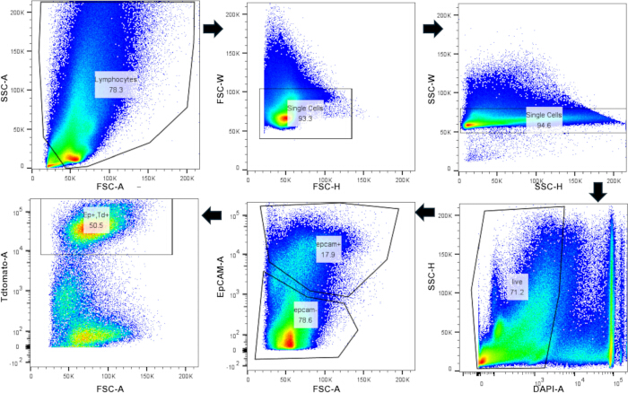
Figure 1: Isolating TdTomato+/CD326 (EpCAM+) cells by flow cytometry. Flow cytometry gating strategy for murine lung single-cell suspension. Please click here to view a larger version of this figure.
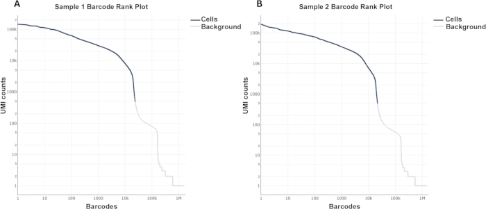
Figure 2: ScRNA-seq Barcode rank plot of murine tumor-bearing lungs. KrasLSL-G12D/+; TdTomato+/- mice lungs were collected 25 weeks after infection with lentivirus-Cre. (A,B) Tumor-bearing lung Barcode rank plot from Cellranger analysis Please click here to view a larger version of this figure.
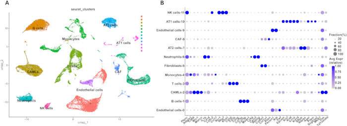
Figure 3: Single-cell transcriptomics analysis of murine tumor-bearing lungs. (A) UMAP projection of whole tumor-bearing lungs datasets. (B) Dot plot of classical genes used for cluster annotations. Please click here to view a larger version of this figure.
| Before optimization | After optimization | |||
| Repeat 1 | Repeat 2 | Repeat 1 | Repeat 2 | |
| Total Cell number | 1.3E+06 | 6.4E+05 | 1.8E+06 | 6.4E+05 |
| Live cells | 9.8E+05 | 5.0E+05 | 1.5E+06 | 5.7E+05 |
| Percentage of live cells | 76% | 77% | 83% | 89% |
Table 1: Tumor-bearing lung single-cell suspension cell viability before and after optimization. The cell concentration and percentage of live cells determined before and after using this protocol.
| Sample 1 | Sample 2 | |
| Estimated Number of Cells | 23,488 | 21,436 |
| Mean Reads per cell | 18,354 | 20,542 |
| Median Genes per Cell | 2,565 | 2,513 |
| Total Genes Detected | 30,893 | 30,556 |
| Median UMI Counts per Cells | 6,864 | 6,583 |
| Valid Barcodes | 95.90% | 95.80% |
| Sequencing Saturation | 26.10% | 28.20% |
| Fraction Reads in Cells | 94.10% | 95.40% |
Table 2: ScRNA-seq quality control analysis by Cell Ranger. The key estimates of the single-cell data obtained from Cell Ranger
Supplementary Table 1: Troubleshooting solutions. Solutions are suggested to address problems that may occur during the digestion process.Please click here to download this file.
Supplementary Table 2: Pre-optimization and post-optimization enzyme cocktail components. The final concentrations of the enzyme cocktail used before and after optimization.Please click here to download this file.
Discussion
This protocol provides a detailed step-by-step procedure for preparing high-quality single-cell suspensions from murine lung tissue. Several critical steps are required to achieve optimal results.
Prior to enzymatic digestion, whole body perfusion must be performed to remove blood from the lungs. This ensures that immune cells detected by scRNA-seq originate from lung tissue rather than circulating blood. During perfusion, it is essential to insert the needle into the left ventricle to prevent the perfusate from entering the pulmonary circulation and damaging lung cells. To preserve cell viability, all buffers, including wash, resuspension, and perfusion solutions, should be pre-chilled on ice. Given the inherent heterogeneity of lung tissue24, samples should be maintained in DMEM/F12 medium before and during digestion, as this medium provides essential nutrients that sustain cell viability.
High cell viability and well-defined cell clusters observed in this protocol confirm that the enzyme combination is both mild and effective. Calcium and magnesium ions are known to promote cell clumping and enhance enzymatic activity25,26. Therefore, Ca+/Mg+ free PBS is used to prepare most buffers to minimize clumping, while DMEM/F12 containing these ions is used as the basal medium for the digestion buffer to maintain balanced enzymatic efficiency.
While previous studies have described dissociating murine lungs or tumor tissues, they often exhibit significant variability or an incomplete description of protease selection and concentration11,15,27. For instance, Han et al.11 used enzyme cocktails without reporting concentration, which makes replication difficult. Conversely, a highly specialized method described by Chen et al.28 is effective for dissociation of Alveolar Type II (AT2) cells but involves a complex workflow, which is impractical if multiple mice need to be collected at the same time.
A critical challenge in lung oncology research is the structural heterogeneity of the tumor-bearing lung, which consists of both fragile normal cells and malignant tumor cells. Standard protocols or commercial kits are often designed for either normal tissue or tumor samples specifically. They either result in incomplete digestion, failing to release cancer cells from the tumor core, or over digestion, which induces significant stress and cell death in the more sensitive normal tissue in tumor-bearing lung samples.
This protocol addresses this by fine-tuning the enzymatic cocktail (Collagenase IV, Dispase II, and DNase I) to achieve a balanced dissociation. This optimization ensures the release of malignant cells while preserving the viability of normal cells. This is supported by flow cytometry analysis, where we successfully identified and resolved two distinct epithelial cells. The epithelial cells are well separated into TdTomato+ or TdTomato- cells. This level of resolution demonstrates that this method is suited for studying the whole tumor-bearing lung microenvironment without sacrificing the integrity of the host tissue. By including all live single cells in the analysis, this protocol enables the study of all relevant cell types within the tumor-bearing lung microenvironment, including cancer-associated macrophage like cells (CAMLs) (Figure 3), which have not been widely reported in previous studies15,16,17.
Following digestion, scRNA-seq analyses demonstrated that this procedure successfully isolates diverse cell types. Enzyme solutions should be freshly prepared on the day of the experiment. All digestion parameters are optimized for a single whole murine lung. When using only a single lobe or multiple lungs, the digestion time should be adjusted to ensure high cell viability and complete tissue dissociation. Ambient RNA contamination and multiplet formation are two major factors that affect scRNA-seq quality29. Although dead cell removal kits are often used to enhance viability, this protocol consistently yields over 80% of live cells after digestion, making this step optional. The barcode rank plot confirmed minimal ambient RNA contamination even without dead cell removal.
The current study validates this methodology within the context of the whole tumor-bearing murine lung. This optimized enzymatic digestion provides a template for studying various other tumor-bearing tissues characterized by similar microenvironments. Furthermore, the high cell viability and preservation of all cell types achieved by this protocol make it suited for advanced sequencing technologies, such as CITE-seq for surface protein mapping and ATAC-seq for epigenetic profiling.
Although this protocol produces high-quality single-cell suspensions at low cost, process-induced cellular stress may still occur during enzymatic digestion. To minimize stress-related gene expression artifacts, a single operator should process no more than four samples simultaneously.
Disclosures
The authors declare that they have no competing interests.
Acknowledgements
This work has been supported by the National Institute of Health, the National Eye Institute (NEI), USA, grants 5R01EY026202, R01EY035333; the National Institute of Dental and Craniofacial Research (NIDCR) grants R01DE031044, 2R01DE014756, and the National Cancer Institute (NCI) grants R01CA271500 and 2R01CA211187.
Materials
| Name | Company | Catalog Number | Comments |
|---|---|---|---|
| DMEM/F12 | Thermo Fisher | 11320033 | Cell culture |
| Collagenase Type IV | Sigma Aldich | C5138 | Digestive enzymes |
| Dispase II | Sigma Aldich | D4693 | Digestive enzymes |
| DNase I | Roche | 11284931001 | Remove DNA |
| Falcon 70 Cell strainer | Falcon | 352350 | Remove undigested tissue |
| Sterile Scissor | FST | 14088-10 | Cut tissues |
| Red blood cell lysis buffer | Sigma Aldich | R7757 | Remove blood cells |
| Heat-inacticated FBS | Phoenix Scientific | PS100 | Protect cells |
| Ca+/Mg+ free PBS | Thermo Fisher | 10010023 | Disolve enzymes/Wash cells |
| Safety-multifly 21G needle | Sarstedt | 3051524 | Whole body perfusion |
| 50 mL syringe | BD | 300866 | Whole body perfusion |
| 6 cm dish | Corning | 430116 | Digest tissues |
| 10 cm dish | Corning | 430167 | Digest tissues |
| Fc block antibody | BD | 553141; RRID:AB_394655 | Flow cytometry |
| DAPI | BD | 564907; RRID:AB_2869624 | Flow cytometry |
| CD326 | BD | 563478; RRID:AB_2738234 | Flow cytometry |
| HIVmPGKCre VSVG | UI Viral Vectore Core | VVC-U of Iowa-7556 | Lentivirous |
| ZE5 (Yeti) flow cytometer | Bio-Rad | flow cytometry analysis | |
| KrasLSL-G12D/+ mouse | Jackson Laboratory | 008179; RRID:IMSR_JAX:008179 | |
| TdTomato reporter mouse | Jackson Laboratory | 007909; RRID:IMSR_JAX:007909 |
References
- Leiter, A., Veluswamy, R. R., Wisnivesky, J. P. The global burden of lung cancer: current status and future trends. Nat Rev Clin Oncol. 20 (9), 624-639 (2023).
- Schabath, M. B., Cote, M. L. Cancer progress and priorities: Lung cancer. Cancer Epidemiol Biomarkers Prev. 28 (10), 1563-1579 (2019).
- Sung, H., et al. Global cancer statistics 2020: GLOBOCAN estimates of incidence and mortality worldwide for 36 cancers in 185 countries. CA Cancer J Clin. 71 (3), 209-249 (2021).
- Yu, H. A., et al. Poor response to erlotinib in patients with tumors containing baseline EGFR T790M mutations. Ann Oncol. 25 (2), 423-428 (2014).
- Cappuzzo, F., et al. MET increased gene copy number and primary resistance to gefitinib therapy in non-small-cell lung cancer. Ann Oncol. 20 (2), 298-304 (2009).
- Maman, S., Witz, I. P. A history of exploring cancer in context. Nat Rev Cancer. 18 (6), 359-376 (2018).
- Croci, D. O., et al. Dynamic cross-talk between tumor and immune cells in orchestrating the immunosuppressive network. Cancer Immunol Immunother. 56 (11), 1687-1700 (2007).
- Dunn, G. P., Old, L. J., Schreiber, R. D. The immunobiology of cancer immunosurveillance and immunoediting. Immunity. 21 (2), 137-148 (2004).
- Mittal, D., Gubin, M. M., Schreiber, R. D., Smyth, M. J. New insights into cancer immunoediting: Elimination, equilibrium and escape. Curr Opin Immunol. 27, 16-25 (2014).
- Papalexi, E., Satija, R. Single-cell RNA sequencing to explore immune cell heterogeneity. Nat Rev Immunol. 18 (1), 35-45 (2018).
- Han, G., et al. An atlas of epithelial cell states and plasticity in lung adenocarcinoma. Nature. 627 (8004), 656-663 (2024).
- van den Brink, S. C., et al. Single-cell sequencing reveals dissociation-induced gene expression in tissue subpopulations. Nat Methods. 14 (10), 935-936 (2017).
- Denisenko, E., et al. Systematic assessment of tissue dissociation and storage biases in single-cell and single-nucleus RNA-seq workflows. Genome Biol. 21 (1), 130(2020).
- Zhao, W., et al. A cellular and spatial atlas of TP53-associated tissue remodeling defines a multicellular tumor ecosystem in lung adenocarcinoma. Nat Cancer. 6 (11), 1857-1879 (2025).
- Marjanovic, N. D., et al. Emergence of a high-plasticity cell state during lung cancer evolution. Cancer Cell. 38 (2), 229-246.e13 (2020).
- Xie, L., et al. PLVAP mediates regulation of the tumour microenvironment in early-stage lung adenocarcinoma. Clin Transl Med. 15 (12), e70532(2025).
- Fernandes, L. M., et al. A single-cell atlas of normal and KRASG12D-malformed lymphatic vessels. JCI Insight. 10 (5), e185181(2025).
- Pirrello, A., Killingsworth, M., Spring, K., Rasko, J. E. J., Yeo, D. Cancer-associated macrophage-like cells as a prognostic biomarker in solid tumors. J Liquid Biopsy. 6, 100275(2024).
- Rudnick, J. A., Debnath, J. Autophagy in host stromal fibroblasts supports tumor desmoplasia. Autophagy. 17 (12), 4497-4498 (2021).
- Nissen, N. I., Karsdal, M., Willumsen, N. Collagens and cancer-associated fibroblasts in reactive stroma. J Exp Clin Cancer Res. 38 (1), 115(2019).
- Wu, J., et al. Transcardiac perfusion of the mouse for brain tissue dissection and fixation. Bio Protoc. 11 (5), e3988(2021).
- Morton, J., Snider, T. A. Guidelines for collection and processing of lungs from aged mice for histological studies. Pathobiol Aging Age Relat Dis. 7 (1), 1313676(2017).
- Pariollaud, M., et al. Circadian disruption enhances HSF1 signaling and tumorigenesis in KRAS-driven lung cancer. Sci Adv. 8 (39), eabo1123(2022).
- Wu, F., et al. Single-cell profiling of tumor heterogeneity and microenvironment in advanced NSCLC. Nat Commun. 12 (1), 2540(2021).
- McKeehan, W. L., Ham, R. G. Calcium and magnesium ions regulate multiplication in normal and transformed cells. Nature. 275 (5682), 756-758 (1978).
- Seltzer, J. L., Welgus, H. G., Jeffrey, J. J., Eisen, A. Z. Role of Ca2⁺ in mammalian collagenase activity. Arch Biochem Biophys. 173 (1), 355-361 (1976).
- Shao, S., et al. Pharmacological expansion of type II alveolar epithelial cells promotes airway repair. Proc Natl Acad Sci U S A. 121 (16), e2400077121(2024).
- Chen, Q., Liu, Y. Isolation and culture of mouse alveolar type II cells. STAR Protoc. 2 (1), 100241(2021).
- Arceneaux, D., et al. A contamination-focused approach for optimizing single-cell RNA-seq experiments. iScience. 26 (7), 107242(2023).
Reprints and Permissions
