This protocol describes a standardized in vitro method for evaluating thermal and surface effects on titanium during Er,Cr:YSGG, and diode laser irradiation using manufacturer-recommended soft-tissue cutting settings. Direct contact thermometry and multimodal surface analysis provide a reproducible workflow for characterizing wavelength-dependent laser titanium interactions.
A subscription to JoVE is required to view this content. Sign in or start your free trial.
Research Article
In Vitro Method for Assessing Laser Titanium Interactions Using Er,Cr:YSGG and Diode Lasers
* These authors contributed equally
In This Article
Summary
Abstract
This study presents a standardized in vitro protocol to evaluate the thermal response and surface alterations of titanium during Er,Cr:YSGG and diode laser irradiation under manufacturer-recommended soft-tissue settings. Custom Grade 4 titanium cylinders with an internal channel enabled direct-temperature measurement via an embedded thermocouple, allowing controlled assessment of laser-induced thermal changes. To ensure geometric consistency and minimize operator-dependent variability, a 3D-printed stabilization system was used to standardize the irradiation angle, sweep trajectory, and tip movement. Surface modifications following laser exposure were quantitatively and qualitatively characterized using complementary techniques, including profilometry for roughness analysis, scanning electron microscopy for micromorphologic evaluation, and atomic force microscopy for nanoscale topographic assessment. Diode-laser irradiation produced power- and time-dependent temperature elevations, with the highest parameter combinations exceeding the clinically accepted 10 °C safety threshold. In contrast, all tested Er,Cr:YSGG conditions remained below this limit, indicating distinct wavelength-dependent thermal behavior. Both laser systems induced significant increases in surface roughness relative to the untreated control surface, while imaging analyses revealed wavelength-dependent differences in micro and nanoscale morphology. Collectively, this protocol provides an experimental framework for systematic investigation of laser-titanium interactions and may support safer, evidence-based parameter selection for laser-assisted soft tissue procedures performed near implant components.
Introduction
Laser-assisted soft tissue procedures are widely employed during second-stage implant surgery and peri-implant mucosal management, where irradiation often occurs in close proximity to titanium components1,2. In such clinical situations, understanding the thermal behavior of titanium and the potential for surface alterations is essential, as excessive temperature elevation or surface disruption may jeopardize peri-implant bone integrity and long-term implant stability3,4. Existing literature provides valuable insights into wavelength-dependent laser titanium interactions, yet methodological variability, such as differences in irradiation geometry, contact mode, angulation, and surface evaluation techniques, poses challenges for establishing reproducible and clinically applicable safety thresholds5,6,7.
Recent studies have increasingly focused on the thermal and surface effects of different laser wavelengths on titanium, particularly in the context of peri-implant soft tissue procedures. Diode lasers have been shown to induce power and time-dependent temperature elevations and surface alterations on titanium, with some settings exceeding clinically relevant thermal thresholds, raising concerns about thermal safety near implant components8,9,10,11. In contrast, erbium-based lasers, including Er,Cr:YSGG systems, exhibit wavelength-specific interactions characterized by strong water absorption and hydrokinetic ablation mechanisms, which may limit excessive heat transfer and surface damage under appropriate conditions12,13,14. Despite these advances, reported outcomes remain heterogeneous due to differences in experimental design and a lack of standardized methodologies.
Previous studies have examined temperature changes9,10,11,15, laser-induced surface modification14,16,17, or specific aspects of titanium optical absorption7, but many experimental models lacked standardized control of parameters such as fiber angulation, tip surface contact, or sweep uniformity. These variations can influence energy delivery and complicate comparisons across studies. Additionally, the use of single-modality imaging in several reports has limited the ability to detect wavelength-dependent micro- and nanoscale alterations on titanium surfaces16,18. A more integrated analytical framework may therefore enhance the interpretability and clinical relevance of laser titanium interaction research12.
To address these methodological gaps, the present study introduces a standardized in vitro protocol to evaluate Er,Cr:YSGG, and diode laser interactions with titanium surfaces under clinically relevant, manufacturer-recommended soft-tissue cutting parameters. It was hypothesized that Er,Cr:YSGG, and diode lasers, when operated under manufacturer-recommended soft-tissue settings, would produce distinct, wavelength-dependent differences in the thermal response and surface modification of titanium. The protocol employs custom-machined Grade 4 titanium cylinders with an internal thermocouple channel, enabling direct contact measurement of baseline and post-irradiation temperatures and minimizing artifacts commonly associated with external probes.
A rigid 3D-printed stabilization system maintains a fixed irradiation angle, controlled handpiece trajectory, and consistent contact mode, reducing operator-dependent variability and ensuring reproducible energy delivery.
A key strength of this protocol is its multimodal evaluation strategy, combining thermal assessment, profilometric roughness analysis, and high-resolution SEM and AFM imaging. This integrated approach enables simultaneous characterization of macroscopic thermal behavior, microscale morphological changes, and nanoscale topographical alterations, offering a more comprehensive assessment than single-parameter techniques commonly used in earlier studies9,10,15,18. By providing a visually demonstrable and methodologically controlled workflow, this protocol establishes a reproducible platform for comparing Er,Cr:YSGG, and diode laser interactions with titanium surfaces.
Overall, the methodology presented here aims to support the development of clinically relevant, wavelength specific safety thresholds for laser-assisted second stage implant exposure and other peri-implant soft tissue procedures10,11,13.
Access restricted. Please log in or start a trial to view this content.
Protocol
Preparation of titanium specimens
Grade 4 titanium cylinders (5 mm × 10 mm, sandblasted large grit acid etched (SLA) surface) were cleaned with 70% ethanol for 30 s. The specimens were placed on a clean surface and air-dried for 10 min. The flat irradiation surface of each cylinder was inspected to confirm the absence of visible debris. Each cylinder was positioned in the 3D-printed stabilization holder, with the flat surface oriented upward and the lateral thermocouple slot accessible. All ethanol waste and any contaminated consumables generated during specimen preparation and cleaning were collected and disposed of in accordance with institutional laboratory waste management guidelines.
Environmental and safety preparation
All experiments were conducted in a controlled laboratory environment at 27 °C. Ambient temperature was continuously monitored throughout the experiments using a digital thermometer positioned adjacent to the experimental setup. Wavelength-appropriate protective eyewear was used during laser application. Reflective objects were removed from the workspace, and laser systems were powered on and allowed to complete internal self-check routines before use. (Figure 1).
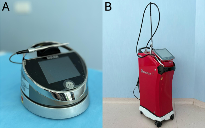
Figure 1: Laser systems used for irradiation. (A) Diode laser. (B) Er,Cr:YSGG laser. Please click here to view a larger version of this figure.
Randomization and group allocation
All titanium cylinders were numbered and randomly allocated into 13 study groups using a computer-generated randomization list, including six Er,Cr:YSGG groups, six diode groups, and one control group. Group assignments and laser parameters are summarized in Table 1. Each specimen was labeled with a unique identification code to ensure traceability throughout the experiment, according to its assigned group.
| Group Designation | Laser System Used | Number of Specimens | Applied Power Setting (W) | Exposure Time (s) |
| D1 | Diode | 8 | 1.2 W | 20 |
| D2 | 8 | 1.2 W | 40 | |
| D3 | 8 | 1.7 W | 20 | |
| D4 | 8 | 1.7 W | 40 | |
| D5 | 8 | 2.2 W | 20 | |
| D6 | 8 | 2.2 W | 40 | |
| E1 | Er,Cr:YSGG | 8 | 2.75 W | 20 |
| E2 | 8 | 2.75 W | 40 | |
| E3 | 8 | 3.75 W | 20 | |
| E4 | 8 | 3.75 W | 40 | |
| E5 | 8 | 4.75 W | 20 | |
| E6 | 8 | 4.75 W | 40 | |
| C | Control | 8 |
Table 1: Overview of the experimental study groups and laser parameters.
Stabilization of specimens in the 3D printed holder
For irradiation, the 3D printed stabilization holder was secured to a rigid tray to prevent movement during laser application. Each titanium cylinder was inserted vertically into its designated slot, with the flat surface facing the irradiation pathway. The holder maintained a fixed irradiation angle of 15°, standardizing the interaction geometry between the laser tip and the titanium surface. The handpiece was guided along the holder channel to ensure a consistent irradiation trajectory across specimens.
Thermocouple placement and baseline temperature measurements
A K-type thermocouple was inserted into the central channel of each titanium cylinder until stable metal to metal contact was achieved (Figure 2). The thermocouple was connected to a digital multimeter set to temperature mode (°C). Baseline temperature was recorded after stabilization, defined as a period of temperature fluctuations below 0.1 °C for 30 s.
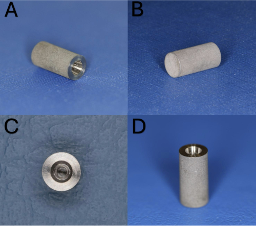
Figure 2: Titanium cylinders with thermocouple channel. (A) Lateral view. (B) Reverse lateral view. (C) Apical view of the thermocouple channel. (D) Coronal view. Please click here to view a larger version of this figure.
Er,Cr:YSGG laser irradiation
Er,Cr:YSGG laser irradiation was performed under continuous air and water spray conditions. The laser tip was positioned in direct contact with the titanium surface at a fixed 15 ° angle. The tip was swept along a 5 mm linear path at a speed of 1 cm/s. Because irradiation was performed under direct contact conditions, the effective laser interaction diameter corresponded approximately to the manufacturer-specified tip diameter of 500 µm. Specimens were irradiated with power–time combinations of 2.75 W, 3.75 W, or 4.75 W for 20 s or 40 s.
Diode laser irradiation
Diode laser irradiation was performed in continuous wave mode using a 400 µm fiber in direct contact with the titanium surface at a fixed angle of 15°. The fiber was swept along a 5 mm linear path at a speed of 1 cm/s. The effective contact footprint on the titanium surface was determined by the fiber diameter used in direct contact mode. Specimens were irradiated with power-time combinations of 1.2 W, 1.7 W, or 2.2 W for 20 s or 40 s.
Temperature recording and calculation of ΔT
Immediately after laser irradiation, the post-irradiation temperature was recorded using the thermocouple connected to the digital multimeter. Temperature change (ΔT) was calculated as the difference between post-irradiation and baseline temperature. Each specimen was irradiated only once, and no repeated laser application was performed on the same specimen.
Profilometric surface roughness measurements
After thermal measurements, the irradiated surfaces were cleaned with oil-free compressed air. Each specimen was mounted on the profilometer stage, and a 2 × 2 mm area was scanned using a stylus force of 4 mN, a scan speed of 0.5 mm/s, and a cutoff length of 0.8 mm. Five scans were performed per specimen, and the mean Ra value was calculated (Figure 3).
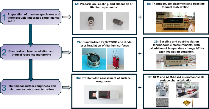
Figure 3: Experimental workflow. Specimen preparation, thermocouple placement, laser irradiation, temperature recording, profilometry, SEM, and AFM analyses. Please click here to view a larger version of this figure.
Access restricted. Please log in or start a trial to view this content.
Results
Temperature analysis
Across all Er,Cr:YSGG groups, temperature change remained below the 10 °C safety threshold, with values ranging between –2.65 °C and +2.20 °C. Power had a significant effect on temperature change (p < 0.001), whereas irradiation duration showed no significant influence (p = 0.898). The lowest temperature was observed in E2 (2.75 W–40 s: –2.65 °C). In contrast, diode laser irradiation produced markedly higher temperature elevations (3.25–15.55 °C), with both power and duration ...
Access restricted. Please log in or start a trial to view this content.
Discussion
This study demonstrated that laser titanium interactions are strongly influenced by wavelength, output power, and the combined effects of power and exposure duration. Er,Cr:YSGG irradiation consistently produced temperature elevations below the clinically accepted 10 °C threshold associated with thermal bone injury3,4, whereas diode laser irradiation showed a progressive thermal load, exceeding this limit at 2.2 W for both 20 s and 40 s. These findings align...
Access restricted. Please log in or start a trial to view this content.
Disclosures
The authors declare that there are no financial or personal conflicts of interest associated with this work. This research was conducted independently within the academic facilities of Akdeniz University. All laser systems and analytical instruments were used solely for scientific and educational purposes as part of institutional research activities. No commercial entity influenced the study design, data acquisition, analysis, or interpretation. The authors alone are responsible for the content and writing of this manuscript.
Acknowledgements
The authors acknowledge the Department of Periodontology at Akdeniz University for providing access to the laser systems, laboratory infrastructure, and imaging facilities required for this study. The authors also thank the technical staff for their assistance with specimen preparation, thermal measurements, and SEM/AFM imaging workflows. No external commercial funding or industry sponsorship contributed to this research.
Access restricted. Please log in or start a trial to view this content.
Materials
| Name | Company | Catalog Number | Comments |
|---|---|---|---|
| 3D-printed stabilization holder | Custom-made | Custom-made / Not applicable | 3D printed stabilization unit designed to fix irradiation angle and handpiece path. |
| Atomic Force Microscope (EzAFM-Compact) | Nanomagnetics Instruments, Oxford, UK | URL: https://www.nanomagnetics-inst.com/product/scanning-probe-microscopy/ezafm | Used for nanoscale surface topography and roughness characterization. |
| Digital Multimeter (Keithley 2000, 6½ Digit) | Keithley Instruments | URL: https://www.tek.com/en/products/keithley/digital-multimeter/keithley-2000-series-6-digit-multimeter-scanning | Used with K-type thermocouple for temperature measurement. |
| Epic Diode Laser (940 nm) | Biolase, Irvine, CA, USA | URL: https://www.biolase.com/products/dental-lasers-soft-tissue/epic-x/ | Continuous-wave diode laser system (940 nm); adjustable 0.5–10 W output; used for soft-tissue mode irradiation. |
| Er,Cr:YSGG Laser System (Waterlase iPlus, 2780 nm) | Biolase, Irvine, CA, USA | URL: https://www.biolase.com/products/dental-lasers-all-tissue/waterlase-iplus-intl/ | Er,Cr:YSGG laser (2780 nm) used with MZ-5 Ziptip, 9 mm; operated in gingivectomy mode with continuous air–water spray. |
| Ethanol, 70% (v/v) | Ulusoy Kozmetik | URL: https://www.ulusoykozmetik.com/urun/tr/105_ulusoy-etil-alkol-70%25C2%25B0 | Specimen surface cleaning |
| EzAFM Control and Analysis Software | Nanomagnetics Instruments, Oxford, UK | URL: https://www.nanomagnetics-inst.com/product/scanning-probe-microscopy/ezafm | Used for AFM control, data acquisition, and image processing. |
| IBM SPSS Statistics 25 | IBM Corp., Armonk, NY, USA | Version 25 | Used for statistical analyses including normality testing, non-parametric comparisons, and two-way ANOVA. |
| K-type Thermocouple | Not specified | Not applicable | K-type thermocouple probe used for temperature acquisition. |
| Microscope Control Software (Quanta FEG 250) | Thermo Fisher Scientific (formerly FEI), Hillsboro, OR, USA | URL: https://www.thermofisher.com | Used for SEM image acquisition and instrument control. |
| MZ-5 Ziptip (9 mm) | Biolase | URL: https://store.biolase.com/products/7200712-pkg-mz5-9mm-ziptips-20-pack-wl-md | Tip used with Er,Cr:YSGG handpiece. |
| Oil-free compressed air | Not specified | Not applicable | Used to remove debris from titanium surfaces. |
| Paraffin Wax | MumveMum (sold via Trendyol) | URL: https://www.trendyol.com/mumvemum/hazir-parafin-1-kg-p-31671380 | Used to coat thermocouple wire to reduce thermal interference. |
| Profilometer (Surftest SJ-201) | Mitutoyo, Tokyo, Japan | URL: https://www.bergeng.com/m m5/downloads/mti/sj201.pdf?srsltid =AfmBOoq2vJN7b4UPc2Yg-aO1 zhsL64p6vFDHSWJ54M_x5gdI8 KkIJgaV | Used for Ra measurements across 2 × 2 mm scanning area. |
| Scanning Electron Microscope (Quanta FEG 250) | Thermo Fisher Scientific (formerly FEI), Hillsboro, OR, USA | URL: https://www.thermofisher.com | SEM imaging at 250×–5000× magnifications. |
| Tips E4, 400 µm, 4 mm | Biolase | URL: https://store.biolase.com/products/7400016-tips-e4-400-µm-4mm-surgical-30-qty | Fiber used for diode laser irradiation. |
| Titanium cylinders (Grade 4, SLA surface, 5×10 mm) | Naxis, Germany | Custom made | Custom-made cylinders with 5 mm internal channel for thermocouple. |
References
- Romanos, G., Nentwig, G. H. Diode laser (980 nm) in oral and maxillofacial surgical procedures: clinical observations based on clinical applications. J Clin Laser Med Surg. 17 (5), 193-197 (1999).
- El Kholey, K. E. Efficacy and safety of a diode laser in second stage implant surgery: A comparative study. Int J Oral Maxillofac Surg. 43 (5), 633-638 (2014).
- Eriksson, A. R., Albrektsson, T. Temperature threshold levels for heat induced bone tissue injury: a vital microscopic study in the rabbit. J Prosthet Dent. 50 (1), 101-107 (1983).
- Eriksson, A. R., Albrektsson, T., Albrektsson, B. Heat caused by drilling cortical bone: temperature measured in vivo in patients and animals. Acta Orthop Scand. 55 (6), 629-631 (1984).
- Kotsakis, G. A., Konstantinidis, I., Karoussis, I. K., Ma, X., Chu, H. Systematic review and meta analysis of the effect of various laser wavelengths in the treatment of peri implantitis. J Periodontol. 85 (9), 1203-1213 (2014).
- Stübinger, S., et al. Effect of Er:YAG, CO2, and diode laser irradiation on surface properties of zirconia endosseous dental implants. Lasers Surg Med. 40 (3), 223-228 (2008).
- Lütjering, G., Williams, J. C. Titanium. , Springer. Berlin Heidelberg. (2007).
- Malmqvist, S., et al. Using 445 nm and 970 nm lasers on dental implants: An in vitro study on change in temperature and surface alterations. Materials. 12 (23), 3934(2019).
- Deppe, H., et al. Thermal effect of a 445 nm diode laser on five dental implant systems: An in vitro study. Sci Rep. 11, 20174(2021).
- Hafeez, M., et al. Thermal effects of diode laser irradiation on titanium implants in different room temperatures: An in vitro study. Photobiomodul Photomed Laser Surg. 40 (8), 554-558 (2022).
- Ahrens, M., et al. Bacterial reduction and temperature increase of titanium dental implant models treated with a 445 nm diode laser: An in vitro study. Sci Rep. 14, 18053(2024).
- Walsh, L. J. The current status of laser applications in dentistry. Aust Dent J. 48 (3), 146-155 (2003).
- Fenelon, T., Bakr, M., Walsh, L. J., George, R. Effects of lasers on titanium dental implant surfaces: a narrative review. Laser Dent Sci. 6 (3), 153-167 (2022).
- Shiba, T., et al. Effect of Er,Cr:YSGG laser irradiation on the surface modification and cell adhesion on titanium discs: An in vitro study. Materials (Basel). 17 (19), 4899(2024).
- Pergolini, D., et al. SEM evaluation of thermal effects produced by a 445 nm diode laser on implant surfaces. Dent J. 11 (6), 148(2023).
- Khalil, M. I., Sakr, H. Implant surface topography following different laser treatments: An in vitro study. Cureus. 15 (5), e38731(2023).
- Ghadiri Zahrani, E., et al. Surface enhancement of titanium Ti 3Al 2.5V through laser remelting process: A material analysis. Micromachines. 15 (12), 1526(2024).
- Block, C. M., Mayo, J. A., Evans, G. H. Effects of the Nd:YAG dental laser on plasma sprayed and hydroxyapatite coated titanium dental implants: surface alteration and attempted sterilization. Int J Oral Maxillofac Implants. 7 (4), 441-449 (1992).
- Tosun, E., et al. Comparative evaluation of antimicrobial effects of Er:YAG, diode, and CO lasers on titanium discs: an experimental study. J Oral Maxillofac Surg. 70 (5), 1064-1069 (2012).
- Matys, J., et al. Thermodynamic effects after diode and Er:YAG laser irradiation of grade IV and V titanium implants placed in bone: An ex vivo study. Biomed Tech (Berl). 61 (5), 499-507 (2016).
- Buyuktarakci, M., Kayar, N. A., Hatipoglu, M. In vitro evaluation of the effects of Er,Cr:YSGG and diode lasers used on titanium cylinder. J Vis Exp. (220), e67955(2025).
- ASM Handbook, Volume 9: Metallography and Microstructures. , ASM International. Materials Park, OH. (2004).
- Kim, H. K., et al. Alterations in surface roughness and chemical characteristics of sandblasted and acid etched titanium implants after irradiation with different diode lasers. Appl Sci. 10 (12), 4167(2020).
- Valente, N. A., et al. Thermodynamic effects of three different diode lasers on an implant bone interface: An ex vivo study with review of the literature. J Oral Implantol. 43 (2), 94-99 (2017).
- Ozgu, I., Ustun, K. Effects of mechanical methods used in peri implantitis treatment on implant surface decontamination and roughness. J Vis Exp. (217), e67778(2025).
Access restricted. Please log in or start a trial to view this content.