Here, we present a standard method for efficiently isolating adipose-derived stem cells and adipocytes, characterized by >90% reduction in processing time, high cell viability, and broad compatibility.
A subscription to JoVE is required to view this content. Sign in or start your free trial.
Method Article
Efficient Isolation of Adipose-derived Stem Cells and Adipocytes from Porcine Adipose Tissue
* These authors contributed equally
In This Article
Summary
Abstract
Adipose-derived stem cells (ADSCs) have emerged as ideal seed cells in regenerative medicine due to their abundant sources, minimally invasive harvesting, multi-lineage differentiation potential, and immunomodulatory properties. Their applications span tissue repair, disease modelling, and cell therapy; however, efficient isolation of high-viability ADSCs remains critical for advancing research and clinical translation. Conventional isolation methods, such as enzymatic digestion combined with mechanical dissociation via pipetting, are limited by lengthy processing times (1–3 h), poor cell viability (often <70%), and substantial batch-to-batch variability, compromising downstream experiments.
Here, we present the SoniConvert system (a mechanical wave-based cell separation system), which combines mechanical wave and enzymatic digestion to address these challenges. The system integrates a microprocessor-based control unit that regulates mechanical wave-mediated dissociation via digital feedback, together with a tissue-specific loosening reagent optimized for adipose tissue (5–15 min incubation). This approach enables rapid conversion of tissue to a single-cell suspension, with mechanical dissociation completed in 3–9 s.
The system offers three advantages: (1) ultra-fast processing, with the core isolation process—from enzymatic digestion to initial cell fraction separation—completed in approximately 30 min (>90% reduction compared to conventional methods); (2) high viability preservation, with trypan blue staining confirming cell viability of 80–95%, exceeding traditional protocols; and (3) broad compatibility, as the resulting single-cell suspensions meet requirements for primary cell culture, flow cytometry, cytotoxicity assays, and adipose organoid construction. By reducing processing time, enhancing cell integrity, and limiting variability, this system provides a practical platform for adipose-related research.
Introduction
Adipose-derived stem cells (ADSCs), a critical subset of mesenchymal stem cells (MSCs) isolated from adipose tissue, exhibit substantial therapeutic potential due to their accessibility, minimally invasive harvesting, robust proliferative capacity, multi-lineage differentiation potential, and potent paracrine and immunomodulatory properties1,2,3,4. These attributes have positioned ADSCs as promising candidates for regenerative medicine, tissue engineering, and immunotherapy. However, the translational utility of ADSCs hinges on the ability to isolate functionally intact, high-purity populations—a process that remains technically challenging. Adipose tissue’s unique biology introduces two primary hurdles: (1) the high lipid content of adipocytes renders them prone to rupture during mechanical processing, releasing free lipids that contaminate the stromal vascular fraction (SVF) and compromise cell viability and yield5,6; and (2) the tissue’s structural complexity, comprising fascia, endothelial cells, immune cells, and blood components7, complicates the separation of adipose-derived cells. These limitations underscore the urgent need for an efficient, rapid, and viability-preserving method to isolate high-quality ADSCs and adipocytes, which are equally critical for downstream applications such as metabolic disease modeling and cell-based therapies.
Several established methods are currently available for isolating adipose-derived cells, including enzymatic digestion, explant culture, and mechanical separation8,9,10. Enzymatic digestion, the most widely adopted approach, employs collagenase to degrade extracellular matrix (ECM) components, releasing encapsulated stem cells. This method typically involves tissue mincing, collagenase digestion (37 °C, 1–2 h), SVF isolation via centrifugation, and adherent culture purification. While effective for ECM breakdown, enzymatic digestion suffers from notable drawbacks: prolonged processing time (3–4 h total), reliance on large sample volumes, and risks of enzymatic cytotoxicity or batch-to-batch variability from animal-derived collagenases—all of which hinder clinical translation. Explant culture, which leverages stem cell migration from tissue fragments, yields cells with preserved viability but suffers from low throughput and delayed cell recovery (7–10 days). Mechanical dissociation, which relies on physical disruption (e.g., oscillation, shearing), reduces processing time but often compromises cell integrity, leading to reduced viability and impure populations. Collectively, these methods fail to reconcile efficiency, purity, and viability—critical criteria for clinical-grade cell isolation. Thus, we propose a novel method for isolating adipose-derived cells (adipocytes and ADSCs) that combines enzymatic digestion with mechanical wave-mediated dissociation11. This approach achieves two key advancements: (1) time efficiency: by combining pre-optimized, animal-free enzyme cocktails with low-frequency mechanical waves, total processing time is reduced from 3–4 h to ~30 min (Figure 1); (2) viability preservation: gentle mechanical dispersion minimizes adipocyte rupture and lipid release. Importantly, the 30 min timeframe refers to the core isolation workflow (digestion, mechanical dissociation, filtration, and initial centrifugation) to obtain crude adipocyte and SVF fractions; optional downstream purification steps, such as Red Blood Cell (RBC) lysis, require additional handling time.
Our optimized adipose cell isolation method demonstrates significant potential for application across multiple research and clinical domains. In fundamental research, the obtained high-viability cells exhibit exceptional performance in adipogenic and osteogenic differentiation studies, providing a reliable cell source for tissue engineering applications. In translational medicine, clinical studies have demonstrated the therapeutic potential of ADSCs in treating conditions such as osteoarthritis12, cardiovascular diseases13, multiple sclerosis14, and inflammatory bowel disease (IBD)/Crohn's disease15. The rapidly isolated ADSCs serve as ideal building blocks for constructing adipose tissue organoids and implementing cell therapy applications, significantly advancing drug screening and disease modeling research. Furthermore, high-quality single-cell suspensions are suitable for single-cell RNA sequencing. In conclusion, this integrated approach establishes a robust technological platform for adipose tissue, supporting both basic research and clinical translation in regenerative medicine.
Access restricted. Please log in or start a trial to view this content.
Protocol
All animal research was conducted in accordance with the Regulations for the Administration of Affairs Concerning Experimental Animals (Ministry of Science and Technology, China, revised in March 2017), and approved by the Animal Ethical and Welfare Committee of Sichuan Agricultural University (Permission number: 20230089). This protocol requires the use of the SoniConvert system, a commercially available device for mechanical wave‑assisted tissue dissociation (hereafter referred to as the mechanical dissociation system). The protocol is optimized for porcine adipose tissue, and its reliance on this specialized equipment is a key limitation.
1. Adipose tissue collection and preparation
- Preoperative disinfection and hair removal
- Euthanize the pig by intravenous overdose of pentobarbital sodium (100 mg/kg body weight). Confirm death before tissue collection.
- Apply sterile gauze soaked in 2% iodophor for spiral disinfection of the porcine dorsum, extending 15 cm from the midline. Allow drying for 30 s.
- Use a sterile razor to shave the hair from a 16 cm × 8 cm area. Ensure all hair is removed from the operative area.
- Wipe the back 2x with 75% ethanol using sterile gauze.
- Adipose tissue collection
- Make a 20 cm transverse incision along the dorsal midline with a sterile scalpel.
- Lift the incision edge with sterile forceps. Dissect along the natural plane between the adipose tissue and dermis to obtain a complete upper layer of back subcutaneous adipose tissue (ULB) sheet approximately 2–3 mm thick.
- Rinse the ULB once in ice-cold sterile saline and place it in a sterile culture dish (Figure 2).
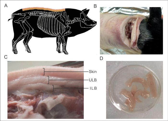
Figure 2: Collection of pig ULB for cell isolation. (A) Illustration of the structure of pig back adipose tissue; (B) Cutting site of Bama minipig; (C) Presentation of back adipose tissue, pig back subcutaneous adipose contains two layers, the upper layer (ULB) and the inner layer (ILB); (D) Collected ULB. Abbreviation: ULB = upper layer of back; ILB = inner layer of back. Please click here to view a larger version of this figure.
Pause point: The collected adipose tissue can be kept in ice-cold sterile saline or D-Hanks solution at 4 °C for a short period before further dissection.
NOTE: The objective is to collect the ULB. Avoid including the inferior layer of back subcutaneous adipose tissue (ILB) during dissection.
- Adipose tissue dissection and cleaning
- Add 10 mL of ice-cold D-Hanks Buffer for every 1.8 g of tissue to the dish and ensure the tissue is fully submerged.
- Identify and excise all visible blood vessels from the tissue by using sterile surgical scissors. Remove any connective tissue and fibrotic components. Discard the excised materials into designated biohazard containers.
NOTE: Use extreme caution when handling sharp instruments; always cut away from the body and dispose of used blades and scissors in sharps containers immediately after use. - Rinse the tissue within the D-Hanks Buffer to remove residual blood and debris.
NOTE: For samples with excessive hematoma, replace the buffer 1–3x as required to achieve visual clarity. Maintain all dissection steps on ice to preserve tissue viability.
- Adipose tissue mechanical dissociation
- Transfer 1.8 g of the rinsed tissue fragments to a sterile 1.5 mL microcentrifuge tube. Add 800 µL of A Type Tissue Dissociation Buffer (A-Buffer) to the tube.
- Using sterile ophthalmic scissors, mince the tissue in the tube into small pieces approximately 1–2 mm3 in size.
NOTE: If significant lipid leakage is observed during or after mincing, wash the tissue again with 1 mL of precooled D-Hanks solution before proceeding to enzymatic digestion to remove lipids from broken adipocytes. This step preserves adipocyte integrity for downstream analysis.
2. Cell isolation and homogenization
- Enzymatic processing
- Add 200 µL of the A Type Tissue Dissociation Agent (A-Type Solution) to a new microcentrifuge tube. Gently rotate and shake the tube by hand 3x to mix the contents and ensure that the adipose fragments are fully immersed in the dissociation solution.
- Verify the tube cap is tightly closed. Place the tube in a 37 °C metal bath and incubate for 5–10 min.
NOTE: Monitor the solution until it becomes turbid and the tissue fragments appear partially digested. The A-Type Solution contains collagenase and neutral protease. Wear nitrile gloves, a laboratory coat, and safety goggles when handling. Avoid skin contact; if exposure occurs, rinse immediately with copious water for 15 min. A-Buffer will not be discarded after adding A-Type Solution.
- Ultrasonic-assisted mechanical dissociation
- Transfer the partially digested adipose tissue fragments together with the surrounding buffer from the microcentrifuge tube into a sterile 1.5 mL microcentrifuge tube designated for the mechanical dissociation system.
- Run the preset program for Adipose Tissue in the mechanical dissociation system to perform mechanical dissociation using a 200 Hz sinusoidal wave.
3. Filtration to obtain a single-cell suspension
- Filtration and centrifugation
- Collect the cell suspension and pass it through a 200 µm cell strainer into a new 50 mL centrifuge tube to remove large debris and undigested tissue clumps.
- Add 5 mL of precooled (4 °C) D-Hanks solution to the strainer. Rinse the strainer with the D-Hanks solution 2x to maximize cell yield.
- Centrifuge the filtered cell suspension at 500 × g for 5 min at 4 °C.
Pause point: After centrifugation, the cell suspension can be processed immediately or kept on ice for a short time before layer separation. - Adipocyte collection: To collect the adipocytes, aspirate and discard the supernatant lipid layer using a sterile pasteur pipette, leaving 1 mL of buffer to protect the adipocyte layer. Collect the second layer for downstream analysis of the adipocytes. Avoid disturbing the third and fourth layers.
- SVF (stromal vascular fraction) collection: After removing the upper layers, gently resuspend the bottom pellet containing the stromal vascular fraction (SVF) in 1 mL of D-Hanks/PBS. Resuspend the cell pellet gently in 1 mL of D-Hanks/PBS to achieve a cell concentration of 2–5 × 10⁶ cells/mL.
NOTE: Ideally, four layers can be observed after centrifuging: the lipid layer at the top; the second layer containing adipocytes; the third is the hydrophilic buffer layer; and the bottom layer is the SVF, which contains stem cells (Figure 1).
- RBC lysis
- NOTE: If the cell pellet exhibits a significant red color, indicating RBC contamination, perform RBC lysis.
Resuspend the cell pellet in 1–3 mL of Red Blood Cell (RBC) lysis buffer using a P1000 pipette with wide-bore tips. Incubate on ice for 15 min with occasional mixing. - Precool all solutions and centrifuge rotors to 4 °C to minimize metabolic activity during processing.
- Centrifuge at 500 × g for 10 min at 4 °C. Aspirate the supernatant completely.
- Wash the cells once more by resuspending the pellet in 5 mL of precooled D-Hanks solution and centrifuge at 500 × g for 5 min at 4 °C. Discard the supernatant.
- NOTE: If the cell pellet exhibits a significant red color, indicating RBC contamination, perform RBC lysis.
- Final resuspension
- Resuspend the final cell pellet in D-Hanks/PBS for downstream applications.
4. Downstream applications and analysis
- SVF cell viability and cell yield assessment
- Prepare the SVF cell suspension obtained in a 1.5 mL microcentrifuge tube. Keep the suspension on ice until use.
- Mix 10 µL of the SVF cell suspension with 10 µL of 0.4% Trypan Blue solution in a sterile 0.5 mL microcentrifuge tube. Gently pipette up and down 3x to ensure uniform mixing.
NOTE: The final concentration of Trypan Blue in the mixture is 0.2%. Avoid introducing air bubbles during mixing, as they may interfere with automated cell counting.
Transfer 10 µL of the stained cell mixture to a counting slide appropriate for the Automated Cell Counter. Ensure the sample fills the chamber completely without overflow. - Insert the counting slide into the Cell Counter. Select the Trypan Blue assay type and adjust the cell concentration range if necessary.
- Press the Count button to initiate automated cell counting. The instrument will capture images of the counting chamber and automatically distinguish live cells from dead cells.
- Record the cell viability percentage and count the total number of viable cells displayed on the instrument screen. Perform three independent measurements for each sample and calculate the mean viability and the cell yield per gram of adipose tissue.
NOTE: Clean the counting slide and the instrument loading port with 70% ethanol after each use to prevent cross-contamination between samples.
- Observation of adipocyte morphology
- Stain the isolated adipocytes with 5 µg/mL Hoechst for nuclei and 1 µM BODIPY for lipids in D-Hanks/PBS for 15 min at 37 °C.
Pause point: After staining, samples should be imaged promptly; if required, they can be kept protected from light at 4 °C for a short time before microscopy. - Use a self-made adipocyte-friendly slide-cover glass system for slide preparation to avoid compressing the cells.
- Observe the nuclei (stained with Hoechst) and lipid droplets (stained with BODIPY) under a fluorescence microscope.
NOTE: Due to the large size (20–300 µm) and high lipid content, adipocytes are fragile, tend to float, and stack into multiple layers, rendering conventional mounting methods unsuitable for clear microscopic observation (Figure 3A). To address this, a homemade adipocyte-friendly mounting system using standard slides and coverslips was developed (Figure 3B).
- Stain the isolated adipocytes with 5 µg/mL Hoechst for nuclei and 1 µM BODIPY for lipids in D-Hanks/PBS for 15 min at 37 °C.
- Purification of stromal vascular components and morphological observation of ADSCs
- Seed the freshly isolated SVF in a T25 cell culture flask containing 5 mL of DMEM supplemented with 10% FBS and 1% penicillin-streptomycin. Incubate at 37 °C with 5% CO₂.
- After 48 h, gently wash the flask 2x with prewarmed PBS to remove non-adherent cells. Add fresh culture medium and continue incubation.
- Replace the culture medium every 2–3 days. Monitor cell morphology and confluence under an inverted microscope.
- When the cells reach 80–90% confluence (typically after 7–10 days), detach the cells using 0.25% trypsin-EDTA for 3–5 min at 37 °C. Neutralize with culture medium and collect the cell suspension.
- Perform Trypan Blue staining to detect the cell viability before FACS detection and sorting.
- Observe the morphology of the purified cultured ADSCs under an optical microscope.
- Filter the collected cell suspension through a 70 µm cell sieve to remove any remaining aggregates. Resuspend the cells in PBS containing 2% FBS for FACS analysis.
- Stain the cells with fluorochrome-conjugated antibodies against CD73, CD90, CD11b, and CD45 for 30 min at 4 °C in the dark.
- Wash the cells 2x with staining buffer and resuspend in PBS containing 2% FBS.
- Perform flow cytometric analysis using a 70 µm nozzle at 20 psi and a flow rate below 3.0.
- Exclude cell debris by gating on the main population using forward-scatter area (FSC-A) and side-scatter area (SSC-A) parameters.
- Remove doublets by FSC-A versus FSC-H gating and select the viable singlet population for downstream analysis.
- Analyze gated cells based on surface marker expression. Use CD73 and CD90 as positive markers and CD11b and CD45 as negative markers to verify the ADSC phenotype.
- Observe the morphology of the enriched and cultured ADSCs under an optical microscope. Characteristic spindle-shaped, fibroblast-like morphology indicates successful ADSC enrichment.
- ADSC adipogenic differentiation
- Seed the third generation of ADSCs at a density of 1 × 10⁴ cells/cm2. When cell confluence reaches approximately 90%, initiate adipogenic induction.
- Prepare the adipogenic induction medium using high-glucose Dulbecco's modified Eagle's medium (DMEM) that contains 10% fetal bovine serum (FBS), 0.5 mM 3-isobutyl-1-methylxanthine (IBMX), 1 µM dexamethasone, 10 µg/mL insulin, and 1 µM rosiglitazone.
- After 3 days of induction, replace the medium with maintenance medium and change the solution every 2 days.
- After 14 days of induction, stain the cells with Oil Red O and observe the lipid droplets under an inverted microscope.
- ADSC osteogenic differentiation
- When cell confluence reaches 80–90%, commence osteogenic induction. Formulate the osteogenic induction medium with high-glucose DMEM that contains 10% FBS, 10 mM β-glycerophosphate, 50 µg/mL ascorbic acid, 10 µM calcitriol, and 1 µM dexamethasone.
- Change the medium every 2–3 days and continue the induction process for approximately 14 days.
- Stain the cells with 2% Alizarin Red S (pH 5.2) and observe them under an inverted microscope.
- Organoid construction and identification
- Prepare a single-cell suspension of ADSCs in complete culture medium and adjust the cell concentration to 2 × 10⁴ cells/20 µL.
- Invert the lid of a 100 mm culture dish and dispense 20 µL drops of the cell suspension onto the inner surface of the lid.
- Add sterile PBS to the bottom of the dish to maintain humidity, cover the lid carefully, and incubate the hanging drops under standard culture conditions.
- Observe the drops daily and allow the cells to aggregate and form spheroids for 6 days.
- Collect the spheroids gently using a pipette and transfer them into prechilled Matrigel on ice.
- Embed the spheroids in Matrigel and plate the mixture into culture plates before incubating it under standard culture conditions.
- Replace the medium regularly and monitor organoid growth until days 18–28, when the structures become compact and mature.
- Fix the organoids with 4% paraformaldehyde, permeabilize them with 0.1% Triton X-100, and block nonspecific binding with 5% BSA.
- Incubate the samples with primary antibodies against CD73, CD90, CD11b, and CD45, and then incubate them with the corresponding fluorescent secondary antibodies.
- Stain the organoids with Hoechst and BODIPY to visualize nuclei and lipid droplets, respectively, and observe the stained samples under a fluorescence microscope.
NOTE: Perform cell sorting and all manipulations involving open cultures, including seeding, feeding, and staining preparation, under strict aseptic conditions in a biosafety cabinet.
Access restricted. Please log in or start a trial to view this content.
Results
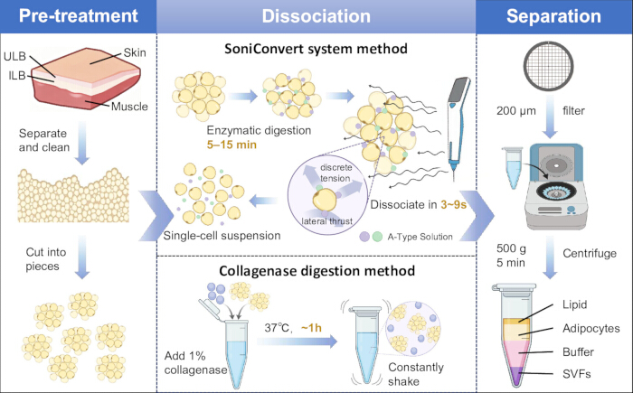
Figure 1: Schematic illustration of the adipose-derived cell isolation process. The diagram depicts the traditional method and the present method as illustrative workflows. Three main steps are involv...
Access restricted. Please log in or start a trial to view this content.
Discussion
The present study introduces a novel integrated approach for isolating adipose-derived cells (mainly adipocytes and ADSCs) that addresses critical limitations of conventional methods. By combining tissue-specific enzymatic loosening with precision mechanical wave dissociation, this system achieves rapid (total processing time ~30 min), high-viability (80–95%), and reproducible cell isolation.
Conventional enzymatic digestion, while widely used, relies on prolonged collagenase incubation ...
Access restricted. Please log in or start a trial to view this content.
Disclosures
Author Ziyi Zhao and Jiaying Yu are affiliated with Chengdu DosSense Biotech Co., Ltd, which manufactures the SoniConvert system (SC-L1W) and the Adipose Tissue Dissociation Kit (TM008) used in this protocol. The other authors declare no competing interests.
Acknowledgements
This work was supported by grants from the National Natural Science Foundation of China (Li M: 32421005 and 32225046, Lu L: 32472857); the Science and Technology Projects of Xizang Autonomous Region of China (XZ202501ZY0147 to M.L.); the China Postdoctoral Science Foundation (Lu L: 2024M763881); the Chongqing Postdoctoral Special Funding (Lu L: 2023CQBSHTB3098); the National/Provincial Undergraduate Training Program on Innovation and Entrepreneurship (Lu L: 202510626001); and the Agricultural Science and Technology Major Project.
Access restricted. Please log in or start a trial to view this content.
Materials
| Name | Company | Catalog Number | Comments |
|---|---|---|---|
| A Type Tissue Dissociation Kit | Docsense | TM001 | Contains A Type Tissue Dissociation Agent (A-Type Solution) and A Type Tissue Dissociation Buffer (A-Buffer). |
| Adipogenic Differentiation Medium Kit | OriCell | GUXMD-90031 | Used for adipogenic differentiation, including high-glucose Dulbecco's modified Eagle's medium (unmodified) and maintenance medium |
| Alizarin Red S | OriCell | A5533ALIR-10001 | Mineralization staining for osteogenic differentiation(pH 5.2) |
| Ascorbic acid | Sigma-Aldrich | A4544 | L-Ascorbic acid, cell culture tested, for osteogenic induction |
| Bama minipigs | Chengdu Dossy Experimental Animals Co.,Ltd | One year old, male, SPF-grade animals maintained under standardized vaccination, deworming, and cleaning procedures.https://www.cd-dossy.cn/animalpl_detail/12606067609851863 04.html | |
| Bio-Rad TC20 Automated Cell Counter | Bio-Rad Laboratories | 1450011 | Automated cell counter for viability and yield assessment |
| BODIPY | Thermo Fisher Scientific | D3922 | Neutral lipid stain |
| BSA (5%) | Sigma-Aldrich | A9418 | Bovine serum albumin, lyophilized powder, for blocking |
| Calcitriol | Sigma-Aldrich | C9756 | 1α,25-Dihydroxyvitamin D3, for osteogenic induction |
| cell sieve (70 μm) | Corning | 352350 | Falcon 70 μm cell strainer, sterile, for FACS sample prep |
| cell strainer (200 μm) | pluriSelect | 43-50200-03 | pluriStrainer, PET mesh, fits 50 mL tubes, sterile |
| centrifuge tube (50 mL) | Corning | 352098 | Falcon 50 mL high clarity conical tube, sterile, polypropylene |
| Counting slide (for Bio-Rad TC20) | Bio-Rad Laboratories | 1450011 | Dual-chamber counting slide for TC20 automated cell counter |
| culture dish (100 mm) | Corning | 430167 | Sterile polystyrene dish, for hanging drop organoid formation |
| Dexamethasone | Sigma-Aldrich | D4902 | ≥98%, for adipogenic and osteogenic induction |
| D-Hanks Balanced Salt Solution or PBS | Gibco | 10010015 | Pre-cooled to 4 °C, pH 7.4. |
| DMEM | Gibco | 11965084 | This is used for fresh isolated SVF cultivation |
| DNase I | Beyotime | D7073 | Optional, recommended if clumping is observed. |
| ethanol | Sigma-Aldrich | 459836 | 75% for surface disinfection. |
| Fetal Bovine Serum (FBS) | Gibco | A4736201 | Tet system approved, US origin, sterile |
| Flow Cytometer | BD Biosciences | BD FACSCanto II | 3 lasers (405 nm, 488 nm, 633 nm), 8 fluorescence channels, for surface marker analysis (CD73, CD90, CD11b, CD45), meets 70  m nozzle requirement at 20 psi m nozzle requirement at 20 psi |
| fluorescence microscope | Olympus | Inverted or upright model capable of FITC and DAPI filter sets. | |
| Fluorescent secondary antibodies | Thermo Fisher Scientific | A-11001 (anti-mouse), A-11008 (anti-rabbit) | Alexa Fluor conjugated, for immunofluorescence |
| Fluorochrome-conjugated antibodies (CD73, CD90, CD11b, CD45) | BD Biosciences | 561254 (CD73), 561970 (CD90), 557321 (CD11b), 555485 (CD45) | For flow cytometric analysis of ADSC phenotype |
| Hoechst | Thermo Fisher Scientific | H3570 | Nuclear stain for live/dead cell assessment. |
| IBMX | Sigma-Aldrich | I5879 | ≥99% (HPLC), for adipogenic induction medium |
| Ice-cold sterile saline | Baxter | 2F7124 | 0.9% Sodium Chloride Injection, USP, for rinsing tissue |
| Insulin | Sigma-Aldrich | I9278 | Recombinant, expressed in yeast, for adipogenic induction |
| iodophor | Sigma-Aldrich | 25104 | 2% disinfectant solution. |
| Matrigel | Corning | 354230 | Growth factor reduced, phenol red-free, for embedding spheroids |
| Metal Bath | Servicebio | SMB-H | Used for incubation at 37 °C. |
| Oil Red O | OriCell | OILR-10001 | Lipid staining for adipocyte differentiation |
| optical microscope | Olympus | Standard brightfield microscope with 10x–40x objectives. | |
| Osteogenic Differentiation Medium Kit | OriCell | GUXMD-90021 | Used for osteogenic differentiation, including high-glucose Dulbecco's modified Eagle's medium (unmodified) and maintenance medium |
| P1000 pipette with wide-bore tips | Beckman Coulter | B01113 | Biomek P1000 Span-8 wide bore tips, sterile |
| Paraformaldehyde (4%) | Thermo Fisher Scientific | J61899 | Aqueous solution, for fixing organoids |
| pasteur pipette | Corning | 7095S-5C | Sterile, disposable. |
| Penicillin-Streptomycin | Gibco | 15070063 | 10,000 U/mL penicillin, 10,000 μg/mL streptomycin, sterile |
| Pentobarbital sodium | Hikma Pharmaceuticals USA Inc. | 24201-010-20 | Injectable, for euthanasia (100 mg/kg body weight, intravenous). ANDA 203619 |
| Primary antibodies (CD73, CD90, CD11b, CD45) | BD Biosciences | 561254 (CD73), 561970 (CD90), 557321 (CD11b), 555485 (CD45) | For organoid immunostaining |
| Red Blood Cell (RBC) Lysis Buffer | Solarbio | R1010 | If required for downstream applications. |
| Refrigerated centrifuge | Eppendorf | 5702R | Used for centrifugation steps at 4 °C. |
| Rosiglitazone | Cayman Chemical | 71740 | ≥98%, PPARγ agonist for adipogenic induction |
| Sharps container | Becton Dickinson | 305270 | Puncture-resistant container for used blades and scissors |
| SoniConvert Single Cell Suspension Preparation System | Docsense | SC-L1W | Used for mechanical dissociation of tissue. |
| Staining Buffer (Flow Cytometry Staining Buffer) | eBioscience (Thermo Fisher Scientific) | 00-4222-57 | 200 mL, ready-to-use, contains FBS and 0.09% sodium azide as preservative, for antibody and cell dilution, surface staining and washing steps in flow cytometry |
| Sterile 1.5 mL Eppendorf tube | Eppendorf | 30121.517 | Safe-Lock microcentrifuge tube, sterile, for tissue mincing |
| Sterile culture dish | Corning | 430167 | 100 mm x 20 mm sterile culture dish, for tissue collection |
| Sterile forceps | Sklar | 96-1719 | Sterile thumb forceps, serrated, for tissue handling |
| Sterile gauze | Dukal Corporation | 7060033 | Sterile gauze pads, for disinfection and wiping |
| Sterile ophthalmic scissors | Sklar | 10023-580 | Vannas-style spring scissors, for mincing tissue into 1-2 mm³ pieces |
| Sterile razor | 3M | 9681 | Surgical prep razor, for hair removal from operative area |
| Sterile scalpel | Integra Miltex | 4-415 | Sterile disposable scalpel, for making incision |
| Sterile surgical scissors | Sklar | 10022-580 | Merit Sterile Iris Scissors, for dissecting blood vessels and connective tissue |
| T25 cell culture flask | Corning | 430639 | Falcon T25 cell culture flask, vented cap, sterile |
| Triton X-100 (0.1%) | Sigma-Aldrich | T8787 | Laboratory grade, for permeabilization |
| Trypan Blue | Gibco, Thermo Fisher Scientific | 15250061 | 0.4% solution for cell viability counting. |
| Trypsin-EDTA (0.25%) | Gibco | 25200056 | 0.25% trypsin, 1 mM EDTA, for detaching ADSCs |
| β-Glycerophosphate | Sigma-Aldrich | G9422 | Disodium salt pentahydrate, for osteogenic induction |
References
- Qin, Y., et al. An update on adipose-derived stem cells for regenerative medicine: where challenge meets opportunity. Adv Sci. 10 (20), 1-27 (2023).
- Wang, L., et al. A review of adipose-derived mesenchymal stem cells’ impacts and challenges: metabolic regulation, tumor modulation, immunomodulation, regenerative medicine and genetic engineering therapies. Front Endocrinol. 16, 1606847(2025).
- Suchanecka, M., et al. Adipose derived stem cells – sources, differentiation capacity and a new target for reconstructive and regenerative medicine. Biomed Pharmacother. 186, 118036(2025).
- Su, H., et al. Bridging the gap: clinical translation of adipose-derived stem cells - a scoping review of clinical trials. Stem Cell Res Ther. 16 (1), 288(2025).
- Alexandersson, I., Harms, M. J., Boucher, J. Isolation and culture of human mature adipocytes using membrane mature adipocyte aggregate cultures (MAAC). J Vis Exp. (156), e60485(2020).
- Attané, C., Estève, D., Moutahir, M., Reina, N., Muller, C. A protocol for human bone marrow adipocyte isolation and purification. STAR Protoc. 2 (3), 100629(2021).
- Emont, M. P., Rosen, E. D. Exploring the heterogeneity of white adipose tissue in mouse and man. Curr Opin Genet Dev. 80, 102045(2023).
- Uguten, M., et al. Comparing mechanical and enzymatic isolation procedures to isolate adipose-derived stromal vascular fraction: a systematic review. Wound Repair Regen. 32 (6), 1008-1021 (2024).
- Villanueva-Carmona, T., Cedó, L., Núñez-Roa, C., Maymó-Masip, E., Vendrell, J., et al. Protocol for the in vitro isolation and culture of mature adipocytes and white adipose tissue explants from humans and mice. STAR Protoc. 4 (4), 102693(2023).
- Cohen, M., et al. Adipose tissue explant culture using PDMS flow chambers: an alternative to static explant culture. Adipocyte. 14 (1), (2025).
- Liu, Y., Zhao, H., Yu, J., et al. A novel laboratory-based strategy for single adipocyte and adipose-derived stem cells extraction for transplantation: an experimental research. Aesthet Plast Surg. 49, 367-379 (2025).
- Tangkanjanavelukul, P., et al. Cartilage regeneration potential in early osteoarthritis of the knee: a prospective, randomized, open, and blinded endpoint study comparing adipose-derived mesenchymal stem cell (ADSC) therapy versus hyaluronic acid. Int J Mol Sci. 26 (17), 8476(2025).
- Qayyum, A. A., et al. Effect of allogeneic adipose tissue-derived mesenchymal stromal cell treatment in chronic ischaemic heart failure with reduced ejection fraction – the SCIENCE trial. Eur J Heart Fail. 25 (4), 576-587 (2023).
- Arab, F. L., et al. Safety and feasibility of intravenous fresh adipose-derived mesenchymal stem cells in secondary progressive multiple sclerosis: phase I/IIa clinical results. Stem Cell Res Ther. 17, 120(2026).
- Sheykhhasan, M., et al. Therapeutic potential and mechanistic insights into adipose-derived stem cells and mesenchymal stem cell-derived exosomes in patients with Crohn’s disease-associated fistulas: challenges and future perspectives. Stem Cell Res Ther. 16, 664(2025).
- Dufau, J., et al. In vitro and ex vivo models of adipocytes. Am J Physiol Cell Physiol. 320 (5), C822-C841 (2021).
- Ghazal, S. K., et al. Adipose tissue-derived stem cells: a comparative review on isolation, culture, and differentiation methods. Cell Tissue Bank. , 1-16 (2021).
- Zhu, R., et al. Isolation methods, proliferation, and adipogenic differentiation of adipose-derived stem cells from different fat depots in bovines. Mol Cell Biochem. , (2023).
- Benitez, G. J., Shinoda, K. Isolation of adipose tissue nuclei for single-cell genomic applications. J Vis Exp. (160), e61230(2020).
- Basatvat, S., Bach, F. C., Barcellona, M. N., et al. Harmonization and standardization of nucleus pulposus cell extraction and culture methods. JOR Spine. 6 (1), e1238(2023).
- Escudero, M., et al. Scalable generation of pre-vascularized and functional human beige adipose organoids. Adv Sci. 2301499, 1-20 (2023).
- Mandl, M., et al. An organoid model derived from human adipose stem/progenitor cells to study adipose tissue physiology. Adipocyte. 11 (1), 164-174 (2022).
Access restricted. Please log in or start a trial to view this content.
Reprints and Permissions
