The study presents a method for in vitro investigation of bacterial infection using tissue stem cell-derived intestinal organoids. The protocol describes how to culture and differentiate organoids in two dimensions, infect them with adherent-invasive Escherichia coli, and assess key readouts, including epithelial barrier breach, infection imaging, and epithelial RNA response.
Method Article
A Two-Dimensional Organoid Culture on Invasin or Basement Membrane Extract to Study Host-Bacteria Interactions
In This Article
Summary
Abstract
Organoid technology provides an experimental in vitro platform for studying human physiology in health and disease. Compared with other in vitro systems, such as cancer-derived cell lines or explants, tissue stem cell (TSC)-derived organoids offer key advantages: they can be expanded indefinitely while maintaining genomic stability and are amenable to CRISPR modification. Since these organoids are generated from normal TSCs, they contain the cell types of their tissue of origin. This makes organoids particularly valuable for investigating interactions between bacteria and differentiated, non-tumoral human cells. Intestinal organoids are grown as two-dimensional polarized monolayers on cell culture inserts coated with Basement Membrane Extract (BME) or invasin, an animal-free, cost-effective alternative, and can be differentiated into major intestinal epithelial cell types for infection with pathogenic adherent-invasive E. coli. Key infection readouts include monitoring epithelial gene expression by Reverse Transcription quantitative PCR (RT-qPCR), visualizing bacteria-epithelium interactions by confocal microscopy, and quantifying epithelial barrier breaching. The protocol can be readily adapted to study other organs and bacterial species, providing a versatile platform for investigating host-microbe interactions in organoids.
Introduction
The intestinal epithelium forms the first line of defense against pathogenic bacteria present in the gut lumen. In vitro models of the intestinal epithelium have been widely used to study how bacteria interact with epithelial cells, how these cells respond to infection, and how bacteria breach the epithelial barrier1. These models are particularly valuable for investigating human-specific pathogens in a controlled, reductionist setting. Historically, in vitro infection biology has relied on tumor-derived cell lines e.g., Caco-2 cells, or on patient-derived explants maintained ex vivo2,3. More recently, intestinal organoids generated from human tissue stem cells have emerged as a powerful tool that combines the advantages of both approaches: like cell lines, organoids can be passaged, expanded, cryopreserved, thawed, and genetically manipulated; and like explants, they are derived from non-tumoral stem cells capable of differentiating into all major epithelial cell types4,5.
When embedded in a matrix such as Matrigel or Basement Membrane Extract (BME), human intestinal organoids typically self-organize into spherical, polarized structures with an apical surface facing inward and a basal surface facing outward. Because most intestinal pathogens access epithelial cells from the apical side, several strategies have been developed to expose this surface to bacteria in organoids4. One approach is micro-injecting bacteria directly into the organoid lumen6. Another involves reversing the polarity of organoids by releasing them from the extracellular matrix, producing "apical-out" organoids in suspension7. A third strategy is to dissociate organoids into single cells and seed them onto cell culture insert inserts to form polarized, two-dimensional monolayers (2D organoid sheets) with the apical side facing the upper compartment8. The first two methods have important limitations: micro-injection is time-consuming and labor-intensive, limiting throughput, and apical-out organoids survive only a few days without matrix support and cannot be differentiated1. In contrast, 2D organoid sheets support high-throughput experimentation (bacteria can simply be added to the upper compartment) and can be maintained for multiple weeks while differentiating into major intestinal epithelial cell types9.
Unlike immortalized cell lines that readily attach to plastic surfaces, organoid-derived cells require plate coating for efficient adhesion. Coating can be performed using diluted BME10, or alternatively using invasin, a bacterial protein that binds integrins on organoid cells11. Invasin is easy to produce in the laboratory, substantially more cost-effective than BME or Matrigel, and fully animal-free.
This protocol describes the establishment and differentiation of intestinal organoids as two-dimensional polarized monolayers on cell culture insert inserts coated with invasin or Basement Membrane Extract (BME). The differentiated organoid sheets are subsequently infected with Adherent-invasive Escherichia coli (AIEC), a pathogenic strain associated with inflammatory bowel disease12. Multiple downstream infection readouts are outlined to evaluate epithelial responses and bacterial translocation. Given that AIEC invades epithelial cells and breaches the intestinal epithelium -- a mechanism thought to contribute to its pathogenicity -- epithelial breaching is quantified by measuring bacteria that reach the basolateral chamber of the cell culture insert following confirmation of barrier integrity by transepithelial/transendothelial electrical resistance (TEER) measurement13. Procedures are also provided for fixing and imaging infected monolayers to visualize intra- and extracellular AIEC, as well as for RNA extraction to assess epithelial transcriptional responses to infection. The complete protocol can be performed in less than two weeks and requires only commonly available laboratory equipment, including a BSL-2 cabinet, a confocal microscope, and a quantitative PCR instrument.
Overall, this protocol provides a cost-efficient, versatile, and accessible method for studying bacterial infection of differentiated human intestinal epithelium in vitro. It can be readily adapted to other organoid systems and other microbial species, making it a useful blueprint for in vitro infection biology studies.
Protocol
The human intestinal organoids were generated from the waste material of patients undergoing colorectal cancer surgery at the University Medical Centre Utrecht (UMCU). Patients provided informed consent for the generation of their organoids, in accordance with protocol UPORT Cancer Biobank 21-042, approved by the local ethics committee for biobanking. Figure 1A illustrates the 8 sections of the protocol . The reagent and equipment details are listed in Table of Materials.
1. Coating of the cell culture insert
- Coating with invasin
- Perform all procedures inside a tissue culture hood under sterile conditions.
- Prepare a 5 µg/mL solution of invasin in sterile phosphate-buffered saline (PBS).
- Add 100 µL of this solution to the upper compartment of each 3 µm pore polyester cell culture insert placed in a 24-well plate.
- Ensure that the membrane is completely covered.
- Incubate overnight at 4 °C.
NOTE: Avoid generating bubbles during dispensing, as bubbles prevent uniform coating.
- Coating with Basement Membrane Extract (Alternative)
- Thaw Basement Membrane Extract on ice.
- Prepare a 10% solution in ice-cold DMEM.
NOTE: Keep the solution cold to prevent premature polymerization. - Add 100 µL of the diluted solution to the upper compartment of each 3 µm pore cell culture insert.
- Ensure complete coverage of the membrane surface.
- Incubate for 1 h at room temperature.
2. Seeding of organoid cells
- Preparing organoids
- Remove culture medium from organoid plates maintained in expansion conditions (3-5 days post-splitting).
- Resuspend organoids in 1 mL of ice-cold DMEM14.
- Transfer the suspension to a 15 mL conical tube containing 4 mL of ice-cold DMEM.
NOTE: Use approximately 1 full well from a 12-well plate to seed 4 cell culture inserts.
- Pelleting organoids
- Centrifuge for 5 min at 500 × g at 4 °C.
- Remove the supernatant completely.
- Dissociating organoids
- Resuspend the pellet in 3 mL of recombinant trypsin substitute.
- Pipette up and down using a P1000 pipette to mechanically disrupt the organoids.
- Incubate at 37 °C for 15-20 min until single cells or small clusters (3-4 cells) are obtained.
NOTE: Do not exceed 20 min incubation. Pipette every 5 min to facilitate dissociation. Insert a P10 tip inside a P1000 tip if further mechanical force is required.
- Washing cells
- Add 5 mL of ice-cold DMEM.
- Centrifuge for 5 min at 500 × g at 4 °C.
- Remove the supernatant.
- Prepare Cell Suspension
- Resuspend cells in Expansion medium (100 µL per cell culture insert; see Table 1 for Expansion medium's composition).
NOTE: Do not include antibiotics. Include Rho kinase inhibitor to prevent anoikis. - Count cells using a cell counter or hemocytometer.
- Adjust to approximately 2 × 10⁶ cells/mL.
- Resuspend cells in Expansion medium (100 µL per cell culture insert; see Table 1 for Expansion medium's composition).
- Seeding the cell culture inserts
- Remove the coating solution from the inserts.
- Add 100 µL of cell suspension (2 × 10⁵ cells) to the upper compartment.
- Add 500 µL of Expansion medium to the lower compartment.
3. Cell culture insert culture and differentiation
- Incubate at 37 °C with 5% CO2 until confluency is reached (approximately 3 days).
NOTE: Refresh medium in both compartments every 4-5 days if required. - Once confluent, wash both compartments with pre-warmed DMEM.
- Add 500 µL Differentiation medium (Table 2) to the lower compartment.
- Add 100 µL Advanced DMEM/F12 supplemented with HEPES and Glutamax (AdvDMEM++) (Table 3) to the upper compartment.
- Incubate for 5 days at 37 °C with 5% CO2.
- Replace media in both compartments every 2 days.
4. Measuring transepithelial/transendothelial electrical resistance (TEER)
- Sterilize TEER electrodes in 70% ethanol for 15 min.
- Rinse electrodes in sterile PBS.
- Insert the short electrode into the upper compartment and the long electrode into the lower compartment.
NOTE: Do not touch the monolayer with the electrode tip. - Record resistance values.
- Measure blank resistance using a cell-free insert containing PBS (100 µL upper, 500 µL lower).
- Calculate TEER: TEER = (R_cell culture insert − R_blank) × surface area (cm²)
NOTE: For 24-well inserts, surface area = 0.33 cm².
5. Infection with AIEC
- Prepare Bacterial Culture
- Inoculate fluorescent AIEC in 3 mL LB medium15.
- Incubate overnight at 37 °C with shaking.
CAUTION: Perform all bacterial procedures in a BSL-2 hood.
- Prepare Inoculum
- Centrifuge 500 µL culture for 5 min at 1500 × g.
- Resuspend in 1 mL AdvDMEM++.
- Measure optical density (OD).
- Dilute to approximately 1 × 10⁸ CFU/mL in AdvDMEM++.
NOTE: OD = 1 corresponds to approximately 1 × 10⁹ CFU/mL.
- Infecting the monolayers
- Replace media in both compartments.
- Add 500 µL Differentiation medium to the lower compartment.
- Add 100 µL bacterial suspension to the upper compartment for the infected condition.
- Add 100 µL AdvDMEM++ to the upper compartment for the non-infected condition.
- Incubate at 37 °C, 5% CO₂.
6. Assessing epithelium breaching
- At 2.5 h post-infection, collect 100 µL from the lower compartment.
- Dilute in PBS and plate on LB agar.
- Add 100 µL fresh Differentiation medium to the lower compartment.
- Incubate plates overnight at 37 °C.
- Repeat sampling at 5 h post-infection.
- Count Colony-Forming Unit (CFU) the following day.
- Calculate CFUs per cell culture insert: CFUs/cell culture insert = colonies × 5 × dilution factor
7. Image Infection
- At 5 h post-infection, remove media and fix with 4% paraformaldehyde.
CAUTION: Paraformaldehyde is toxic and volatile. Handle inside a fume hood while wearing gloves. - Fix for 30 min.
- Wash three times with PBS.
- Excise the membrane insert and place cells facing upward in a well plate.
- Stain with fluorescent phalloidin for 1-2 h.
- Wash three times with PBS.
- Stain nuclei with DAPI for 10 min.
- Wash three times with PBS.
- Mount on a glass slide with mounting medium and apply a coverslip.
- Seal with nail polish.
- Acquire confocal images of DAPI, actin, and bacteria channels.
- Acquire a z-stack spanning basal to apical surfaces with 1 µm intervals.
- Open images in Fiji.
- Select Image → Z Project → Average Intensity.
- Select Image → Stacks → Orthogonal Views to visualize cross-sections.
8. Analyzing epithelial response
- At 5 h post-infection, remove media.
- Add 350 µL lysis buffer to the upper compartment.
- Pipette to lyse cells and transfer to an RNase-free tube on ice.
- Perform RNA extraction according to kit instructions.
- Measure RNA concentration and normalize samples.
- Perform reverse transcription.
- Perform quantitative PCR following the manufacturer's instructions (primer sequences for TNFα and the reference gene β-actin are provided in Table 4).
Results
After reaching confluency, intestinal cells on the cell culture insert displayed a mosaic-like pattern (Figure 1B) without visible gaps in the monolayer. Following differentiation, epithelial tightness was assessed by measuring transepithelial electrical resistance (TEER) (Figure 1C). TEER values above 500 Ω·cm² were consistent with the formation of a tight epithelial monolayer¹⁶.
The quantity of AIEC detected in the basolateral compartment increased over time, reflecting bacterial breaching of the epithelial barrier (Figure 2A,B). Absence of AIEC in the lower compartment would indicate failure of bacterial translocation.
The workflow for visualizing infection is summarized in Figure 3A. Confocal imaging demonstrated the formation of a polarized monolayer, with nuclei localized basally and an apical enrichment of the actin network (Figure 3B). Higher magnification revealed both intracellular AIEC and extracellular bacteria attached to the apical membrane (Figure 3C).
RT-qPCR analysis showed upregulation of TNFα expression in response to AIEC infection (Figure 4A,B), confirming activation of an epithelial inflammatory response.
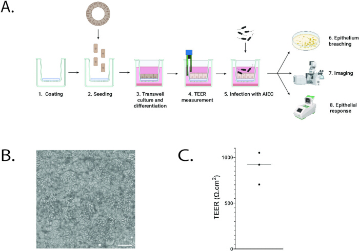
Figure 1: Overview of the protocol. (A) Schematics of the protocol. (B) Bright field image of intestinal organoid cells cultured and differentiated on an invasin-coated cell culture insert. Scale bar: 50 µm. (C) Transepithelial/transendothelial electrical resistance (TEER) of intestinal organoids grown and differentiated on invasin-coated cell culture inserts. Each dot shows an independent experiment. Please click here to view a larger version of this figure.
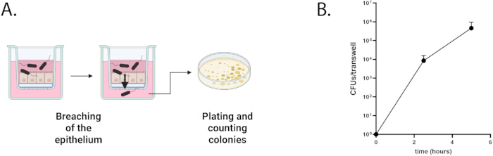
Figure 2: Assessing epithelium breaching (A) Schematics of the protocol. (B) Quantification of Colony-Forming Units (CFUs) of AIEC over time in the bottom compartment of the cell culture insert. Please click here to view a larger version of this figure.
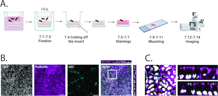
Figure 3: Imaging infection. (A) Schematics of the protocol. (B) Confocal imaging of intestinal organoids infected with fluorescent AIEC for 5 h. Grey: DAPI. Magenta: phalloidin. Cyan: AIEC. Scale bar: 50 µm. Orthogonal views of the merged image are displayed on the top and on the right. (C) High magnification of the white square in B. Grey: DAPI. Magenta: phalloidin. Cyan: AIEC. Scale bar: 10 µm. Orthogonal views are displayed on the right. Intracellular AIEC (*) and extracellular AIEC attached to the apical membrane of epithelial cells ($) are highlighted. Please click here to view a larger version of this figure.
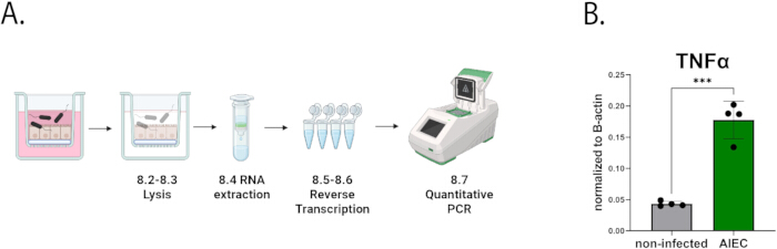
Figure 4: Analyzing epithelial response. (A) Schematics of the protocol. (B) Expression of TNFα by intestinal organoids after 5 h of infection with AIEC (green) versus non-infected control (grey). TNFα expression was normalized to B-actin expression using the formula 2-(Cq(TNFα)-Cq(β-actin)). n=4 replicates. Statistical analysis was performed using an unpaired Student's t-test. ***P < 0.001. Please click here to view a larger version of this figure.
| Component | Concentration |
| Adv DMEM | 1X final |
| HEPES | 1% v/v |
| Glutamax | 1% v/v |
| Wnt surrogate | 0.5 nM |
| R-spo1 UPE | 2% v/v |
| Noggin UPE | 1% v/v |
| B27 without vitamin A | 2% v/v |
| N-acetyl cysteine | 1.25 mM |
| Nicotinamide | 10 µM |
| hEGF | 50 ng/mL |
| A83-01 (TGF-β inhibitor) | 0.5 µM |
| SB202190 (p38 inhibitor) | 3 µM |
| Prostaglandin E2 | 1 µM |
| Rho kinase inhibitor | 10 µM |
Table 1: Expansion medium for human intestinal organoids. Composition of the Expansion medium used for maintenance and proliferation of human intestinal organoids.
| Component | Concentration |
| Adv DMEM | 1X final |
| HEPES | 1% v/v |
| Glutamax | 1% v/v |
| R-spo1 UPE | 2% v/v |
| Noggin UPE | 1% v/v |
| B27 without vitamin A | 2% v/v |
| NAC | 1.25 mM |
| hEGF | 50 ng/mL |
Table 2: Differentiation medium for human intestinal organoids. Composition of the Differentiation medium used to promote epithelial maturation of human intestinal organoid monolayers.
| Component | Concentration |
| Adv DMEM | 1X final |
| HEPES | 1% v/v |
| Glutamax | 1% v/v |
Table 3: Adv DMEM++ for infection. Composition of Advanced DMEM/F12 medium supplemented with HEPES and Glutamax (Adv DMEM++) used during bacterial infection assays.
| Name | Sequence |
| TNFα F | CTCTTCTGCCTGCTGCACTTTG |
| TNFα R | ATGGGCTACAGGCTTGTCACTC |
| β-actin F | TGACGTTGACATCCGTAAAG |
| β-actin R | GAGGAGCAATGATCTTGATCT |
Table 4: Primers for qPCR of TNFα. Shows the primer sequences used for quantitative PCR analysis of TNFα using β-actin as a reference gene.
Discussion
This study established a protocol to investigate bacterial infection of human intestinal epithelial cells using TSC-derived organoids cultured on cell culture insert inserts. Inserts with 3 µm pores enabled efficient cell attachment and bacterial translocation, whereas 0.4 µm pores do not permit AIEC passage, consistent with the approximate 1 µm size of E. coli. No translocation of organoid cells through 3 µm pores was observed, likely due to their epithelial and non-invasive phenotype.
Several experimental variables were critical for reproducibility, including efficient cell attachment, appropriate seeding density, and complete confluency prior to infection. Organoid-derived epithelial cells do not adhere to uncoated plastic, making coating essential. BME provided consistently high attachment efficiency. Invasin coating yielded comparable results and represents a cost-effective alternative11, although it must be produced in-house. Collagen coating has also been proposed as an alternative17.
Bacterial growth conditions strongly influence infection efficiency. E. coli strains grow well in DMEM; however, residual antibiotics can inhibit proliferation. Under optimal conditions, infected cultures usually display increased turbidity compared to controls, and bacterial growth can be confirmed by plating apical compartment contents.
The imaging strategy enabled visualization of both intra- and extracellular bacteria by targeting AIEC expressing a fluorophore under a constitutive promoter (e.g., using the plasmid pMB1-spect-PthrC-eGFP). Alternative methods enable selective detection of intracellular bacteria, including differential immunofluorescence staining18 or reporters expressed at defined stages of invasion19. Gentamicin protection assays provide an additional quantitative approach20.
This model is intentionally reductionist and focuses on direct interactions between bacteria and the intestinal epithelium. It enables the investigation of infection in a differentiated epithelial monolayer without additional in vivo cell types. The differentiation medium predominantly induced enterocyte and goblet cell differentiation21 but can be modified to alter epithelial composition22,23. System complexity can be increased by co-culturing immune or stromal cells in the basal compartment24.
The system does not recapitulate the three-dimensional architecture of the intestinal mucosa. Organ-on-chip platforms overcome some structural limitations25 but require greater technical complexity and resources. Furthermore, the model is best suited for acute infections, as bacterial overgrowth limits experiments to approximately 8 h. More complex systems, such as microfluidic platforms enabling continuous medium exchange, are required to study chronic infections in vitro26.
Similar cell culture insert-based organoid infection systems have been applied across multiple tissues and pathogens, including stomach organoids infected with Helicobacter pylori27,28, intestinal organoids infected with Shigella29, airway organoids infected with Pseudomonas aeruginosa10, and urothelial organoids infected with Enterococcus faecalis30. This protocol, therefore, provides a versatile framework for investigating epithelial-bacterial interactions across organ systems, provided that pathogen-specific growth requirements are considered.
Organoid technology provides physiologically relevant human models to investigate host-pathogen interactions in non-transformed epithelial cells4,5. Given the increasing burden of antibiotic-resistant infections and their association with chronic inflammatory diseases and cancer, such in vitro systems represent valuable tools for mechanistic and translational research.
Disclosures
H.C. is inventor on several patents related to organoid technology. H.C.'s full disclosure is given at https://www.uu.nl/staff/JCClevers/.
Acknowledgements
The authors thank Utrecht Portal for Organoid Technology (UPORT), in particular Anneta Brousali, Jorieke Salij, and Arnold Binda, as well as the patients and surgeons of the University Medical Center Utrecht for providing colon tissues. The authors also thank Nicolas Barnich for sharing the AIEC strain LF82. This work was supported by a CRUK grant OPTIMISTIC (C10674/A27140). TG was funded by the European Union's Horizon 2020 research and innovation program under the Marie Sklodowska-Curie grant agreement 101107988. Schematics were created using BioRender (https://www.biorender.com/).
Materials
| Name | Company | Catalog Number | Comments |
|---|---|---|---|
| A83-01 (TGF-β inhibitor) | Tocris | 2939 | |
| Adherent-invasive Escherichia coli LF82 | kind gift of Nicolas Barnich | ||
| Advanced DMEM/F12 | Invitrogen | 12634-010 | |
| B27 without vitamin A | Invitrogen | 12587010 | |
| cDNA kit | Applied Biosystems | 4387406 | The manual is available here: https://documents.thermofisher.com/TFS-Assets/LSG/manuals/MAN 0017977_highcap_cDNA_RT_UG.pdf |
| Cell counter | DeNovix | H2796 | |
| Cell counter | Leica | Stellaris | |
| Coverslips 24 mm x 50 mm | Menzel-Gläzer | BB024050S1 | |
| Cultrex Basement Membrane Extract (BME) | R&D Systems, Bio-Techne | 3533-001-02 | |
| DAPI | Sigma-Aldrich | 10236276001 | |
| Disposable Scalpel Sterile N° 10 | Swann Morton | 3033838 | |
| DMEM | Gibco | 11965092 | |
| Fiji | Fiji | Fiji is open access and can be downloaded here: https://imagej.net/software/fiji/downloads | |
| Glutamax | Gibco | 35050-061 | |
| hEGF | Peprotech | AF-100-15 | |
| HEPES | Gibco | 15630-080 | |
| Invasin | custom-made according to Wijnakker et al. PNAS 2024 | ||
| LB | Thermofisher | CM0996 | |
| LB Agar | Thermofisher | CM1116 | |
| Malassez chamber | KOVA | 87144E | |
| Microscope slides | Skycraft | 18901 | |
| Milli cell Voltohmmeter (TEER measurement) | Merck | MERS00002 | |
| mounting medium | Vectors Laboratories | Vectashield H-1000-10 | |
| N-Acetyl Cystein | Sigma-Aldrich | A9165 | |
| Nicotinamide | Sigma-Aldrich | N0636 | |
| Noggin UPE | IPA Therapeutics | N002 | |
| ParaFormaldehyde solution 4% | Sigma-Aldrich | 1.00496 | |
| Phalloidin 647 | Sigma-Aldrich | 65906-10NMOL | |
| plasmid pMB1-spect-PthrC-eGFP | Addgene | 107408 | |
| Prostaglandine E2 | Tocris | 2296 | |
| qPCR machine | Biorad | C1000-Touch | |
| Rho kinase inhibitor | AbMole | M1817 | |
| RNA extraction kit | Qiagen | 74534 | The manual is available here: https://www.qiagen.com/us/resources/resourcedetail?id=bf60203 f-291d-48ae-8206-86b0696b28c0& lang=en |
| R-spo3 UPE | IPA Therapeutics | R001 | |
| SB202190 (p38 inhibitor) | Sigma-Aldrich | S7076 | |
| Spectinomycin | Sigma-Aldrich | S9007 | |
| Suspension plate for organoid culture | Greiner | 665102 | |
| SYBR green | Biorad | 170-8887 | the manual is available here: https://www.bio-rad.com/webroot/web/pdf/lsr/literature/4106212.pdf |
| Thermocycler | Biorad | T100 | |
| Cell culture insert | Transwells 24w 3 µm pores, Greiner | 662631 | |
| Triton X-100 | Sigma-Aldrich | T8787 | |
| TrypLE | Gibco, Thermo Fisher Scientific | 12605-010 | |
| Wnt surrogate | U-Protein Express | custom order | https://www.life-sciences-europe.com/organisation/protein-express-immunoprecise-group-utrecht-netherlands-west-europe-2001-30404.html |
References
- McCoy, R., et al. In vitro Models for Investigating Intestinal Host-Pathogen Interactions. Adv Sci. 11 (8), 2306727(2023).
- Sambuy, Y., De Angelis, I., Ranaldi, G., Scarino, M. L., Stammati, A., Zucco, F. The Caco-2 cell line as a model of the intestinal barrier: influence of cell and culture-related factors on Caco-2 cell functional characteristics. Cell Biol Toxicol. 21 (1), 1-26 (2005).
- Gustafsson, J. K., Ermund, A., Johansson, M. E. V., Schütte, A., Hansson, G. C., Sjövall, H. An ex vivo method for studying mucus formation, properties, and thickness in human colonic biopsies and mouse small and large intestinal explants. Am J Physiol Gastrointest Liver Physiol. 302 (4), G430-G438 (2012).
- Puschhof, J., Pleguezuelos-Manzano, C., Clevers, H. Organoids and organs-on-chips: Insights into human gut-microbe interactions. Cell Host Microbe. 29 (6), 867-878 (2021).
- Clevers, H. Modeling Development and Disease with Organoids. Cell. 165 (7), 1586-1597 (2016).
- Kim, M., Fèvre, C., Lavina, M., Disson, O., Lecuit, M. Protocol for live imaging of bacteria-cell interactions in genetically modified mouse small intestinal organoids. STAR Protoc. 5 (1), 102773(2024).
- Co, J. Y., Margalef-Català, M., Monack, D. M., Amieva, M. R. Controlling the polarity of human gastrointestinal organoids to investigate epithelial biology and infectious diseases. Nat Protoc. 16 (11), 5171-5192 (2021).
- Moon, C., VanDussen, K. L., Miyoshi, H., Stappenbeck, T. S. Development of a primary mouse intestinal epithelial cell monolayer culture system to evaluate factors that modulate IgA transcytosis. Mucosal Immunol. 7 (4), 818-828 (2014).
- VanDussen, K. L., et al. Development of an enhanced human gastrointestinal epithelial culture system to facilitate patient-based assays. Gut. 64 (6), 911-920 (2015).
- Pleguezuelos-Manzano, C., et al. Dual RNA sequencing of a co-culture model of Pseudomonas aeruginosa and human 2D upper airway organoids. Sci Rep. 15 (1), 2222(2025).
- Wijnakker, J. J. A. P. M., et al. Integrin-activating Yersinia protein Invasin sustains long-term expansion of primary epithelial cells as 2D organoid sheets. Proc Natl Acad Sci U S A. 122 (1), e2420595121(2025).
- Darfeuille-Michaud, A., et al. Presence of adherent Escherichia coli strains in ileal mucosa of patients with Crohn's disease. Gastroenterology. 115 (6), 1405-1413 (1998).
- Darfeuille-Michaud, A., et al. High prevalence of adherent-invasive Escherichia coli associated with ileal mucosa in Crohn's disease. Gastroenterology. 127 (2), 412-421 (2004).
- Pleguezuelos-Manzano, C., Puschhof, J., van den Brink, S., Geurts, V., Beumer, J., Clevers, H. Establishment and culture of human intestinal organoids derived from adult stem cells. Curr Protoc Immunol. 130 (1), e106(2020).
- Anilionyte, O., Liang, H., Ma, X., Yang, L., Zhou, K. Short, auto-inducible promoters for well-controlled protein expression in Escherichia coli. Appl Microbiol Biotechnol. 102 (16), 7007-7015 (2018).
- Chen, S., Einspanier, R., Schoen, J. Transepithelial electrical resistance (TEER): a functional parameter to monitor the quality of oviduct epithelial cells cultured on filter supports. Histochem Cell Biol. 144 (5), 509-515 (2015).
- In, J. G., Foulke-Abel, J., Clarke, E., Kovbasnjuk, O. Human colonoid monolayers to study interactions between pathogens, commensals, and host intestinal epithelium. J Vis Exp. (146), e59357(2019).
- Quereda, J. J., et al. A dual microscopy-based assay to assess Listeria monocytogenes cellular entry and vacuolar escape. Appl Environ Microbiol. 82 (1), 211-217 (2016).
- Schulte, M., Grotheer, L., Hensel, M. Bright individuals: Applications of fluorescent protein-based reporter systems in single-cell cellular microbiology. Mol Microbiol. 121 (3), 605-617 (2024).
- Sharma, A., Puhar, A. Gentamicin protection assay to determine the number of intracellular bacteria during infection of human TC7 intestinal epithelial cells by Shigella flexneri. Bio-protocol. 9 (13), e3292(2019).
- Sato, T., et al. Long-term expansion of epithelial organoids from human colon, adenoma, adenocarcinoma, and Barrett's epithelium. Gastroenterology. 141 (5), 1762-1772 (2011).
- Fujii, M., et al. Human intestinal organoids maintain self-renewal capacity and cellular diversity in niche-inspired culture conditions. Cell Stem Cell. 23 (6), 787-793 (2018).
- He, G. -W., et al. Optimized human intestinal organoid model reveals interleukin-22-dependency of paneth cell formation. Cell Stem Cell. 29 (9), 1333-1345 (2022).
- Ding, L., et al. Toll-like receptor 9 pathway mediates Schlafen⁺-MDSC polarization during Helicobacter-induced gastric metaplasias. Gastroenterology. 163 (2), 411-425 (2022).
- Alonso-Roman, R., et al. Organ-on-chip models for infectious disease research. Nature Microbiol. 9 (4), 891-904 (2024).
- Jalili-Firoozinezhad, S., et al. A complex human gut microbiome cultured in an anaerobic intestine-on-a-chip. Nature Biomed Eng. 3 (7), 520-531 (2019).
- Vllahu, M., et al. Inflammation promotes stomach epithelial defense by stimulating the secretion of antimicrobial peptides in the mucus. Gut Microbes. 16 (1), 2390680(2024).
- Aguilar, C., et al. et al.Helicobacter pylori shows tropism to gastric differentiated pit cells dependent on urea chemotaxis. Nature Commun. 13 (1), 1-14 (2022).
- Llanos-Chea, A., et al. Bacteriophage therapy testing against Shigella flexneri in a novel human intestinal organoid-derived infection model. J Ped Gastroenterol Nutr. 68 (4), 509-516 (2019).
- Horsley, H., Dharmasena, D., Malone-Lee, J., Rohn, J. L. A urine-dependent human urothelial organoid offers a potential alternative to rodent models of infection. Sci Rep. 8 (1), 1238(2018).
Reprints and Permissions
