This case report describes a diabetic patient who developed acute foot drop after spinal anesthesia. Diagnostic evaluation using MRI, electrophysiological studies, and ultrasound suggested irritation of the L5 nerve root leading to secondary peroneal neuropathy. Conservative treatment and rehabilitation resulted in significant recovery over three months.
Case Report
Case Report and Literature Review of Spinal Anesthesia-Related Peroneal Nerve Injury Complicated with Diabetic Foot Peripheral Neuropathy
In This Article
Erratum Notice
Important: There has been an erratum issued for this article. View Erratum Notice
Summary
Abstract
Perioperative peripheral nerve injury (PPNI) is rare but potentially disabling. In patients with diabetic peripheral neuropathy (DPN), the threshold for nerve injury is lowered due to microvascular compromise and impaired myelin repair. We report a 48-year-old male with type 2 diabetes and chronic foot ulcers who developed immediate left foot drop following low spinal anesthesia for debridement. Electrophysiology revealed conduction block and focal demyelination of the peroneal nerve at the fibular head; high-frequency ultrasound showed no structural abnormality, while lumbar magnetic resonance imaging (MRI) demonstrated a punctate T2 hyperintensity in the left L5 nerve root. Symmetrical sensory deficits supported underlying DPN. The findings suggested L5 nerve root irritation related to spinal anesthesia, resulting in secondary peroneal nerve dysfunction in the setting of diabetic peripheral neuropathy. Conservative management—including neuroprotection, ankle-foot orthosis, glycemic control, offloading, and early rehabilitation—led to significant recovery: dorsiflexion improved from 0/5 to 4/5 by 3 months, with electrophysiological evidence of remyelination. This case underscores the need for a stratified diagnostic approach (rule out central, then clarify peripheral) and highlights that functional nerve injury can occur without compressive findings on imaging. Early multimodal intervention enables favorable outcomes even in vulnerable diabetic patients.
Introduction
Post-spinal anesthesia nerve injury has a low overall incidence rate, but it has a significant impact on disability and medical disputes. Therefore, authoritative guidelines emphasize the importance of standardized positioning, avoiding local compression, stabilizing hemodynamics, and early postoperative nerve assessment1,2,3,4. The peroneal nerve is located superficially at the head of the fibula and crosses a fibrous tunnel, making it a high-incidence site for lower limb mononeuropathy5. It is highly sensitive to traction and external forces5. In the context of spinal anesthesia, immediate postoperative foot drop is not common and can be easily confused with central events or L5 radiculopathy, sciatic neuropathy6,7. For patients with diabetes, regional anesthesia is not contraindicated, but long-term diabetes-induced diabetic peripheral neuropathy (DPN) significantly alters the pathological and physiological state of nerve tissue. The core pathological basis includes microvascular impairment leading to reduced blood supply, oxidative stress-mediated damage to nerve fibers, and decreased myelin repair capacity8,9,10,11. Recent mechanistic studies further elucidate that hyperglycemia drives the hexosamine biosynthetic pathway, leading to aberrant O-GlcNAc modification of key proteins. This post-translational modification has been identified as a critical driver in diabetic complications: it exacerbates peripheral neuropathy by impairing Schwann cell function and mitochondrial dynamics12 and contributes to vasculopathy by promoting inflammation and inhibiting endothelial repair13. These changes significantly lower the tolerance threshold of nerve tissue to perioperative hypoperfusion, mechanical stimulation (such as puncture operations), and drug toxicity, thereby amplifying the damaging effects of such stimuli.
Clinical studies have shown that diabetic patients have a higher incidence of PPNI during the perioperative period, and the recovery speed of nerve function after injury is slower, with a higher risk of residual sequelae. Therefore, for diabetic patients who develop foot drop after spinal anesthesia, adopting a "rule out central lesions first, then clarify the location and nature of peripheral nerve injury" stratified diagnostic strategy is particularly crucial: at the central level, lumbar spine MRI is used to rule out spinal cord injury, epidural hematoma/abscess; at the peripheral level, electrophysiology provides typing and prognostic information, while high-resolution ultrasound helps to exclude rupture/occupancy and is used for longitudinal follow-up, providing a basis for treatment planning and longitudinal follow-up14,15,16,17,18,19.
Traditional approaches often rely on delayed clinical observation, which may miss the critical window for differentiating reversible conduction blocks from axonal degeneration. In contrast, we propose a structured diagnostic algorithm that prioritizes early multimodal assessment. Specifically, lumbar MRI should be performed within 48 h of symptom onset to rule out compressive lesions such as epidural hematoma or abscess. Electrodiagnostic studies (nerve conduction studies/electromyography) are recommended between days 7 and 14, allowing sufficient time for Wallerian degeneration to manifest if axonal injury occurred, thereby accurately assessing conduction block versus denervation. Preoperative risk stratification for diabetic patients must include baseline neurological examinations and HbA1c optimization. This proactive strategy significantly improves diagnostic accuracy by distinguishing central radiculopathy from peripheral mononeuropathy within the first week, optimizing the timing for neuroprotective interventions compared to delayed approaches.
This article presents a case of peroneal mononeuropathy following spinal anesthesia in the context of DPN, in accordance with the CARE (Guidelines for Case Reports), focusing on the stratified diagnostic pathway, imaging and electrophysiological evidence, treatment strategy and outcome, and discussing its rarity and association with DPN.
Case Presentation:
A 48-year-old male (168 cm, 64 kg) with a 12-year history of type 2 diabetes (HbA1c 7.6%), status post renal transplantation one year prior, and chronic bilateral foot sensory loss presented with a non-healing left foot ulcer for over three months. He had no history of lumbar stenosis or genu valgum. His medications included linagliptin, insulin, tacrolimus, mycophenolate mofetil, prednisone, aspirin, diuretics, losartan/hydrochlorothiazide, and atorvastatin. Preoperative ankle-brachial index was 0.96, ruling out critical limb ischemia.
On the day of surgery, spinal anesthesia was administered with the patient in the lateral decubitus position. The procedure involved technical adjustments to the needle trajectory and interspace selection to achieve successful dural puncture, although the exact sequence of interspace attempts could not be fully reconstructed from the retrospective medical records. The final successful puncture was achieved at the L4–5 interspace using a midline approach with a 25-gauge Quincke needle. Crucially, during the needle insertion process at the final level (or during the adjustment maneuvers), the patient reported acute back pain radiating to the left lower limb accompanied by a distinct electric shock-like sensation. The operator adjusted the needle angle/position slightly to alleviate the paresthesia before proceeding. Clear cerebrospinal fluid (CSF) reflux was confirmed prior to injection. There was no resistance during administration of 0.75% ropivacaine (1.5 ml) and 10% glucose solution (0.5 ml), and the patient reported no pain during the injection. The sensory block extended to the T8 dermatome. The patient was subsequently turned to the supine position. Intraoperative vital signs remained stable.
Immediately after surgery (within 1 h), the patient developed an inability to dorsiflex the left foot and tenderness at the fibular head; plantar flexion was preserved. Neurological examination revealed dorsiflexion and toe extension graded 0/5 according to the Medical Research Council (MRC) scale, decreased pinprick sensation in the peroneal nerve distribution, and a positive Tinel’s sign at the fibular head. Lumbar MRI, electromyography (EMG), and ultrasound examinations were subsequently performed on day 7 to evaluate the etiology (see Results section).
Diagnosis, Assessment, and Plan:
Based on the clinical presentation, we suspected acute iatrogenic L5 nerve root injury manifesting as peroneal nerve dysfunction, exacerbated by underlying diabetic neural vulnerability.
To confirm the diagnosis and rule out other etiologies, a stratified diagnostic workup was planned: Lumbar MRI was ordered to exclude central lesions such as epidural hematoma or abscess; high-frequency ultrasound was utilized to assess the structural integrity of the peroneal nerve at the fibular head; and electrophysiological studies were scheduled to differentiate between conduction block and axonal degeneration. Differential diagnoses considered included diabetic peripheral neuropathy (typically symmetric), direct surgical trauma, and positional compression.
The management strategy focused on general neuroprotection, posture management to avoid nerve traction, the application of an ankle-foot orthosis (AFO) for gait support, and contracture prevention. A rehabilitation plan involving nerve gliding exercises and tibialis anterior muscle training was initiated, alongside staged analgesia, neurotrophic treatment, and strict glycemic control. Diabetic foot offloading care was implemented. Electrophysiological reassessment was scheduled at 6–8 weeks to monitor for remyelination. Surgical intervention was reserved for cases that showed no significant clinical improvement by 3–6 months or for those with secondary compressive pathologies identified upon re-evaluation.
Protocol
This study was conducted in accordance with the Declaration of Helsinki. Ethical approval was waived by the Ethics Committee of The First Affiliated Hospital, Zhejiang University School of Medicine, Liangzhu Branch, due to the case report's retrospective nature. Written informed consent was obtained from the patient.
1. Immediate postoperative neurological assessment
- Motor strength (dorsiflexion, plantar flexion), sensation (pinprick, light touch), and reflexes (patellar and Achilles reflexes) in both lower limbs were evaluated within 1 h of surgery. Motor strength was graded using the MRC scale. Sensory testing was performed using a sterile pin for pinprick and a cotton wisp for light touch.
- The presence of foot drop, Tinel’s sign, or localized tenderness at the fibular head was documented.
2. Stratified diagnostic workup (within 7 days)
- Lumbar spine MRI was performed using a 1.5 T scanner with T2-weighted imaging sequences (sagittal slice thickness: 5 mm) to exclude central lesions.
- High-resolution ultrasound of the common peroneal nerve at the fibular head was conducted using a 12–15 MHz linear probe. The patient was positioned supine with the leg slightly internally rotated. Scanning was performed in both transverse and longitudinal orientations.
- Nerve conduction studies and electromyography were obtained. Stimulation sites included the ankle and below/at/above the fibular head for the peroneal nerve, with recording at the extensor digitorum brevis muscle. Filter settings were set at 2 Hz to 10 kHz. Skin temperature was maintained above 32 °C throughout the procedure.
3. Multimodal conservative management
- An ankle-foot orthosis (AFO) was initiated for gait support and contracture prevention.
- Neurotrophic agents (methylcobalamin, vitamin B complex) and neuropathic pain medication (e.g., pregabalin) were administered.
- Diabetic foot offloading was implemented using a total contact cast or removable walker.
- Physical therapy consisted of daily nerve gliding exercises and progressive resistance training for dorsiflexors. The progression protocol involved increasing the resistance band tension weekly based on tolerance.
4. Follow-up and re-evaluation
- Muscle strength (MRC scale) and functional gait (observed walking pattern) were assessed at 6 weeks and 3 months.
- Electrophysiological studies were repeated at 6–8 weeks to monitor remyelination.
- Surgical evaluation (e.g., neurolysis) was planned if no improvement in strength or electrophysiology was observed by 3–6 months.
Results
Diagnostic findings
Lumbar MRI (1.5 T) revealed a punctate T2 hyperintensity in the region of the left L5 nerve root on sagittal images (Figure 1), consistent with focal intraneural injury or edema. Axial sections did not visualize this hyperintense signal but effectively ruled out large compressive hematomas or disc herniations (Figure 2).
High-frequency ultrasound showed normal morphology and echogenicity of the peroneal nerve at the fibular head, with no focal thickening, compression, or rupture.
Electrophysiological studies demonstrated slowed motor conduction velocity and a relative conduction block (>50% reduction in compound muscle action potential (CMAP) amplitude between distal and proximal stimulation sites) at the fibular head segment. Distal CMAP amplitude was low. The tibial nerve parameters were normal. These findings supported a diagnosis of demyelinating conduction block secondary to proximal radiculopathy.
Clinical recovery timeline
From postoperative days 1 to 7, the patient received oral pregabalin, methylcobalamin, and vitamin B complex. Rehabilitation involving neuromuscular electrical stimulation and simple ultrasound therapy was initiated. At 6 weeks postoperatively, dorsiflexion strength improved to 3/5, and pain was significantly reduced.
At 3 months, dorsiflexion strength reached 4/5, allowing the patient to ascend and descend stairs independently. Tenderness at the fibular head had disappeared. Follow-up electromyography showed improved conduction velocity and increased distal CMAP, indicating remyelination. The foot ulcers had epithelialized without recurrence (Figure 3). The timeline of events is summarized in Figure 4.
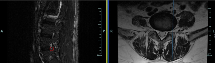
Figure 1: Sagittal MRI detection of focal L5 nerve root injury. The left panel displays a sagittal view where a discrete punctate T2 hyperintensity (indicating focal intraneural injury) is clearly visible within the left L5 nerve root, highlighted by a red circle. The right panel shows the corresponding axial slice at this level. Anatomical orientation is marked as: A (Anterior), P (Posterior), L (Left), and R (Right). Please click here to view a larger version of this figure.
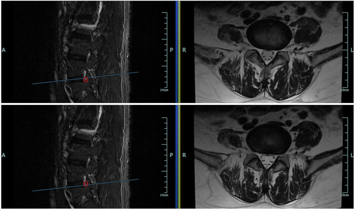
Figure 2: Geometric explanation for the absence of the lesion on axial imaging. The figure presents two sagittal–axial image pairs (top and bottom rows) that illustrate the slice-selection geometry. In each row, the left image shows a sagittal view with the focal hyperintense lesion marked (red circle). The blue line indicates the position of the corresponding axial slice displayed in the right image. In both examples, the axial plane passes adjacent to, but does not intersect, the lesion, explaining its absence on axial imaging. Anatomical orientation is indicated as: A (Anterior), P (Posterior), L (Left), and R (Right). Please click here to view a larger version of this figure.
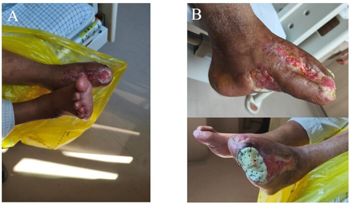
Figure 3: Clinical photographs of the affected foot. (A) Day 7 postoperatively showing plantar-flexed foot position consistent with impaired dorsiflexion. (B) At 3 months, improved active dorsiflexion of the foot following treatment. Please click here to view a larger version of this figure.
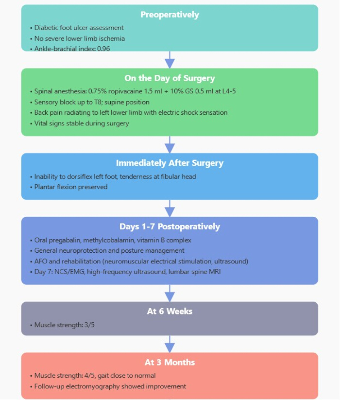
Figure 4: Perioperative–follow-up timeline. The figure illustrates anesthesia method, key events (puncture-induced radicular pain, immediate foot drop), diagnostic milestones (MRI, ultrasound, EMG on day 7), and muscle strength recovery (dorsiflexion 4/5 at 3 months). Please click here to view a larger version of this figure.
Discussion
Perioperative peroneal nerve injury has a low overall incidence rate, but it can cause significant functional impairment20. Therefore, the ASA and ASRA guidelines both emphasize the identification and prevention of variable-risk sources, including standardized positioning, avoiding local compression, and early nerve assessment1,2. The peroneal nerve is located superficially at the fibular head and crosses a fibrous tunnel, making it highly sensitive to traction and external forces, and a high-incidence site for lower limb mononeuropathy5. Postoperative peroneal nerve lesions following spinal anesthesia are relatively rare, and the literature suggests that they often need to be differentiated from central or radicular events6,7. In this case, the lumbar spine MRI did not reveal any central injuries such as spinal cord contusion or epidural space-occupying lesions, but only showed a punctate high signal on T2-weighted imaging in the left L5 nerve root (indicating mild inflammatory edema); high-resolution ultrasound showed intact continuity of the left lower limb peroneal nerve without swelling, ruling out focal compression or space-occupying lesions; electrophysiological studies showed slowed motor conduction velocity, relative conduction block, low amplitude of distal CMAP, decreased peroneal nerve sensory wave, and normal tibial nerve parameters at the fibular head segment. Finally, the cause of injury is considered to be a demyelinating conduction block in the fibular head segment of the common peroneal nerve, which is secondary to L5 nerve root injury. The underlying mechanism is considered to be anesthesia-related transient neurotoxicity or mechanical injury, rather than typical positional compression8,9.
The uniqueness of this patient lies in his long-term diabetes and post-renal transplantation status. Diabetic peripheral neuropathy (DPN) forms a vulnerable neural baseline state through microvascular impairment, oxidative stress, and decreased myelin repair capacity10. The long-term use of immunosuppressants after renal transplantation further weakens nerve protection and repair functions; along with perioperative anesthesia-related factors, this results in a superimposed effect. The patient experienced radiating pain and electric shock sensations in the left lower limb during the puncture, indicating that the L5 nerve root may have been slightly mechanically irritated by the puncture needle. We identify the immediate recognition of this paresthesia and the subsequent cessation of needle advancement as the single most critical procedural step determining diagnostic clarity and therapeutic success. Had the operator ignored this warning sign or continued injection, the mechanical irritation could have progressed to severe axonal loss. The 0.75% ropivacaine used, although a low-toxicity local anesthetic, may exert a toxic effect on the perineurium of DPN patients (in whom perineurial permeability is increased). This model of a vulnerable neural baseline combined with perioperative stress explains the phenomenon of mononeuropathy in the absence of imaging findings of typical compression and is consistent with the findings of negative ultrasound results but positive EMG and MRI results.
In terms of diagnosis, for postoperative foot drop following spinal anesthesia, the assessment pathway of "rule out central, then clarify peripheral" should be followed: lumbar spine MRI should be prioritized to exclude spinal cord, nerve root, and epidural lesions6,7; high-resolution ultrasound should be used to quickly screen for anatomical abnormalities of the peroneal nerve (such as rupture, space-occupying lesions)14,15,16; electrophysiological studies can distinguish between conduction block and axonal degeneration14,15,16,17,18. Our structured diagnostic algorithm, which prioritized early multimodal assessment (MRI <48h, EMG days 7–14), illustrates the potential advantages over traditional "wait-and-see" strategies or isolated clinical observation. While conventional approaches often delay advanced imaging until conservative management fails—risking missed windows for reversible compression—our method rapidly distinguished between axonal degeneration and reversible conduction block in this case, thereby avoiding unnecessary interventions and accelerating targeted rehabilitation. This case suggests that early multimodal assessment may be beneficial for similar high-risk patients.
Decision-making is increasingly guided by a function-preserving, graded strategy distilled from evidence on compressive, neoplastic, and traumatic peripheral neuropathies: when features indicate predominant demyelinating block and imaging reveals no mass or scar tether, standardized conservative care and rehabilitation are prioritized; if no reinnervation trajectory is evident on serial strength testing and follow-up electrodiagnostic studies (EDX)/high-resolution ultrasound (HRUS) within 3–6 months, if progressive atrophy or refractory pain emerges, or if ultrasonography/MRI shows compressive change, evaluation for precise decompression/neurolysis is warranted. To assist clinicians in managing similar complex cases, we propose specific troubleshooting guidance: (1) For ambiguous diagnostic findings: If MRI is negative for compression but motor deficits persist beyond 48 h, do not assume functional recovery is imminent; immediately proceed to electrodiagnostic studies to differentiate between conduction block (favorable prognosis) and axonal degeneration (poor prognosis). (2) For delayed recovery: In diabetic patients, if no clinical improvement (e.g., return of muscle strength) is observed by 6 weeks, repeat EMG/NCS is mandatory to assess for reinnervation signs; absence of reinnervation potentials at this stage should prompt a re-evaluation for occult compressive lesions or consideration of surgical consultation. For nerve-sheath tumors, traumatic neuromas, or clear axonal discontinuity, a pathway of lesion-directed surgery with microsurgical reconstruction (including autograft as indicated) and staged rehabilitation is applied21,22. Contemporary series and reviews provide practical thresholds for reassessment timing, procedure selection, and functional follow-up, which informed our “6–8 weeks recheck → 3–6 months surgical review” milestones in this case21,22,23,24,25,26,27. In patients with diabetic foot, adherence to IWGDF off-loading principles can proceed in parallel with neural rehabilitation to improve tissue perfusion and reduce the risk of recurrence, supporting overall functional recovery28.
This study has several limitations regarding methodology and generalizability. First, as a single-case retrospective report, our findings are subject to selection bias and limited statistical power, restricting broad generalizability to all diabetic populations. Second, the retrospective data collection inherently restricted our ability to detail the precise granular mechanics of the anesthesia technique. Third, the use of a 1.5T MRI may lack the sensitivity of higher-field scanners (3.0T) for detecting subtle neural changes, potentially leading to false-negative findings on axial imaging. Finally, we could not definitively separate mechanical trauma from potential local anesthetic neurotoxicity, though the clinical presentation favors a mechanical etiology exacerbated by DPN.
Finally, it is important to emphasize the insights at the case level: For patients with underlying diseases such as diabetic peripheral neuropathy (DPN), baseline neurological function should be evaluated preoperatively; during anesthesia, priority should be given to selecting safe interspaces, using fine-needle puncture, and paying attention to patient feedback to avoid the accumulation of high-concentration local anesthetics and mechanical injury. For foot drop after spinal anesthesia in the context of diabetic foot, the inherent perception that it is caused by positional compression should be challenged, and MRI, HRUS, and EDX should be combined to distinguish between peripheral and central injuries. After confirming nerve injury, early implementation of nerve protection, rehabilitation, and metabolic management optimization can usually result in significant functional recovery within 3 months. For patients with stagnant recovery and evidence suggesting axonal degeneration or compression, the time window for surgical decompression and re-evaluation should be seized. Clinically, attention should be paid to the whole-process prevention and control of such rare complications, and precise assessment and intervention should be adopted to improve patient prognosis1,14,15,16,17,18,26. Future research and clinical applications should focus on two key directions: First, prospective multicenter studies are needed to define the optimal diagnostic timing window and cost-effectiveness of early MRI/EMG protocols specifically for high-risk diabetic populations undergoing neuraxial anesthesia. Second, clinical trials should investigate perioperative neuroprotective strategies (e.g., specific antioxidant regimens or optimized glycemic control protocols) to raise the threshold of nerve vulnerability in patients with pre-existing DPN, potentially preventing such iatrogenic injuries.
Disclosures
The authors have nothing to disclose.
Acknowledgements
We gratefully acknowledge the patient for his/her cooperation throughout the diagnostic and follow-up process, and for consenting to the publication of this case report to benefit future clinical practice.
Materials
| Name | Company | Catalog Number | Comments |
|---|---|---|---|
| 1.5 Tesla MRI Scanner | Siemens Healthineers | MAGNETOM Avanto Fit | 1.5T superconducting system. |
| Electrophysiology System | Nihon Kohden | MEB-2300K | Supports standard NCS/EMG protocols with filters (2 Hz–10 kHz) and limb temperature monitoring. |
References
- Practice advisory for the prevention of perioperative peripheral neuropathies 2018: An updated report by the American Society of Anesthesiologists Task Force on Prevention of Perioperative Peripheral Neuropathies. Anesthesiology. 128 (1), American Society of Anesthesiologists Task Force on Prevention of Perioperative Peripheral Neuropathies. 11-26 (2018).
- Neal, J. M., et al. The second ASRA practice advisory on neurologic complications associated with regional anesthesia and pain medicine: Executive summary 2015. Reg Anesth Pain Med. 40 (5), 401-430 (2015).
- Chui, J., Murkin, J. M., Posner, K. L., Domino, K. B. Perioperative peripheral nerve injury after general anesthesia: A qualitative systematic review. Anesth Analg. 127 (1), 134-143 (2018).
- Welch, M. B., et al. Perioperative peripheral nerve injuries: A retrospective study of 380,680 cases during a 10-year period at a single institution. Anesthesiology. 111 (3), 490-497 (2009).
- Mizuno, J., Takahashi, T. Factors that increase external pressure to the fibular head region, but not medial region, during use of a knee-crutch/leg-holder system in the lithotomy position. Ther Clin Risk Manag. 11, 255-261 (2015).
- Nirmala, B., Kumari, G. Foot drop after spinal anaesthesia: A rare complication. Indian J Anaesth. 55 (1), 78-79 (2011).
- Goyal, V. K., Mathur, V. Foot drop: An iatrogenic complication of spinal anesthesia. Braz J Anesthesiol. 68 (4), 412-415 (2018).
- Kopp, S. L., Jacob, A. K., Hebl, J. R. Regional anesthesia in patients with preexisting neurologic disease. Reg Anesth Pain Med. 40 (5), 467-478 (2015).
- Levy, N., Lirk, P. Regional anaesthesia in patients with diabetes. Anaesthesia. 76 (Suppl 1), 127-135 (2021).
- Feldman, E. L., et al. Diabetic neuropathy. Nat Rev Dis Primers. 5 (1), 41(2019).
- Koning, S., et al. The impact of diabetes mellitus on foot perfusion measured by ICG NIR fluorescence imaging. Diabetes Res Clin Pract. 214, 111772(2024).
- Qi, B., et al. Metformin attenuates O-GlcNAc modification to improve renal function via AMPK/mTOR signaling in diabetic nephropathy. J Biol Chem. 301 (12), 110909(2025).
- Qi, B., et al. O-linked β-N-acetylglucosamine modification in diabetic foot ulcer pathogenesis. Burns Trauma. 13, tkaf044(2025).
- Marciniak, C., Armon, C., Wilson, J., Miller, R. Practice parameter: Utility of electrodiagnostic techniques in evaluating patients with suspected peroneal neuropathy: An evidence-based review. Muscle Nerve. 31 (4), 520-527 (2005).
- Fortier, L. M., et al. An update on peroneal nerve entrapment and neuropathy. Orthop Rev (Pavia). 13 (2), 24937(2021).
- Kim, J. Y., Song, S., Park, H. J., Rhee, W. I., Won, S. J. Diagnostic cutoff value for ultrasonography of the common fibular neuropathy at the fibular head. Ann Rehabil Med. 40 (6), 1057-1063 (2016).
- Symanski, J. S., Ross, A. B., Davis, K. W., Brunner, M. C., Lee, K. S. Ultrasound for traumatic nerve injury, entrapment neuropathy, and imaging-guided perineural injection. Radiographics. 42 (5), 1546-1561 (2022).
- Zaottini, F., et al. High-resolution ultrasound of peripheral neuropathies in rheumatological patients: An overview of clinical applications and imaging findings. Front Med (Lausanne). 9, 984379(2022).
- Dong, Y., et al. Imaging diagnosis in peripheral nerve injury. Front Neurol. 14, 1250808(2023).
- Ahmad, F. U., Pandey, P., Sharma, B. S., Garg, A. Foot drop after spinal anesthesia in a patient with a low-lying cord. Int J Obstet Anesth. 15 (3), 233-236 (2006).
- Dong, Y., Lu, H. Editorial: Surgical treatment of peripheral neuropathic pain, peripheral nerve tumors, and peripheral nerve injury. Front Neurol. 14, 1266638(2023).
- Xu, G., et al. Advancements in autologous peripheral nerve transplantation care: A review of strategies and practices to facilitate recovery. Front Neurol. 15, 1330224(2024).
- Zhou, H., et al. Clinical characteristics and management experience of schwannoma in extremities: Lessons learned from a 10-year retrospective study. Front Neurol. 13, 1083896(2022).
- Yang, H., et al. Traumatic neuromas of peripheral nerves: Diagnosis, management and future perspectives. Front Neurol. 13, 1039529(2022).
- Zhou, H. Y., Jiang, S., Ma, F. X., Lu, H. Peripheral nerve tumors of the hand: Clinical features, diagnosis, and treatment. World J Clin Cases. 8 (21), 5086-5098 (2020).
- Singhal, N., Sethi, P., Jain, J. K., Agarwal, S. Spinal subdural hematoma with cauda equina syndrome: A complication of combined spinal epidural anesthesia. J Anaesthesiol Clin Pharmacol. 31 (2), 244-245 (2015).
- Lu, H., Chen, L., Jiang, S., Shen, H. A rapidly progressive foot drop caused by the posttraumatic intraneural ganglion cyst of the deep peroneal nerve. BMC Musculoskelet Disord. 19 (1), 298(2018).
- Bus, S. A., et al. Guidelines on offloading foot ulcers in persons with diabetes (IWGDF 2023 update). Diabetes Metab Res Rev. 40 (3), e3647(2024).
Reprints and Permissions
Erratum
Formal Correction: Case Report and Literature Review of Spinal Anesthesia-Related Peroneal Nerve Injury Complicated with Diabetic Foot Peripheral Neuropathy
Posted by JoVE Editors on 5/29/2026. Citeable Link.
This corrects the article 10.3791/70690