This study applied bidirectional two-sample Mendelian randomization to assess the causal relationship between Helicobacter pylori (H. pylori) antibodies and iron-deficiency anemia (IDA). H. pylori OMP antibody was causally linked to elevated IDA risk in European individuals.
Research Article
Causal Relationship Between Helicobacter pylori OMP Antibodies and Iron-Deficiency Anemia: A Mendelian Randomization Study
In This Article
Summary
Abstract
Previous observational studies have suggested a potential causal relationship between Helicobacter pylori (H. pylori) infection and iron-deficiency anemia (IDA). However, the evidence for causal inference remains contentious, as observational studies are prone to confounding factors that may bias the results. Additionally, the mechanisms underlying the association between H. pylori infection and IDA remain incompletely elucidated. To address these gaps and delve deeper into the nature of the relationship between H. pylori and IDA, this study conducted a bidirectional two-sample Mendelian randomization (MR) analysis, which is less susceptible to confounding and reverse causality compared to traditional observational designs. Inverse-variance weighted (IVW) was used as the primary analytical method, with weighted median, MR-Egger regression, weighted mode, and simple mode as supplementary approaches. Seven common serum H. pylori antibodies were selected as exposure factors for the forward MR analysis. A series of sensitivity analyses was conducted to assess the reliability of the main MR assumptions and ensure the robustness of the study results. Reverse MR analysis was further performed to assess the potential reverse causality, and the results confirmed that there is no reverse causal relationship between IDA and H. pylori infection. Genetically predicted serum levels of H. pylori outer membrane protein (OMP) antibodies were positively associated with an increased risk of IDA (odds ratio [OR] = 1.077, 95% CI 1.010–1.148, P = 0.02194), while no causal association was observed for other antibodies. Reverse MR excluded reverse causality between IDA and H. pylori infection. This study first performed antibody-stratified two-sample MR analysis for seven H. pylori antibodies, suggesting that elevated OMP antibody levels increase IDA susceptibility and providing reliable causal evidence to enrich relevant research.
Introduction
Anemia is quantitatively defined as a decrease in the number of circulating erythrocytes or functionally defined as a condition characterized by an insufficient count of red blood cells (oxygen carriers) to meet an individual’s metabolic demands1. Iron deficiency anemia (IDA) is one of the most prevalent forms of anemia, accounting for approximately 25% of all anemia cases. It is pathologically characterized by depleted body iron stores, impaired hemoglobin synthesis, and a consequent reduction in erythrocyte count. The primary etiological factors include insufficient dietary iron intake, increased physiological iron demand, excessive blood loss, and dysfunctions in iron absorption, transport, and utilization2. Patients with IDA exhibit diminished oxygen-carrying capacity, clinically manifesting as fatigue, weakness, dizziness, and pallor. In severe cases, IDA may precipitate serious complications such as palpitations, chest pain, heart failure, and compromised immune function, and in extreme instances, it can even be fatal.
Helicobacter pylori (H. pylori) is a Gram-negative pathogen residing in the human gastric mucosal layer. Infection is mostly acquired in early childhood and may remain lifelong in the absence of clinical intervention3,4. As one of the most successful human pathogens, H. pylori colonizes more than half of the global population and represents the most prevalent bacterial infection worldwide5. The prevalence of H. pylori infection is markedly higher in developing regions, with reported rates reaching up to 80%6. Although H. pylori infection most commonly induces asymptomatic chronic gastritis, affected individuals are at increased risk of developing peptic ulcer disease, gastric mucosa‑associated lymphoid tissue (MALT) lymphoma, and gastric cancer7,8. Notably, H. pylori was classified as a Group I carcinogen by the World Health Organization’s International Agency for Research on Cancer as early as 19949. A diverse array of proteins contributes critically to the pathogenesis of H. pylori infection, most notably cytotoxin‑associated gene A (CagA), outer membrane proteins (OMPs), immunoglobulin G (IgG), urease (UREA), vacuolating cytotoxin (VacA), and catalase. To date, accumulating evidence suggests that H. pylori infection may represent a potential etiology of unexplained iron deficiency anemia in adults10,11. This contentious topic has prompted numerous clinical investigations worldwide; however, the existing evidence remains inconsistent, with no definitive consensus yet reached. Mendelian randomization (MR), an analytical approach that leverages genetic variants to infer causal associations between exposures and outcomes, has emerged as a robust methodological tool to mitigate biases and confounding inherent to observational studies12. This study used Mendelian randomization to examine the causal association between H. pylori infection and iron-deficiency anemia.
Protocol
This study was conducted using publicly available GWAS summary-level data. All original source studies had passed ethical review and obtained institutional review board approval prior to data release. All participants in the original cohorts provided signed informed consent. Since the present research used only de-identified, aggregated, published data, additional ethical review and participant informed consent were not required.
Mendelian randomization design
Mendelian randomization (MR)13, which uses single-nucleotide polymorphisms (SNPs) as instrumental variables (IVs), is a method of causal inference based on genetic variation. This method works by leveraging the natural influence of randomly allocated genotypes on phenotypes to deduce how genetic biological factors affect disease development. Given that genetic variants are randomly distributed and emerge prior to disease occurrence, MR is recognized as a natural randomized controlled trial (RCT). Distinct from traditional research designs, it effectively eliminates biases arising from reverse causality and confounding variables that may undermine the validity of the results. Figure 1 presents the overall workflow of this Mendelian randomization (MR) investigation. Genetic variants were adopted as instrumental variables (IVs) to perform the MR analysis in the present research. Three key assumptions underpin the validity of this MR design14: (1) relevance assumption: genetic variants must be strongly correlated with the exposure of interest. (2) independence assumption: genetic variants should not be linked to any confounders that could influence the causal pathway between exposure and outcome. (3) exclusion-restriction assumption: genetic variants impact the outcome solely via the exposure. The reliability of the MR analysis depends entirely on these three foundational prerequisites.
Exposure GWAS data source
Suitable genetic variants were screened and adopted as instrumental variables for H. pylori infection; all included participants had a uniform European genetic background, thereby lowering confounding bias attributable to ethnic differences. By retrieving H.pylori-related phenotypes from the IEU OpenGWAS database, this study confirmed that the seven exposure indicators applied in this MR analysis were all derived from one original investigation, which involved 8,735 UK Biobank participants consisting of 55.9% females and 44.1% males, with a median recruitment age of 58 years (interquartile range: 51–64 years) and assessed seropositivity across 13 pathogen-related biomarkers, among which anti-H. pylori IgG seropositivity covered 8,735 individuals with 9,170,312 SNPs, H. pylori CagA antibody involved 985 individuals with 9,165,056 SNPs, H. pylori CAT antibody involved 1,558 individuals with 9,167,570 SNPs, H. pylori GroEL antibody involved 2,716 individuals with 9,172,299 SNPs, H. pylori OMP antibody involved 2,640 individuals with 9,167,440 SNPs, H. pylori UREA antibody involved 2,251 individuals with 9,170,248 SNPs, and H. pylori VacA antibody involved 1,571 individuals with 9,178,635 SNPs (Table 1).
Outcome: GWAS data sources
Relevant genetic loci linked to IDA were retrieved from a published GWAS dataset, comprising 12,317 cases and 468,624 controls with a total of 24,180,477 genetic variants; all enrolled individuals were of European ethnic origin (Table 1).
Selection of IVs
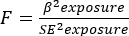
Instrument selection. Due to the small sample size, no SNPs met the criteria when the p-value threshold was set to 5 × 10⁻8. Therefore, this study adjusted the threshold to 5 × 10⁻6. This adjusted threshold has been widely used in previous MR studies, which confirms its rationality and reliability15. To eliminate the confounding effects of linkage disequilibrium (LD), stringent filtering parameters were implemented in this study. Since LD prevents the complete independent segregation of different genes, Mendel's law of independent assortment does not apply to all genetic variants. The LD clumping procedure was performed with explicit criteria (r2 < 0.001, genetic distance 10,000 kb) to minimize analytical bias induced by LD. In addition, consistency checks were performed on the effect directions between the effect allele (EA) and the other allele (OA). During this process, SNP loci with palindromic structures were excluded to reduce analytical bias arising from issues such as incorrect allelic pairing. Finally, the strength of the association between continuous or discrete genetic instrumental variables (IVs) and continuous exposure factors was quantified using the F-statistic derived from linear regression models. Generally, an F-statistic ≥ 10 is considered sufficient to effectively avoid causal estimation bias caused by weak instrumental variables. This study assessed the strength of instrumental variables (IVs) using the formula
 (1)
(1)
βexposure = Beta coefficient for exposure (effect size of SNP on exposure).
SEexposure = Standard error of the beta estimate for exposure.
F = F-statistic (measure of instrumental variable strength).
Using the aforementioned methods, this study identified 59 SNPs.
Two-sample MR and sensitivity analyses
Within the two-sample Mendelian randomization framework, multiple analytical methods were applied to explore the causal link between H. pylori antibodies and IDA, encompassing inverse-variance weighted (IVW), MR-Egger regression, weighted median, weighted mode, and simple mode approaches. The IVW approach was used as the primary analysis, assuming that all SNPs were valid but vulnerable to horizontal pleiotropy13. The MR-Egger intercept test was employed to identify pleiotropy, where P > 0.05 indicates no significant difference, thus suggesting the absence of pleiotropy. Additionally, sensitivity analyses were conducted, utilizing MR-PRESSO to detect and correct for the impact of outliers in the data17. The primary IVW and MR-Egger methods were evaluated for heterogeneity. Cochran’s Q-statistic was employed to ascertain whether IVs exhibited heterogeneity, with a P > 0.05 indicating there was no heterogeneity. The leave-one-out analysis was employed to ensure that the results remain uninfluenced by individual-biased SNPs18. Statistical analyses and data visualizations were conducted using R version 4.3.2. Mendelian randomization analyses were performed with the TwoSampleMR package (version 0.6.8) and the MRPRESSO package (version 1.0). Key analytical functions included: TwoSampleMR::harmonise_data(), TwoSampleMR::mr(), and RPRESSO::mr_presso().
Results
Genome-wide significant SNPs with P < 5 × 10⁻6 were selected. Then, SNPs in linkage disequilibrium (r2 < 0.001 within a window size of 10,000 kb) and unreconciled palindromic SNPs were eliminated, leaving 59 SNPs of the antibody levels of H. pylori IgG, CagA, VacA, UREA, CAT, OMP, and GroEL (Table 2). Subsequent MR-PRESSO analysis confirmed the absence of outliers among these SNPs. Following the removal of palindromic SNPs, five MR analytical approaches were applied to evaluate the causal association between H. pylori antibody markers and IDA. Analytical results showed that H. pylori OMP antibody was significantly associated with IDA susceptibility using the IVW approach (OR = 1.077, 95% CI: 1.010 – 1.148, P = 0.02194) (Table 2). The scatter plot depicts the estimated effect sizes of individual SNPs on H. pylori outer membrane protein (OMP) antibody levels and iron deficiency anemia (IDA) (Figure 2). To further confirm the robustness and validity of the MR results, a series of comprehensive control and validation analyses (robustness checks) were systematically performed, including heterogeneity testing, horizontal pleiotropy assessment, and leave-one-out sensitivity analysis, as detailed below. First, the MR-Egger and inverse variance-weighted (IVW) methods were employed to evaluate heterogeneity among the instrumental variables (IVs) (Table 3). Cochran's Q test revealed no significant heterogeneity across the IVs (MR-Egger Q = 8.78, P = 0.27; IVW Q = 8.72, P = 0.19), indicating that the impact of heterogeneity on the causal effect estimate was negligible. Accordingly, the fixed-effects IVW model was used to calculate the effect size in this study. Additionally, the horizontal pleiotropy of instrumental variables was evaluated via the MR-Egger intercept test and MR-PRESSO global test (Table 3). The MR-Egger intercept test showed no evidence of horizontal pleiotropy among the IVs (P > 0.05). Similarly, the MR-PRESSO global test detected no outlier SNPs or horizontal pleiotropy (P > 0.05). Finally, leave-one-out analysis was conducted, and the results demonstrated no substantial changes in the overall causal effect estimate (Figure 3), suggesting that the causal effect of H. pylori OMP antibody levels on iron deficiency anemia was driven by the collective contribution of these eight SNPs rather than the influence of any single SNP. To rule out the possibility of reverse causality, this study further performed reverse MR analysis, which revealed no significant causal effect of IDA on H. pylori OMP antibody levels (Table 4). In summary, the key findings of this study are as follows: (1) Among seven H. pylori antibodies (IgG, CagA, VacA, UREA, CAT, OMP, GroEL), only OMP antibody levels were significantly associated with an increased risk of IDA (IVW OR = 1.077, 95% CI 1.010–1.148, P = 0.02194). (2) Reverse MR analysis excluded the possibility of reverse causality, confirming that IDA does not affect H. pylori OMP antibody levels. (3) Comprehensive robustness checks (heterogeneity testing, horizontal pleiotropy assessment, leave-one-out analysis, and outlier detection) confirmed the validity and reliability of this causal association, with no evidence of heterogeneity, horizontal pleiotropy, or single-SNP bias. These findings have important implications for understanding the etiological relationship between H. pylori infection and IDA: they suggest that H. pylori infection, specifically through elevated OMP antibody levels, may be a causal risk factor for IDA. This provides a potential mechanistic link between H. pylori and iron metabolism disorders, and highlights the need for further research to explore the underlying biological pathways (e.g., OMP-mediated iron sequestration or mucosal damage) that may contribute to IDA development. Additionally, these results may have clinical implications, as targeted intervention for H. pylori infection (especially in populations with elevated OMP antibodies) could potentially reduce the risk of IDA.
DATA AVAILABILITY:
The present study is based on freely available summary statistics from genome-wide association studies. Data regarding H. pylori antibodies and IDA are from IEU Open GWAS (IEU Open GWAS project (mrcieu.ac.uk)). Raw data are provided in the supplementary appendix.
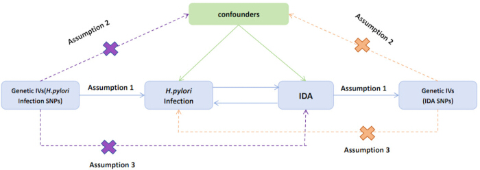
Figure 1: The workflow of the Mendelian randomization (MR) study. Abbreviations; SNP = single nucleotide polymorphism; IDA = iron deficiency anemia. Please click here to view a larger version of this figure.
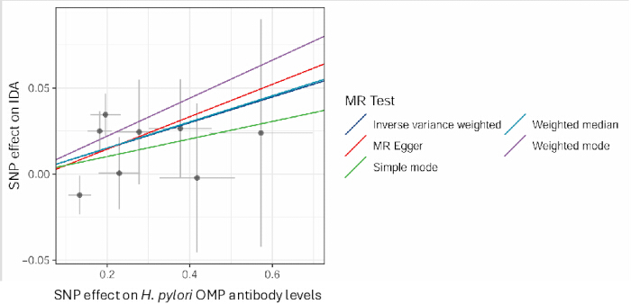
Figure 2: Scatter plot. The horizontal axis shows SNP effects on H. pylori OMP antibody (exposure), and the vertical axis shows SNP effects on IDA (outcome). Perpendicular bars at each point represent the 95% CI. Lines of different colors indicate causal estimates from different MR methods. Abbreviations; SNP = single nucleotide polymorphism; CI = confidence interval; MR = Mendelian randomization. Each scatter point denotes an SNP locus. Please click here to view a larger version of this figure.
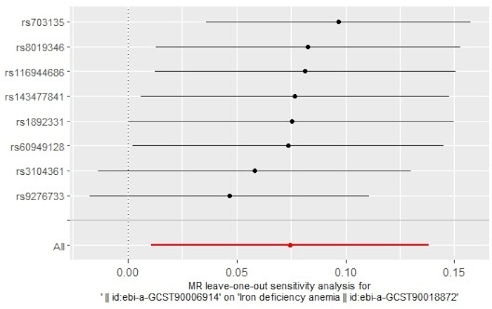
Figure 3: Leave-one-out Plot. Each black dot shows the leave-one-out IVW causal effect estimate of each remaining SNP, with horizontal black lines indicating the corresponding 95% CIs. The red dot represents the overall IVW pooled causal effect estimate of all SNPs, and the horizontal red line denotes its 95% CI. Abbreviations; SNP = single nucleotide polymorphism; MR = Mendelian randomization; IVW = inverse variance weighted method. Please click here to view a larger version of this figure.
| Exposure and Outcome | Sample size | Ancestry | Year | Numbers of SNPs | GWAS ID |
| Anti-H. pylori IgG | 8735 | European | 2020 | 9,170,312 | ebi-a-GCST90006910, https://opengwas.io/datasets/ebi-a-GCST90006910 |
| H. pylori CagA antibody | 985 | European | 2020 | 9,165,056 | ebi-a-GCST90006911,https://opengwas.io/datasets/ebi-a-GCST90006911 |
| H. pylori CAT antibody | 1558 | European | 2020 | 9,167,570 | ebi-a-GCST90006912,https://opengwas.io/datasets/ebi-a-GCST90006912 |
| H. pylori GroEL antibody | 2716 | European | 2020 | 9,172,299 | ebi-a-GCST90006913,https://opengwas.io/datasets/ebi-a-GCST90006913 |
| H. pylori OMP antibody | 2640 | European | 2020 | 9,167,440 | ebi-a-GCST90006914,https://opengwas.io/datasets/ebi-a-GCST90006914 |
| H. pylori UREA antibody | 2251 | European | 2020 | 9,170,248 | ebi-a-GCST90006915,https://opengwas.io/datasets/ebi-a-GCST90006915 |
| H. pylori VacA antibody | 1571 | European | 2020 | 9,178,635 | ebi-a-GCST90006916,https://opengwas.io/datasets/ebi-a-GCST90006916 |
| IDA | 480941 | European | 2021 | 24180477 | ebi-a-GCST90018872,https://opengwas.io/datasets/ebi-a-GCST90018872 |
Table 1: Characteristics of exposure and outcome. Abbreviations; SNP: single-nucleotide polymorphisms; IDA: iron deficiency anemia
Table 2: The result of the MR analysis. Abbreviations; SNP = single-nucleotide polymorphisms; CI = confidence interval; IDA = iron deficiency anemia. Please click here to download table 2.
| Exposure | Outcome | Heterogeneity | Pleiotropy | ||
| Q statistic (IVW) | p | MR-Egger intercept | p | ||
| H. pylori OMP antibody | IDA | 8.78 | 0.27 | 8.72 | 0.19 |
Table 3: Sensitivity analysis results. Abbreviations; IVW = inverse variance weighted method; IDA = iron deficiency anemia.
| Exposure | outcome | N.SNPs | Methods | OR (95% CI) | p |
| IDA | Anti-H. pylori IgG | 2 | Inverse variance weighted | 1.03 (0.92–1.18) | 0.54 |
| IDA | H.pylori CagA antibody | 2 | Inverse variance weighted | 1.94(0.89–4.20) | 0.09 |
| IDA | H.pylori CAT antibody | 2 | Inverse variance weighted | 0.82 (0.43–1.56) | 0.54 |
| IDA | H.pylori GroEL antibody | 2 | Inverse variance weighted | 0.74 (0.46–1.17) | 0.2 |
| IDA | H.pylori OMP antibody | 2 | Inverse variance weighted | 1.55 (0.96–2.51) | 0.07 |
| IDA | H.pylori UREA antibody | 2 | Inverse variance weighted | 1.18 (0.54–2.61) | 0.67 |
| IDA | H.pylori VacA antibody | 2 | Inverse variance weighted | 0.74 (0.40–1.37) | 0.34 |
Table 4: The result of reverse MR validation. Abbreviations; SNP = single-nucleotide polymorphisms; CI = confidence interval; IDA = iron deficiency anemia.
Discussion
Iron deficiency anemia (IDA) significantly impairs patients' work capacity and increases mortality, thereby constraining socioeconomic development. In most epidemiological surveys across both developing and developed countries, the overall mortality rate of IDA is generally underestimated19,20. The world health organization (WHO) explicitly recommends that researchers and clinicians thoroughly investigate the etiology of IDA and develop targeted therapeutic strategies, as timely intervention not only restores patients' health but also enhances national productivity21. IDA is induced by multiple complex factors, including insufficient iron intake, chronic blood loss, chronic diseases, malabsorption, hemolysis, or a combination of these2. Among these potential etiologies, whether H. pylori infection contributes to the development and progression of IDA remains controversial11,22,23. H. pylori infection is a highly prevalent global microbial disease, affecting over 50% of the global population; infection rates reach 70–90% in parts of Africa, Mexico, South America, and Central America24,25. This bacterium is a well-established major causative agent of digestive diseases such as peptic ulcers and gastric cancer26,27, and recent studies have also linked it to various extragastric conditions28,29,30. Current research evidence on the link between H. pylori infection and IDA is still insufficient. Accordingly, this study sought to elucidate the causal association between the two conditions.
A study first reported that H. pylori eradication exerts a beneficial therapeutic effect on refractory IDA, suggesting a potential link between H. pylori and IDA31. A large-scale U.S. population-based study enrolling 7,462 children, adolescents, and adults demonstrated that H. pylori infection is an independent risk factor for IDA, with infected individuals having a significantly increased risk of developing IDA (OR = 2.6, 95% CI: 1.5–4.6)32. Subsequent observational studies and meta-analyses have further validated this association: a previous meta-analysis integrating 15 observational investigations and 5 randomized controlled trials (RCTs) demonstrated a notable association between H. pylori infection and IDA, with a pooled OR of 2.22 (95% CI: 1.52–3.24, P < 0.0001). Moreover, following eradication therapy, patients exhibited a mean increase in hemoglobin of 4.06 g/L (95% CI: -2.57–10.69, P = 0.01) and a mean increase in serum ferritin of 9.47 μg/L (95% CI: -0.50–19.43, P < 0.0001)33. Study confirmed that approximately 60% of patients with H. pylori-associated IDA achieve significant anemia remission and marked elevations in key iron metabolic indices (e.g., serum ferritin, transferrin saturation) after H. pylori eradication33. These clinical findings collectively support a pathogenic role of H. pylori in IDA development, yet the underlying molecular mechanism bridging H. pylori OMP function and host iron depletion remains poorly elucidated.
Multiple well-recognized biological mechanisms underlie the initiation and exacerbation of IDA by H. pylori infection, with emerging molecular evidence highlighting the central role of outer membrane proteins (OMPs) in bacterial iron acquisition and the disruption of host iron homeostasis. The classic pathological pathway primarily involves gastric mucosal injury: H. pylori colonization induces damage to gastric epithelial cells, triggers chronic gastritis and mucosal atrophy, and suppresses gastric acid secretion. Gastric acid is indispensable for the release of dietary iron and subsequent intestinal absorption; thus, hypochlorhydria directly impairs iron bioavailability, leading to serum iron and transferrin saturation levels that meet the diagnostic criteria for IDA34. Concurrently, chronic inflammatory responses further downregulate the expression of duodenal iron transporters, limiting iron absorption, while exfoliation of damaged mucosal cells exacerbates intestinal iron loss—creating a dual iron-depleting effect characterized by reduced uptake and excessive consumption35,36. In addition to causing gastric mucosal damage, H. pylori competes directly with the host for iron acquisition through outer membrane proteins and other iron metabolism-regulatory proteins. Specifically, the bacterium expresses surface OMPs, such as lactoferrin-binding proteins and transferrin-binding proteins, which specifically recognize host iron carriers to sequester iron for bacterial proliferation. This process directly depletes systemic iron reserves, thereby triggering absolute iron deficiency35,37.
Additionally, infection-induced pro-inflammatory cytokines upregulate hepcidin expression, which mediates the internalization and degradation of intestinal iron transporters, blocks iron release from macrophages, and induces systemic iron sequestration—further exacerbating the anemic state38,39. Advancements in microbial molecular biology have clarified the functional characteristics of H. pylori iron metabolism-related proteins, particularly the OMP family, which provides a direct biological basis for interpreting the primary study findings40. Although canonical siderophores and their dedicated receptors have not been identified in H. pylori, the bacterium encodes a panel of key regulatory and transport proteins, including ferric uptake regulator (Fur), high-affinity ferrous iron transporter (FeoB), ferric citrate transporter (FecA), and non-heme iron-containing ferritin (Pfr)41. As a central transcriptional regulator, Fur senses ambient iron availability and modulates the expression of iron-responsive genes to maintain bacterial iron homeostasis42,43.
Notably, the OMP Frp family—comprising FrpB1, FrpB2, and FrpB3—possesses conserved β-barrel membrane structures and hemoglobin-binding motifs, with distinct expression patterns in response to different human iron sources (e.g., heme and hemoglobin)44. Under iron-limited conditions, FecA and Frp family OMPs are significantly upregulated to enhance bacterial iron-capturing capacity, whereas only minimal OMP expression is retained under iron-replete conditions to sustain basic metabolic demands45. These MR results, which reveal a causal association between H. pylori OMP antibody markers and IDA risk, align closely with this biological paradigm: elevated OMP-related immune responses reflect active H. pylori colonization and enhanced OMP-mediated iron sequestration, which directly disrupts host iron balance and increases IDA susceptibility.
Although the observed effect size of OMP antibodies on IDA is relatively modest (OR = 1.08), a small but statistically significant association is biologically and clinically meaningful in this context. As a chronic, cumulative metabolic disorder, IDA is often driven by long-term subtle perturbations in host iron homeostasis rather than strong acute triggers. Even a slight elevation in OMP antibody levels reflects persistent H. pylori colonization and sustained competitive iron acquisition at the gastric mucosal level, which can gradually exhaust systemic iron reserves and elevate long-term IDA risk. Such minor effect magnitudes are common in serological biomarker and chronic infectious disease-related analyses, and still carry valuable predictive etiological and public health implications, rather than being regarded as merely a trivial statistical finding.
Notably, the pathogenic association between H. pylori infection and IDA exhibits prominent geographical heterogeneity, which introduces inherent limitations to the generalizability of these findings, as they are derived exclusively from a European population. Such heterogeneity is largely attributed to interregional differences in H. pylori strain virulence, population dietary structure, and genetic background46. H. pylori isolates from East Asia are predominantly CagA-positive, mostly of the highly virulent EPIYA-D subtype, whereas Western strains have a higher proportion of low-virulence EPIYA-C subtypes47.
Dietary iron source patterns also modify disease susceptibility: plant-based iron, the major dietary iron source in East Asian populations, is highly dependent on gastric acid for absorption, rendering individuals more vulnerable to IDA when H. pylori infection suppresses acid secretion. In contrast, Western populations have a higher intake of animal-based iron, whose absorption is less susceptible to hypochlorhydria-mediated impairment. Additionally, inconsistent IDA diagnostic thresholds for serum ferritin and variable H. pylori detection methodologies across databases may introduce residual confounding. Methodologically, future studies could adopt multi-population two-sample MR designs, conduct stratified analyses by strain genotype and dietary pattern, and incorporate multi-omics data to validate the OMP-centered pathogenic pathway.
From a clinical perspective, H. pylori eradication offers distinct advantages over conventional single iron supplementation for the management of IDA. Specifically, combined H. pylori eradication and iron supplementation achieves superior therapeutic efficacy compared with iron monotherapy, particularly in idiopathic and refractory IDA cases46,48. Eradication therapy targets the fundamental etiology of H. pylori-related IDA, avoids gastrointestinal adverse events associated with long-term iron supplementation, and reduces long-term medical costs. Patients with persistent H. pylori infection also exhibit a poorer therapeutic response to iron supplementation alone, whereas prior bacterial eradication markedly improves iron repletion efficiency49. In East Asian regions with high H. pylori prevalence, routine H. pylori screening in IDA patients and standardized eradication for seropositive individuals hold substantial public health and clinical value. Collectively, this MR analysis of individuals of European ancestry confirms a robust causal association between circulating H. pylori OMP antibody levels and incident IDA risk, reinforcing the clinical rationale for incorporating H. pylori eradication into IDA therapeutic regimens.
Nevertheless, this study has limitations: it is constrained by population restriction, which limits cross-ethnic generalizability, and no significant correlations were observed between other H. pylori antibody subtypes and IDA risk. Large-scale multicenter studies encompassing diverse ethnic cohorts are therefore warranted to clarify the strain-specific pathogenic effects of H. pylori on IDA, validate the OMP-driven iron acquisition mechanism identified herein, and support the development of individualized prevention and targeted treatment strategies for patients with IDA.
Disclosures
The authors have no relevant financial or non-financial interests to disclose.
Acknowledgements
The authors would like to thank all participants and investigators who contributed to the GWAS data. This work was supported by grants from the Linyi People's Hospital Innovation Team Development Project (LYSRMYY-KCTD-008).
Materials
| Name | Company | Catalog Number | Comments |
|---|---|---|---|
| R (v4.3.2) | Software | R Core Team | https://www.r-project.org |
| TwoSampleMR (v0.6.8) | R package | MRC Integrative Epidemiology Unit (IEU) | https://mrcieu.github.io/TwoSampleMR/ |
| MR-PRESSO (v1.0) | R package | Verbanck et al. (2018) | https://github.com/rondolab/MR-PRESSO |
| OpenGWAS database | Database | MRC IEU | https://gwas.mrcieu.ac.uk |