Here, we present three uniportal endoscopic protocols for lumbar decompression. These techniques include interlaminar microdiscectomy, interlaminar laminectomy, and transforaminal microdiscectomy.
Method Article
Uniportal Endoscopic Techniques for Lumbar Decompression: Interlaminar Microdiscectomy, Interlaminar Laminectomy, and Transforaminal Microdiscectomy
329 views
⸱
September 11th, 2026
In This Article
Summary
Abstract
Endoscopic spine surgery enables targeted neural decompression with the least amount of tissue disruption to paraspinal structures compared to other spine surgery techniques. Though with minimal visualization, endoscopic spine surgery can be challenging. This article presents a stepwise overview of three key uniportal lumbar endoscopic techniques: interlaminar microdiscectomy, interlaminar laminectomy, and transforaminal microdiscectomy. Through integrated operative footage, the videos demonstrate indications, portal trajectory, and critical anatomic landmarks unique to each approach.
The interlaminar microdiscectomy technique is illustrated for median and paramedian disc herniations, emphasizing safe ligamentum flavum separation, dural protection, and direct fragment removal under endoscopic visualization. The interlaminar laminectomy segment details the management of central and lateral recess stenosis, highlighting stepwise bone removal, decompression of traversing and exiting roots, and controlled hemostasis within a limited corridor. The transforaminal microdiscectomy sequence demonstrates access through Kambin's triangle, foraminoplasty, and targeted fragment excision without violating the spinal canal. Operative tips focus on portal alignment, irrigation control, and avoidance of nerve injury, supplemented by discussion of ergonomic hand positioning and intraoperative troubleshooting.
Together, these video demonstrations provide a comprehensive visual reference for surgeons transitioning to endoscopic lumbar surgery. By standardizing procedural steps and emphasizing anatomical orientation, this method article aims to enhance reproducibility, expand technical proficiency, and promote the safe adoption of endoscopic decompression for lumbar disc and canal pathology.
Introduction
Endoscopic disc surgery was founded in the 1970s and represents a trend toward minimally invasive surgical approaches1. Neurosurgery has been at the forefront of technological advancement, with each generation introducing new tools that improve surgical accuracy, efficiency, and clinical outcomes2. The combination of endoscopic visualization with external monitors has expanded the adoption of this surgical approach from lumbar to thoracic and cervical pathologies3.
Endoscopic spine surgery continues to gain popularity due to its precision and minimally invasive approach to common spinal conditions like disc herniation. These approaches have expanded to tumor resection, infections, and segmental instability4. The technique offers several advantages, which include minimal tissue damage, decreased length of stay, and improved patient outcomes. The endoscopic approach is done through careful dissection, serial dilation, and placement of a working cannula5. Once in position, the endoscope is advanced while soft tissue is dissected until the proper trajectory and anatomical landmarks are visualized.
Research demonstrates positive outcomes for patients, with recent studies showing comparable or even superior clinical results when compared to traditional open microdiscectomy5. One limitation of endoscopic approaches cited by surgeons is the difficulty in visualizing anatomy and confirming proper location, trajectory, and depth perception6. These challenges are especially true early in training and during independent practice when there are limited anatomical landmarks or guides for decision making.
The primary objective and educational purpose of this manuscript are twofold. First, our goal is to provide a reproducible, step-by-step visual guide for endoscopic techniques used for lumbar decompression. Second, to improve procedural standardization while validating the efficacy of endoscopic spine surgery. We present three key techniques with step-by-step guidance and corresponding visual documentation. These techniques include: interlaminar microdiscectomy, interlaminar laminectomy, and transforaminal microdiscectomy. This manuscript fills important gaps in the literature by providing a clear visualization of each technique, procedural standardization with defined anatomical landmarks, and training utility for surgeons looking to refine their endoscopic approaches. Our research combines technical description with visual evidence, which we believe serves both as validation of the techniques and as a practical guide for their implementation.
Protocol
This protocol follows the guidelines of the human research ethics committee of the University of California and was conducted in accordance with the Declaration of Helsinki.
1. Interlaminar Laminectomy (Figure 1)
- Find the intralaminar space.
- Identify the inferior edge of the cranial lamina. This is the starting point for the procedure.
- Perform Ipsilateral laminotomy of L4.
- Drill the inferior edge of the cranial lamina, proceeding laterally-to-medially and extending cranially while undercutting the base of the spinous process.
- Protect the dura safely by staying external to the ligamentum flavum until bone removal is complete. Avoid drilling centrally without orientation.
- Find the midline of flavum.
- Identify the midline of the ligamentum flavum. These landmark guides safe flavectomy and neural decompression.
- Perform contralateral drilling.
- On the contralateral side, drill and thin the superior medial edge of the caudal lamina until the ligamentum flavum is exposed along the laminar margins.
- Perform continuous irrigation to clear out bone dust, improve visualization, and minimize thermal injury.
- Detach the contralateral flavum.
- Using a Kerrison rongeur, identify the insertion points of the ligamentum flavum and detach it from the caudal bony edges.
- Work carefully to separate the ligament from the bone without traction on the dura.
- Perform ipsilateral drilling.
- Perform additional drilling on the ipsilateral lamina as needed to enlarge the working space and ensure complete ligamentum flavum detachments.
- Detach flavum from the ipsilateral side.
- Using a Kerrison rongeur, detach the ligamentum flavum from the cranial bony edges.
- In cases of hypertrophy or calcification, use careful sub-ligamentous dissection to avoid dural tears.
- Remove flavum.
- Use Pituitary rongeurs to grasp and remove the freed ligamentum flavum, typically working from caudal to cranial, to open the epidural space.
- Expose epidural fat with a distinct shimmer to confirm safe dissection into the epidural compartment.
- Complete laminectomy.
- Gently mobilize neural elements with a blunt dissector and confirm that the thecal sac and nerve roots are freely mobile, indicating sufficient decompression.
- Control epidural bleeding with electrocautery coagulation or thrombin.
- After hemostasis is achieved, withdraw the working cannula and close the skin incision with a single absorbable suture and skin glue.
2. Interlaminar microdiscectomy (Figure 2)
- Orientation and visualization
- Establish interlaminar access via a work cannula and identify orientation and visibility through cauterization and irrigation.
- Improve surgical field visibility and hemostasis through cauterization.
- Remove soft tissue.
- Dissect soft tissue and paraspinal muscles using bipolar cautery and pituitary graspers to establish a clear working corridor. Continue until the ligamentum flavum is exposed. At L5/S1, the interlaminar window is usually wide enough not to require drilling to gain access to the disc space.
- Resect the ligamentum flavum
- Carefully incise and resect the ligamentum flavum. Expand the defect in the flavum and expose the dura and nerve root with micro-rongeurs and Kerrison punches.
- Widen the corridor through the flavum as deemed necessary with Kerrison punches.
- Mobilize neural structures
- Identify the lateral edge of the nerve root and the dural sac. Probe and remove the perineural membrane with Micro-rongeurs. Recognize the dural sac and nerve root as they come into clear view.
- Insert a blunt probe into the neuroforamen to mobilize the nerve root. Gently retract the nerve with a nerve root retractor, and carefully probe in all directions to confirm the nerve is free from surrounding tissue. Establish a clear view of the disc herniation.
- Open annulus fibrosus and remove disc.
- Seat the cannula deeper into the interlaminar window, retract the nervous structures, and expose the disc herniation.
- Remove the herniated disc material with forceps in a piecemeal fashion to decompress the neural elements.
- Search for residual herniation.
- Rotate the cannula to visualize all angles of the disc space. Identify and remove any residual herniated material with forceps to ensure complete decompression.
- Confirm adequate decompression.
- Confirm adequate decompression by ensuring relaxation and free movement of the nerve root, together with pulsation of the dural sac. This signals the endpoint of the procedure.
- Control bleeding with ablation and hemostatic agents. Conduct a final inspection, confirm the nerve is decompressed and that no bleeding remains.
3. Transforaminal microdiscectomy (Figure 3)
- Establish transforaminal orientation and optimize visualization.
- Establish transforaminal access and identify orientation and visibility through cauterization and irrigation.
- Cauterize bleeding vessels to improve surgical field visibility while achieving hemostasis.
- Debulk and confirm anatomy.
- Debulk surrounding tissue and identify critical landmarks, which include: the caudal pedicle, superior articular process, disc material, and traversing nerve root.
- Clear identification prevents nerve injury and dural tears.
- Perform annular release and discectomy.
- Release fibrotic adhesions anchoring the disc fragment to neural structures using endoscopic rongeurs.
- Mobilize nerve structures and free them from adhesions and fibrotic tissue with blunt probes or endoscopic pen fields.
- Once mobilized, remove fragments with endoscopic Kerrison punches. Cylce between these tools as necessary.
- Confirm adequate decompression.
- Perform final decompression. Two endpoints confirm success: pulsation of the dural sac indicates compression is relieved, and free movement of the nerve root without tethering confirms adequate decompression.
- Confirm via a blunt probe passing above and below the nerve root.
- Achieve hemostasis and perform final checks.
- Control bleeding with radiofrequency ablation and hemostatic agents.
- Conduct a final inspection, confirm the nerve is decompressed, and obtain hemostasis.
Results
The study includes three patients who underwent a minimally invasive, endoscopic approach to achieve lumbar decompression secondary to disc herniation. All three patients experienced relief of symptoms as documented in progress notes. Intraoperative visualization via the endoscope confirmed decompression of the thecal sac and/or nerve root.
For the interlaminar laminotomy, the interlaminar space was identified, followed by laminotomy. The ligamentum flavum was detached and subsequently removed. The thecal sac was then freed, and full decompression was visualized. These steps were performed bilaterally (Figure 1).
For the interlaminar microdiscectomy, the surrounding soft tissue was removed. Once the ligamentum flavum was visualized, an opening was made and widened. Once the thecal sac was visualized, it was protected by the working cannula. The disc herniation was then removed, and decompression of the thecal sac was confirmed visually (Figure 2).
For the transforaminal microdiscectomy, Kambin's triangle was entered. Surrounding soft tissue was dissected and removed. The annulus and disc herniation were visualized and removed. The nerve root was then inspected to ensure proper decompression (Figure 3).
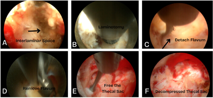
Figure 1: Interlaminar laminotomy. Endoscopic interlaminar laminotomy showing identification of the interlaminar space, removal of the ligamentum flavum, and bilateral decompression of the thecal sac. Please click here to view a larger version of this figure.
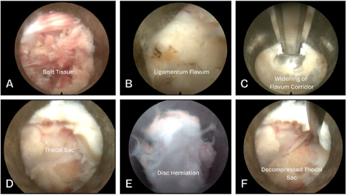
Figure 2: Interlaminar microdiscectomy. Endoscopic interlaminar microdiscectomy showing ligamentum flavum fenestration, thecal sac protection with the working cannula, and removal of the disc herniation with confirmed decompression. Please click here to view a larger version of this figure.
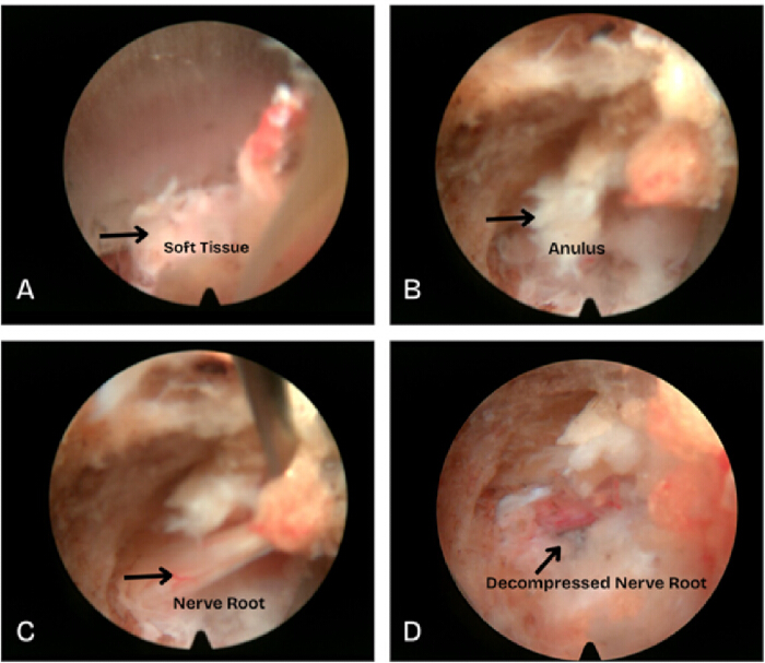
Figure 3: Transforaminal microdiscectomy. Endoscopic transforaminal microdiscectomy via Kambin's triangle showing visualization and removal of the disc herniation with inspection of the decompressed nerve root. Please click here to view a larger version of this figure.
Discussion
Lumbar disc herniation accounts for 5% of lower back disorders and remains one of the most common indications for spine surgery7. In patients with radicular pain and no severe neurological deficits, conservative management is advised for 4–6 weeks. For patients with progressive or persistent symptoms despite conservative non-operative therapy, microdiscectomy is indicated. Microdiscectomy is traditionally performed via an open approach, but can now be done via a tubular or endoscopic approach4. When comparing endoscopic microdiscectomy to the open approach, long-term 5-year data show no significant differences in pain, functional scores, or reoperation rates8. Compared to other approaches, endoscopic microdiscectomy is the most minimally invasive. The endoscopic technique is gaining popularity due to positive outcomes showing faster return to work, shorter hospital stays, and fewer infections when compared to open techniques9.
The complication rates for endoscopic microdiscectomy range from 9%–10%, with the most common complications being recurrence (6.6%) and reoperation (4.3%)10. There is a learning curve associated with this approach of about 20–50 cases5. An added benefit is that once proficiency is achieved, surgical time decreases by 50%11. When comparing open and endoscopic techniques head-to-head, they are equivalent regarding patient pain and functionality scores12. A recent meta-analysis demonstrates that endoscopic microdiscectomy also had lower reoperation rates compared to open surgery13. There is clear evidence that endoscopic approaches have perioperative advantages, including less blood loss, immediate postoperative pain relief, faster return to work, and less exposure to radiation12.
Patient selection is key in achieving favorable outcomes. Patients under 45 years old, those with symptoms less than 12 months, and those with isolated radiculopathy without significant back pain typically have improved outcomes14. Poor candidates include patients with spinal instability, more severe presentations like cauda equina syndrome, or those with fibrotic adhesions7.
There are disadvantages to this technique that warrant further attention. First, there is a learning curve, requiring 20–50 cases to become competent. Second, although there is good visualization of anatomy once the working cannula and endoscope are in place, reaching that point can be difficult and requires considerable skill. Third, once in the operative space, there is no visualization of the surrounding anatomy for reference; all necessary work must be done within the cannula. These challenges highlight the need for clear procedural documentation and visual guidance.
Several technical variations of interlaminar decompression are essential to mention. The first is the contralateral approach, in which the surgeon works from the opposite side of the dominant pathology. This trajectory can improve the surgeon's degrees of freedom and provide a more direct line to the target with better ergonomics. Kim et al. found it advantageous for asymmetric spinal stenosis, where a deviated spinous process or hypertrophic facet restricts ipsilateral access15. The timing and method of ligamentum flavum removal may vary as well. The flavum is often left intact until bony decompression is complete, where it protects the underlying neural elements from the drill and other sharp instruments before finally being detached16. The ligament can then be taken en bloc or piecemeal17. Some surgeons report en bloc resection as the more effective and safer option, but the evidence remains limited and requires more studies18.
Surgeons should be prepared to manage the complications specific to these techniques. One example includes Incidental dural tears, which are handled according to their size and characteristics. Small tears are often managed conservatively or with hemostatic agents, whereas larger defects (>10 mm) may require endoscopic patch repair or conversion to an open procedure19,20. Control of irrigation is another important surgical variable to consider. Continuous irrigation maintains the working field and supports hemostasis, but sustained high pressure carries its own risks. Formal thresholds have not been established, although current recommendations favor keeping irrigation pressure below 30 mmHg21. Reported complications include headache, neck pain, seizures, autonomic dysreflexia, and intracranial hypertension22,23. Uncontrolled irrigation should be avoided. The risks associated with irrigation should be a part of the preoperative discussion with the patient. Moreover, the surgeon should consider irrigation-related injury when a patient shows an unexpected neurological deficit during recovery.
Endoscopic spinal surgery delivers good results, but it carries real risks that deserve a frank accounting. Complications run local and systemic, some immediate and some delayed. The ones that matter most in practice are injury to neural structures, dural and other structural tears, residual pain, and persistent neurological deficits24.
The learning curve is steep, and most surgeons never touch these techniques in residency, which is exactly why a step-by-step visual reference is important. Cost is the next barrier. The instrumentation is expensive, and developing or underserved regions often cannot access the tools at all. Case selection matters too. A down-migrated fragment, a calcified herniation, or accompanying central stenosis can still favor a conventional open approach. Endoscopic techniques also make it harder to maintain normal curvature and restore biomechanical alignment, and revising the segment is more difficult if a second operation becomes necessary24.
The purpose of this manuscript is to demonstrate the current technique and procedural steps necessary to conduct a proper lumbar decompression. The protocol provides a series of approaches to decompress the spine following a disc herniation. Interlaminar microdiscectomy allows for the removal of disc herniation through the interlaminar window. Interlaminar laminectomy involves the removal of bone and is also useful for disc herniation. Transforaminal microdiscectomy allows for lateral access to the foramen and disc space, providing an alternative trajectory for decompression.
New Innovations are further expanding the capabilities of endoscopic approaches. These include larger working channels, expandable cages, 3D endoscopy, robotics, and augmented reality3. Overall, we believe these three endoscopic approaches offer effective treatment for disc herniation while offering minimal tissue damage and fast recovery. We believe this technique will continue to evolve moving forward.
Disclosures
Dr. Richard Price is involved in teaching and consulting for Joimax, Stryker, and VB Spine. All other authors have nothing to disclose.
Materials
| Name | Company | Catalog Number | Comments |
|---|---|---|---|
| C-arm mobile fluoroscopy unit (OEC) | GE HealthCare | https://www.gehealthcare.com/products/surgical-imaging | Intraoperative lumbar fluoroscopy; OEC model per institution |
| ChloraPrep | BD (Becton Dickinson) | 930815 | Skin antiseptic; Chlorhexidine gluconate and isopropyl alcohol surgical prep |
| Dermabond Advanced | Ethicon (Johnson & Johnson) | DNX12 | Tissue adhesive; Topical skin closure |
| Diamond burr, ball tip (abrasor) | Joimax | JSBDA323534C | Endoscopic bone removal; 3.5 x 320 mm, also 4.5 x 265 mm (JSBDA274544C) |
| Endoscopic camera head (1788) with coupler | Stryker | 1788 | Endoscopic video |
| High-speed drill and shaver handpiece (Shrill) | Joimax | https://www.joimax.com/en/shrill/ | Bone and soft-tissue removal; drives the diamond burr |
| iLESSYS Pro | Joimax | https://www.joimax.com/en/ilessys/ | Interlaminar endoscopic system; Working-channel endoscope and instrument set (rongeurs, Endo-Kerrison punches); interlaminar approach |
| Irrigation tubing set (Versicon) | Joimax | JTSB350D | Single-use; for Versicon JISP3000 irrigation pump |
| Jamshidi (11 G x 6 in) | BD (Becton Dickinson) | DJ6011X | Bone access needle; Percutaneous bony access |
| Surgiflo | Ethicon (Johnson & Johnson) | 2994 | Hemostatic matrix with thrombin (Flowable hemostatic matrix used with recombinant thrombin [Recothrom]) |
| Transforaminal access kit with 3 reamers (TESSYS) | Joimax | TDAK0020 | Serial dilators and reamers for foraminoplasty |
| Transforaminal endoscopic system (TESSYS) | Joimax | https://www.joimax.com/en/tessys/ | Working-channel endoscope and instrument set (graspers, cutters, Endo-Kerrison punches); transforaminal approach |
| Vaporflex | Joimax | JVP32024 | Bipolar radiofrequency probe; Ball tip; 320 mm and 275 mm (JVP28024); hemostasis and tissue ablation |
| Vicryl Plus | Ethicon (Johnson & Johnson) | VCP864D | Absorbable suture, polyglactin 910 (3-0, 18 in); Subcutaneous and skin closure |
References
- Heo DH, Kim HS, Whang YH, Lee DC, Lim K. History of endoscopic spine surgery: where did it all begin? Development of indications and techniques. Spine J. 2026;26(3):467-478.
- Vaishnav AS, Othman YA, Virk SS, Gang CH, Qureshi SA. Current state of minimally invasive spine surgery. J Spine Surg. 2019;5(Suppl 1):S2-S10.
- Hahn BS, Park JY. Incorporating new technologies to overcome the limitations of endoscopic spine surgery: navigation, robotics, and visualization. World Neurosurg. 2021;145:712-721.
- Ahn Y. A historical review of endoscopic spinal discectomy. World Neurosurg. 2021;145:591-596.
- Kapetanakis S, Chatzivasiliadis M, Gkantsinikoudis N, Pazarlis K. Full-endoscopic lumbar discectomy: a review of the surgical techniques, indications and anatomical considerations. J Clin Med. 2025;14:8961.
- Kwon B, Moon A. Advances in endoscopic lumbar spine surgery: a comprehensive review of the techniques used for the treatment of lumbar disc herniations and spinal stenosis and lumbar spinal fusion. Spine J. 2026;26(3):457-466.
- Sayed D, et al. The American Society of Pain and Neuroscience (ASPN) evidence-based clinical guideline of interventional treatments for low back pain. J Pain Res. 2022;15:3729-3832.
- Overdevest GM, et al. Tubular discectomy versus conventional microdiscectomy for the treatment of lumbar disc herniation: long-term results of a randomised controlled trial. J Neurol Neurosurg Psychiatry. 2017;88(12):1008-1016.
- Yong JH, Wang E, Chin BZ, Hey HWD. Full-endoscopic versus microscopic lumbar discectomy for lumbar disc herniation: a systematic review and meta-analysis of 4,186 cases. Spine J. 2026;26(5):981-995.
- Rajamani PA, et al. A 2-year outcomes and complications of various techniques of lumbar discectomy: a multicentric retrospective study. World Neurosurg. 2021;156:e319-e328.
- Olinger C, et al. Initial learning curve after switching to uniportal endoscopic discectomy for lumbar disc herniations. Eur Spine J. 2023;32:2694-2699.
- Chang H, et al. Comparison of full-endoscopic foraminoplasty and lumbar discectomy (FEFLD), unilateral biportal endoscopic (UBE) discectomy, and microdiscectomy (MD) for symptomatic lumbar disc herniation. Eur Spine J. 2023;32:542-554.
- Di L, et al. A systematic review and meta-analysis of preoperative characteristics and postoperative outcomes in patients undergoing endoscopic spine surgery: Part I endoscopic microdiscectomy. J Clin Med. 2025;14:6757.
- Jiang X, Zhou X, Xu N. Clinical effects of transforaminal and interlaminar percutaneous endoscopic discectomy for lumbar disc herniation: a retrospective study. Medicine (Baltimore). 2018;97:e13417.
- Kim JS, et al. Unilateral bi-portal endoscopic decompression via the contralateral approach in asymmetric spinal stenosis: a technical note. Asian Spine J. 2021;15:688-700.
- Kim HS, Wu PH, Jang IT. Lumbar endoscopic unilateral laminotomy for bilateral decompression outside-in approach: a proctorship guideline with 12 steps of effectiveness and safety. Neurospine. 2020;17(Suppl 1):S99-S109.
- Li C, et al. En bloc resection of the ligamentum flavum for bilateral decompression in unilateral biportal endoscopic transforaminal lumbar interbody fusion: a 2-year follow-up study. J Orthop Surg Res. 2024;19:815.
- Park CW, Oh JYL. Biportal endoscopic en bloc removal of the ligamentum flavum for spinal stenosis: nuances for the "butterfly" technique. Asian Spine J. 2024;18(4):587-593.
- Trathitephun W, et al. Intraoperative management of iatrogenic durotomy in endoscopic spine surgery: a systematic review. Neurospine. 2024;21(3):756-766.
- Kim HS, et al. Incidental durotomy during endoscopic stenosis lumbar decompression: incidence, classification, and proposed management strategies. World Neurosurg. 2020;139:e13-e22.
- Liu D, Mobbs RJ. Risk analysis of neurological deterioration associated with fluid insufflation in uniportal spine endoscopy: a case series and literature review. Int J Spine Surg. 2025;19(3):279-287.
- Vargas RAA, et al. Durotomy- and irrigation-related serious adverse events during spinal endoscopy: illustrative case series and international surgeon survey. Int J Spine Surg. 2023;17(3):387-398.
- Farshad M, et al. Intra-, epidural and intracranial pressure changes during interlaminar endoscopy, with and without dural tear. Neurospine. 2025;22(2):583-591.
- Kim HS, et al. Complications and limitations of endoscopic spine surgery and percutaneous instrumentation. Indian Spine J. 2020;3:78-85.
Reprints and Permissions
