This prospective observational study describes the development and internal validation of a predictive model using first-stage transabdominal and transperineal ultrasound parameters combined with maternal clinical characteristics to facilitate early identification of women at risk for prolonged second-stage labor.
A subscription to JoVE is required to view this content. Sign in or start your free trial.
Research Article
Development and Internal Validation of a Predictive Model for Prolonged Second-Stage Labor Using First-Stage Data: A Prospective Observational Study
55 views
⸱
September 11th, 2026
* These authors contributed equally
In This Article
Summary
Abstract
Prolongation of the second stage of labor is associated with adverse maternal and neonatal outcomes, making early risk identification clinically important. This study assessed whether combining transabdominal and transperineal ultrasound parameters obtained during the first stage of labor could predict a prolonged second stage. This prospective observational study enrolled primiparas who underwent transabdominal and transperineal ultrasound during the active first stage of labor. Fetal position, angle of progression (AOP), head–pubic symphysis distance, head–perineum distance, and maternal clinical characteristics were recorded. Participants were classified into normal (≤120 min) and prolonged (>120 minutes) second-stage groups. Least absolute shrinkage and selection operator (LASSO) regression was used to select key predictors, and multivariable logistic regression identified independent predictors for model development. Predictive performance was evaluated using receiver operating characteristic (ROC) curve analysis, calibration, and decision curve analysis (DCA) during internal validation. The analysis included 159 primiparas (95 normal and 64 prolonged). Compared with the normal group, the prolonged group had higher incidences of fetal distress (31.2% vs 15.8%, p = 0.035) and low Appearance, Pulse, Grimace, Activity, and Respiration (Apgar) scores (70.3% vs 36.8%, p < 0.001). LASSO regression identified four key predictors—maternal age, gestational body mass index increase, neonatal birth weight, and AOP—all of which remained independent predictors (p < 0.05). The predictive model achieved an area under the ROC curve of 0.927 and demonstrated good discrimination, calibration, and clinical utility in internal validation. First-stage ultrasound parameters combined with maternal clinical characteristics may facilitate the early identification of high-risk primiparas and support intrapartum management.
Introduction
Prolonged labor has long been a major concern in obstetric practice. In particular, prolongation of the second stage not only increases maternal pain but also contributes to adverse neonatal outcomes1. According to the latest guidelines issued by the World Health Organization (WHO), the normal duration of the second stage of labor is ≤120 min. In other words, a second stage lasting >120 min is considered prolonged. The global incidence of prolonged second stage is estimated to be 3%–8%1,2. Such prolongation increases the risks of maternal infection, cesarean section (C-section), postpartum hemorrhage, fetal distress, low Appearance, Pulse, Grimace, Activity, and Respiration (Apgar) scores, and perinatal mortality1. Previous studies have demonstrated that maternal age, gestational body mass index (BMI) increase, and neonatal birth weight are associated with a prolonged second stage of labor; however, their predictive value remains limited3. Therefore, identifying more effective predictors is of considerable importance for facilitating timely clinical interventions, optimizing labor progression, reducing the occurrence of prolonged labor, and improving maternal and neonatal outcomes.
In recent years, ultrasound imaging has become increasingly integrated into obstetric care, particularly in intrapartum management, because it enables real-time, objective assessment of fetal position, orientation, and descent3,4. Evidence suggests that intrapartum ultrasound plays an important role in predicting labor progression, evaluating fetal status, and guiding clinical decision-making regarding the mode of delivery5. Nevertheless, important gaps remain in the current literature. First, relatively few studies have specifically investigated the use of ultrasound parameters to predict a prolonged second stage of labor. Second, although fetal head position, fetal position, and their spatial relationship to the maternal pelvis during the first stage of labor may directly influence the duration of the second stage6, studies evaluating these first-stage ultrasound parameters in relation to second-stage prolongation remain limited.
Against this backdrop, the present prospective observational study collected delivery data from a single center during the first stage of labor. Ultrasound parameters were obtained using transabdominal and transperineal scanning, including head–symphysis distance (HSD), angle of progression (AOP), and head–perineum distance (HPD). Unlike previous ultrasound-based studies that primarily focused on predicting immediate delivery outcomes or the need for operative intervention during the second stage of labor, the present study specifically investigated the early prediction of prolonged second-stage labor using first-stage ultrasound parameters. These parameters were combined with maternal demographic and clinical characteristics to evaluate their predictive value for prolonged second-stage labor. This approach addresses an important knowledge gap because no previous study has integrated multidimensional first-stage ultrasound findings with maternal clinical characteristics into a predictive model for second-stage prolongation. Accordingly, a predictive model was developed and internally validated. This study is expected to provide clinicians with an objective tool for the early identification of primiparas at risk of prolonged second-stage labor during the first stage of labor, thereby supporting more individualized intrapartum management and improved maternal and neonatal outcomes.
Access restricted. Please log in or start a trial to view this content.
Protocol
This study was conducted in accordance with the Declaration of Helsinki and approved by the Ethics Committee of Yuyao Maternal and Child Health Hospital (Yuyao Second People’s Hospital) (approval No. YMCH-EC-2023KT-0012). All participants provided written informed consent.
Study Participants
Primiparous women with spontaneous onset of labor who successfully achieved vaginal delivery were included. This enabled precise recording of the second-stage duration required for the predictive model. The eligibility criteria were as follows: (1) singleton pregnancy with a gestational age of ≥37 weeks; (2) cephalic presentation; and (3) informed consent obtained from both the participant and her family. The exclusion criteria included the following: (1) absolute contraindications to trial of labor identified during the active phase (e.g., placenta previa, vasa previa, cord prolapse); and/or (2) secondary uterine inertia (defined as failure of cervical dilation or fetal descent to progress for ≥4 h despite oxytocin augmentation or amniotomy). Women undergoing induction of labor were also excluded.
Notably, women who required emergency surgical intervention (C-section or instrumental delivery) due to acute fetal distress during the second stage were managed according to clinical guidelines but were not included in this analysis in order to preserve the completeness of the labor duration records.
The sample size was calculated based on the results of a preliminary trial (n = 30, in which the incidence of second-stage prolongation was 20%). With α set at 0.05 and β at 0.20, assuming an estimated incidence of second-stage prolongation of approximately 20%, a minimum of 150 participants was required to detect a significant predictor with an odds ratio (OR) ≥ 2.5. A total of 159 women were included in the final analysis.
Acquisition of Clinical Data
Maternal variables included age, gestational body mass index (BMI) increase (defined as the difference between pre-pregnancy BMI and pre-delivery BMI), gestational diabetes, gestational hypertension, mental state (assessed using the Self-Rating Anxiety Scale, with a score ≥50 indicating anxiety), uterine inertia (specifically referring to a clinically assessed hypotonic contraction pattern, defined as uterine contractions occurring at intervals >5 min or lasting <30 s, distinguished from the American College of Obstetricians and Gynecologists [ACOG] definition of labor arrest), and parity. Neonatal variables included birth weight (kg), fetal distress (defined as recurrent late or variable decelerations on cardiotocography or a baseline fetal heart rate of <110 beats/min or >160 beats/min), and Apgar score (with a 1-min score of <7 classified as low).
Gestational diabetes was diagnosed according to the International Association of Diabetes and Pregnancy Study Groups criteria (fasting glucose 5.1–6.9 mmol/L or 1 h ≥10.0 mmol/L or 2 h 8.5–11.0 mmol/L on the 75 g Oral Glucose Tolerance Test at 24–28 weeks). Gestational hypertension was defined according to ACOG guidelines (systolic blood pressure ≥140 mmHg or diastolic blood pressure ≥90 mmHg measured twice ≥4 h apart after 20 weeks of gestation, without proteinuria). Fetal distress on cardiotocography was defined as recurrent late decelerations (≥50% of contractions), recurrent variable decelerations lasting >60 s, prolonged deceleration lasting 2–10 min, or a baseline fetal heart rate of <110 or >160 beats/min for ≥10 min (National Institute for Health and Care Excellence [NICE] criteria). Uterine inertia (hypotonic contractions) was defined as contraction intervals >5 min or contraction duration of <30 s over 30 min, as assessed by palpation or tocodynamometry. Mental state (anxiety) was assessed using the Self-Rating Anxiety Scale at admission (cervical dilation < 4 cm), with a raw score of ≥50 (standardized index) classified as abnormal. A low Apgar score was defined as a 1-min score of <7 (WHO).
Because this was an observational study, labor management followed routine clinical practice without a standardized protocol. However, epidural analgesia (yes/no, timing, duration), oxytocin augmentation (yes/no, total dose, duration), artificial rupture of membranes (yes/no, cervical dilation at the procedure), and mode of delivery (spontaneous vaginal delivery only, according to the inclusion criteria) were systematically recorded. No significant interaction between these variables and the primary model predictors was observed.
Transabdominal and Transperineal Ultrasound Examination at the Early First Stage of Labor
Ultrasound scanning was conducted at the beginning of the active phase of labor (cervical dilation ≥4 cm). The parturient was placed in the supine position with a 15° left lateral tilt after bladder emptying. For transperineal scanning, the thighs were slightly abducted and flexed. All images were captured at the peak of a uterine contraction, which was identified by clinical palpation in conjunction with external tocodynamometry (cardiotocography) to confirm the peak of each contraction. Each measurement was repeated during three separate contractions, with the mean value used for analysis. Ultrasound examinations were performed using a portable diagnostic ultrasound system (see Table of Materials). For transabdominal imaging, a convex probe (2–5 MHz) was used; for transperineal imaging, a high-frequency linear or small-footprint convex probe (5–8 MHz) was used. Imaging settings were standardized (depth 6–10 cm, gain optimized, and a single focal zone positioned at the pubic symphysis).
For fetal position assessment, a transabdominal transverse scan identified the fetal orbit, cerebellum, and thalamic midline relative to the maternal pelvis to determine occiput anterior (OA), transverse, or posterior position. For transperineal measurements, the transducer was covered with a sterile sheath and placed on the perineum in the midsagittal plane without excessive pressure. Anatomical landmarks included the entire pubic symphysis, the fetal skull contour, and the anorectal junction.
The following parameters were measured (Figure 1A–D): angle of progression (AOP), defined as the angle between the long axis of the pubic symphysis and a line extending from its inferior edge to the deepest point of the fetal skull; midline angle (MLA), defined as the angle between the fetal skull midline (falx cerebri) and the maternal pelvic anteroposterior axis; head–symphysis distance (HSD), defined as the distance from the inferior edge of the pubic symphysis to the nearest point on the fetal skull; and head–perineum distance (HPD), defined as the shortest distance from the outer fetal skull to the perineal skin7.
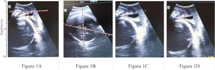
Figure 1. Ultrasound measurements obtained during the first stage of labor. (A) Measurement of the angle of progression (AOP), defined as the angle between the long axis of the pubic symphysis and a line extending from its inferior edge to the deepest point of the fetal skull. (B) Measurement of the midline angle (MLA), defined as the angle between the fetal skull midline (falx cerebri) and the maternal pelvic anteroposterior axis. (C) Measurement of the head–symphysis distance (HSD), defined as the distance from the inferior edge of the pubic symphysis to the nearest point on the fetal skull. (D) Measurement of the head–perineum distance (HPD), defined as the shortest distance from the outer surface of the fetal skull to the maternal perineal skin. Please click here to view a larger version of this figure.
Each measurement was performed three times during three separate contraction peaks, and the average value was used for analysis. All examinations were conducted by three experienced sonographers (each with ≥10 years of experience in obstetric ultrasound), who completed standardized training in intrapartum transperineal ultrasound measurements before the study. Competency was confirmed by achieving an intraclass correlation coefficient (ICC) of ≥0.90 against a gold-standard assessor (senior author). Inter-observer and intra-observer reproducibility were assessed during a pilot phase involving 10 patients (not included in the main study), yielding ICCs ranging from 0.89 to 0.96 for all parameters (AOP, HSD, HPD, and MLA).
Image acceptance required clear visualization of the pubic symphysis, fetal skull contour, and anorectal junction. Images with excessive transducer pressure or poorly visualized anatomical landmarks were discarded and reacquired. Sonographers were blinded to group assignment (normal versus prolonged second stage) and to clinical data during image acquisition and measurement. Data were double-entered with range checks, and out-of-range values triggered image re-review. Missing data (<5%) were handled using complete-case analysis after confirming that the data were missing at random (Little’s missing completely at random test, p > 0.05).
Recording and Grouping of Labor Duration
Throughout the labor process, the course of labor for each participant was carefully documented. The first stage of labor was defined as the period from the onset of regular uterine contractions to complete cervical dilation (10 cm). The active phase of labor was defined as cervical dilation ≥4 cm accompanied by intensified, regular uterine contractions. The second stage of labor was defined as the period from complete cervical dilation (10 cm) to complete fetal expulsion. Ultrasound examinations were performed during the active phase of the first stage of labor to assess fetal head position, fetal position, and related ultrasound parameters. According to the World Health Organization (WHO) guidelines, a normal second stage of labor should not exceed 120 min2. To maintain a standardized research threshold for all primiparas in this study, participants were categorized according to this 120-min criterion into two groups: a normal second-stage group (≤120 min) and a prolonged second-stage group (>120 min).
Statistical Analysis
All statistical analyses were performed using SPSS version 26.0 (RRID:SCR_002865) and R version 4.1.0 (RRID:SCR_001905) (see Table of Materials). The following R packages were used: glmnet (version 4.1-2) for least absolute shrinkage and selection operator (LASSO) regression, rms (version 6.2-0) for nomogram construction and calibration, pROC (version 1.18.0) for receiver operating characteristic (ROC) analysis, and rmda (version 1.6) for decision curve analysis (DCA). Normality of continuous variables was assessed using the Shapiro–Wilk test to guide the choice of descriptive statistics and comparison methods. Normally distributed data were presented as mean ± standard deviation (x̄ ± s) and compared using the independent-samples t-test, which is robust to mild deviations from normality when sample sizes are adequate. Non-normally distributed data were presented as the median with interquartile range (M [P25, P75]) and compared using the Mann–Whitney U test, which evaluates whether one distribution tends to have larger values than the other rather than strictly testing differences in medians. Categorical variables were presented as counts and percentages (n [%]) and compared using the chi-square (χ2) test or Fisher’s exact test when the expected frequency was <5. All tests were two-tailed, and a p-value <0.05 was considered statistically significant. Variables with p <0.1 in the univariate analysis were entered into the LASSO regression for variable selection. LASSO regression was performed using the glmnet package with 10-fold cross-validation to determine the optimal regularization parameter (λ). The one-standard-error criterion (the largest λ within one standard error of the minimum cross-validation error) was applied to select the most parsimonious set of predictors with non-zero coefficients. Before LASSO fitting, all continuous predictors (maternal age, gestational BMI increase, AOP, HSD, HPD, and neonatal birth weight) were centered and standardized to z-scores using the default glmnet setting (standardize = TRUE) so that coefficients were penalized equally regardless of measurement units. The selected variables were subsequently entered into a multivariable logistic regression model (enter method) to calculate adjusted odds ratios (aORs) and 95% confidence intervals (CIs). Model significance was assessed using the likelihood ratio test.
Based on the logistic regression coefficients, a predictive model was constructed as Logit(P) = β0 + Σ(βiXi). This model was visualized as a nomogram using the rms package, in which each predictor was assigned a point value proportional to its regression coefficient, and the total points were mapped to predicted probabilities. Discrimination was evaluated by plotting the ROC curve and calculating the area under the curve (AUC) with its 95% CI using the pROC package (bootstrap method with 1,000 resamples). The optimal cutoff probability was determined by maximizing the Youden index (sensitivity + specificity − 1), with the corresponding sensitivity and specificity reported. Calibration was assessed using a calibration curve generated from 1,000 bootstrap resamples (rms package), supplemented by the Hosmer–Lemeshow goodness-of-fit test (a non-significant p-value indicates good calibration). Clinical utility was evaluated using DCA with the rmda package, which calculates the net benefit across a range of threshold probabilities (0%–100%). Net benefit was defined as (true positives / n) − (false positives / n) × (threshold probability / [1 − threshold probability]). The overall study workflow is described above and illustrated in Supplementary Figure 1. This model is exploratory; the predictor neonatal birth weight refers to actual weight measured after delivery, and clinical prediction would require model refitting using estimated fetal weight (EFW) obtained before delivery.
Access restricted. Please log in or start a trial to view this content.
Results
Study Population and Group Assignment
A total of 159 parturients were included in the final analysis. Of these, 95 had a normal second-stage duration, whereas 64 experienced a prolonged second stage (Table 1). Compared with the normal group, the prolonged group had significantly higher incidences of fetal distress and low Apgar scores (both p < 0.05). No significant difference in perinatal mortality was observed between the groups (p > 0.05).
Access restricted. Please log in or start a trial to view this content.
Discussion
With continuous improvements in obstetric management, prolonged second-stage labor has become an increasingly important factor affecting maternal and neonatal outcomes. Previous studies have shown that prolonged second-stage labor is associated with increased labor complexity, higher rates of cesarean delivery, and lower neonatal Apgar scores1,2. These findings highlight the clinical importance of optimizing perinatal management and implementing strategies for th...
Access restricted. Please log in or start a trial to view this content.
Disclosures
Conflict of Interest:
The authors declare no competing financial or non-financial interests related to this work.
Acknowledgements
The authors thank the nursing and medical staff of the Department of Obstetrics at Yuyao Maternal and Child Health Hospital (Yuyao Second People’s Hospital) for their assistance with participant recruitment and data collection. This work was supported by the 2024 Yuyao Municipal Health Science and Technology Program (Grant No. 2024YYB03).
Access restricted. Please log in or start a trial to view this content.
Materials
| Name | Company | Catalog Number | Comments |
|---|---|---|---|
| Portable diagnostic ultrasound system | Mindray | CAU-01040644 | Portable diagnostic ultrasound system used for transabdominal and transperineal imaging; see Protocol for imaging settings |
| Convex ultrasound probe (2–5 MHz) | Mindray | C5-2 | Used for transabdominal ultrasound imaging |
| High-frequency linear or small-footprint convex ultrasound probe (5–8 MHz) | Mindray | 75L38EA | Used for transperineal ultrasound imaging |
| R software | R Foundation for Statistical Computing | Version 4.1.0 | Statistical computing software (RRID:SCR_001905) |
| SPSS software | IBM Corp. | Version 26.0 | Statistical analysis software (RRID:SCR_002865) |
| Self-Rating Anxiety Scale | Published questionnaire (Zung Self-Rating Anxiety Scale) | Not applicable | Anxiety assessment questionnaire used at admission |
References
- American College of Obstetricians and Gynecologists, Society for Maternal-Fetal Medicine. Obstetric Care Consensus No. 1: Safe prevention of the primary cesarean delivery. Obstet Gynecol. 2014;123(3):693-711. doi:10.1097/01.AOG.0000444441.04111.1d.
- World Health Organization. WHO Guidelines Approved by the Guidelines Review Committee. World Health Organization; Geneva; 2015.
- Miyamoto K, et al. Prolonged second stage of labor in delivery using epidural analgesia is a risk factor for postpartum urinary retention. J Obstet Gynaecol Res. 2024;50(3):424-429. doi:10.1111/jog.15867.
- Kahrs BH. Ultrasound before operative vaginal delivery: Why and how. Minerva Obstet Gynecol. 2021;73(1):67-73. doi:10.23736/S2724-606X.20.04667-5.
- Yano E, et al. Anatomical identification of ischial spines applicable to intrapartum transperineal ultrasound based on magnetic resonance imaging of pregnant women. J Matern Fetal Neonatal Med. 2022;35(25):9736-9741. doi:10.1080/14767058.2022.2051007.
- Kahrs BH, Eggebø TM. Intrapartum ultrasound in women with prolonged first stage of labor. Am J Obstet Gynecol MFM. 2021;3(6):100427. doi:10.1016/j.ajogmf.2021.100427.
- Lee NMW, et al. Implementation of sonopartogram: Multicenter feasibility study. Ultrasound Obstet Gynecol. 2024;64(2):214-221. doi:10.1002/uog.27634.
- Saucedo AM, et al. First and second stage risk factors associated with perineal lacerations. Matern Child Health J. 2024;28(7):1228-1233. doi:10.1007/s10995-024-03919-1.
- Zhang M, et al. Risk factors for episiotomy during vaginal childbirth: A retrospective cohort study in Western China. J Evid Based Med. 2018;11(4):233-241. doi:10.1111/jebm.12316.
- Friedman EA, Cohen WR. The active phase of labor. Am J Obstet Gynecol. 2023;228(5 Suppl):S1037-S1049. doi:10.1016/j.ajog.2021.12.269.
- Kalache KD, et al. Transperineal ultrasound imaging in prolonged second stage of labor with occipitoanterior presenting fetuses: How well does the angle of progression predict the mode of delivery? Ultrasound Obstet Gynecol. 2009;33(3):326-330. doi:10.1002/uog.6294.
- Chan YT, et al. Relationship between intrapartum transperineal ultrasound measurement of angle of progression and head-perineum distance with correlation to conventional clinical parameters of labor progress and time to delivery. J Matern Fetal Neonatal Med. 2015;28(12):1476-1481. doi:10.3109/14767058.2014.958459.
- Chor CM, et al. Prediction of labor outcome using serial transperineal ultrasound in the first stage of labor. J Matern Fetal Neonatal Med. 2019;32(1):31-37. doi:10.1080/14767058.2017.1369946.
- Maged AM, et al. Measurement of the fetal occiput-spine angle during the first stage of labor as predictor of the progress and outcome of labor. J Matern Fetal Neonatal Med. 2019;32(14):2332-2337. doi:10.1080/14767058.2018.1432589.
- Gimovsky AC, Berghella V. Evidence-based labor management: Second stage of labor (part 4). Am J Obstet Gynecol MFM. 2022;4(3):100548. doi:10.1016/j.ajogmf.2021.100548.
- Barak O, et al. The routine use of intrapartum ultrasound in clinical decision-making during the second stage of labor: Does it have any impact on delivery outcomes? Gynecol Obstet Invest. 2018;83(1):9-14. doi:10.1159/000455847.
- Beck R, et al. Intrapartum sonography of fetal head in second stage of labor with neuraxial analgesia: A literature review and possible medicolegal aftermath. Eur Rev Med Pharmacol Sci. 2019;23(8):3159-3166. doi:10.26355/eurrev_201904_17673.
- Li N, et al. Risk factors and clinical prediction of shoulder dystocia in non-macrosomia. Zhonghua Fu Chan Ke Za Zhi. 2015;50(1):17-21. doi:10.3760/cma.j.issn.0529-567X.2015.01.005.
- Carvalho Neto RH, et al. Assessment of the angle of progression and distance perineum-head in the prediction of type of delivery and duration of labor using intrapartum ultrasonography. J Matern Fetal Neonatal Med. 2021;34(14):2340-2348. doi:10.1080/14767058.2019.1666818.
- Haberman S, et al. A novel partogram for stages 1 and 2 of labor based on fetal head station measured by ultrasound: A prospective multicenter cohort study. Am J Perinatol. 2021;38(S01):e14-e20. doi:10.1055/s-0040-1702989.
- Pan WL, et al. Accuracy of non-invasive methods for assessing the progress of labor in the first stage: A systematic review and meta-analysis. BMC Pregnancy Childbirth. 2022;22(1):608. doi:10.1186/s12884-022-04938-y.
- Ghi T, Dall'Asta A. Sonographic evaluation of the fetal head position and attitude during labor. Am J Obstet Gynecol. 2024;230(3 Suppl):S890-S900. doi:10.1016/j.ajog.2022.06.003.
- Nassr AA, et al. Intrapartum ultrasound measurement of angle of progression at the onset of the second stage of labor for prediction of spontaneous vaginal delivery in term singleton pregnancies: A systematic review and meta-analysis. Am J Obstet Gynecol. 2022;226(2):205-214. https://doi.org/10.1016/j.ajog.2021.07.031.
- Hans R, Reddy D, Shetty J. Serial intrapartum ultrasound to predict vaginal delivery using angle of progression and head-progression distance in term nulliparous women. Eur J Obstet Gynecol Reprod Biol. 2025;293:18-23.
Access restricted. Please log in or start a trial to view this content.