This protocol describes a cost-effective workflow for mono- and multiplex immunofluorescence (IF) of adherent two-dimensional cell cultures, reducing experimental costs through inexpensive glass coverslips, standard 24-well plates, and low-volume staining conditions that minimize antibody consumption, while providing a representative approach for protein localization with standard fluorescence microscopy.
Method Article
A Cost-Effective Workflow for Mono- and Multiplex Immunofluorescence in Adherent Two-Dimensional Cell Cultures on Glass Coverslips
In This Article
Summary
Abstract
Immunofluorescence (IF) microscopy is a widely used technique for visualizing protein localization and co-distribution at single-cell resolution while preserving cellular morphology and subcellular organization. The goal of this protocol is to present a practical and cost-effective workflow for mono- and multiplex immunofluorescence staining of adherent 2D cell cultures using standard laboratory materials. The method relies on culturing cells on inexpensive glass coverslips placed in conventional 24-well plates and implements multiplexing through host-species separation of primary antibodies, spectrally distinct fluorophores, and robust control conditions, including no-primary, no-antibody, single-stain, and crosstalk controls to ensure interpretability of multiplex IF. Low-volume staining steps are used to reduce reagent and antibody consumption without compromising signal quality. This workflow avoids the need for specialized imaging plates or chambers and is compatible with routine widefield or confocal fluorescence microscopy. Overall, this protocol provides an accessible and practical immunofluorescence workflow for representative assessment of protein localization and co-distribution in adherent cell cultures. Adaptation to other cell types or antibody panels requires target-specific optimization, including antibody validation, fixation and permeabilization conditions, fluorophore selection, and compatibility with microscope and filter sets.
Introduction
Immunofluorescence (IF) microscopy is widely used to visualize the subcellular localization, relative abundance, and colocalization of proteins in fixed cells, thereby linking molecular markers to cellular morphology and subcellular context1,2. In contrast to bulk readouts (e.g., immunoblotting) or dissociative single-cell assays (e.g., flow cytometry), IF preserves cellular spatial organization and is particularly useful for assessing cell-to-cell phenotypic heterogeneity and changes in subcellular protein distribution at single-cell resolution3,4,5. As experimental questions increasingly require simultaneous assessment of multiple markers, multiplex IF enables the concurrent detection of two or more targets within the same specimen, commonly by combining primary antibodies raised in different host species with spectrally distinct fluorophores6 (Figure 1), using direct and/or indirect detection strategies (Figure 2). However, multiplexing introduces key technical challenges, especially antibody cross-reactivity and spectral crosstalk, that can compromise interpretability unless addressed through careful panel design and rigorous control conditions7,8.
The overall goal of this method is to provide a practical, low-cost workflow that is broadly applicable to immunofluorescence-validated antibodies and optimized for routine imaging of adherent 2D cultures. Multiplexing is implemented through host-species separation, spectrally distinct fluorophores, and control groups that verify background levels and staining performance9,10. By using 12 mm glass coverslips in standard 24-well plates rather than specialized chambered slides, this workflow avoids a dedicated, higher-cost consumable in favor of materials that are standard stock in most cell culture laboratories. The workflow also uses substantially less staining solution per unit growth area (150 µL for ≈1.13 cm2, or ≈133 µL/cm2) than comparable chambered coverslip formats11,12, which typically require 300–500 µL/cm2, corresponding to an approximately 2- to 4-fold reduction in per-area antibody and reagent consumption. Accordingly, the protocol supports qualitative multi-target assessment of marker localization and co-distribution using standard widefield or confocal microscopes, along with cytoskeletal and nuclear counterstains. This workflow is intended for conventional fixed-cell multiplex IF based on host-species separation and spectral distinction, and is not designed for iterative or high-dimensional multiplex approaches.
Protocol
This study did not involve human participants or animal subjects. All experiments were performed using the D17 canine osteosarcoma cell line, an established cell line obtained from a public cell bank (Banco de Células do Rio de Janeiro, Cat. No. 0276). No ethical approval was therefore required.
1. Cell line maintenance
- Maintain adherent cells under standard culture conditions (37 °C, 5% CO₂, humidified incubator) using the appropriate complete medium for the cell line used.
NOTE: As a representative example, D17 canine osteosarcoma cells (Banco de Células do Rio de Janeiro [BCRJ], Cat. No. 0276) are maintained in Dulbecco's Modified Eagle Medium (DMEM) supplemented with 10% fetal bovine serum (FBS) and 1% antibiotic-antimycotic, subcultured at 70–80% confluence using trypsin-EDTA(ethylenediaminetetraacetic acid), and used within a validated passage range (e.g., passages 5–20). - Confirm mycoplasma-negative status before experiments using a validated mycoplasma detection assay.
2. Preparation of 24-well plate and glass coverslips
NOTE: Coverslip surfaces should be cleaned by immersion in 70% ethanol (5 min, repeated 3×) prior to sterilization by either method described below; after sterilization, coverslips should be stored in a closed container at room temperature until use.
- Place clean 12 mm round glass coverslips in a glass Petri dish (or coverslip holder) and autoclave. Autoclave a pair of fine-tipped metal forceps as well.
- Alternative: Clean coverslips and forceps with 70% ethanol, allow to air-dry, and surface-decontaminate by ultraviolet (UV) exposure (30 min) inside a biosafety cabinet prior to use. Store sterilized coverslips in a closed container at room temperature until use.
- In a biosafety cabinet, use the sterile forceps to transfer one sterile coverslip into each well designated for the experimental groups in a 24-well tissue culture plate.
- Optional: coat coverslips with adhesion-promoting coatings following the manufacturer’s instructions or established laboratory guidelines, if required for the cell type used.
NOTE: Coating options (poly-L-lysine, collagen I, or fibronectin) and their working concentrations are listed in the Table of Materials. For most adherent cell lines cultured on glass, uncoated, sterilized coverslips are sufficient; coating components may be antigenic, so the selected coating should not cross-react with any antibodies in the panel. - If coverslips were coated, aspirate the excess coating solution, rinse once with sterile phosphate-buffered saline (PBS), and allow coverslips to air-dry in the biosafety cabinet before cell seeding.
3. Experimental groups
- Include a panel of controls appropriate to the multiplex design.
NOTE: A no-antibody control (neither primary nor secondary antibody) reveals intrinsic autofluorescence, and a no-primary control (secondary antibody cocktail without any primary) assesses non-specific secondary binding and background across all channels simultaneously; because this protocol uses strictly spectrally distinct secondary antibodies, this single control evaluates each secondary independently within its respective channel, serving the same purpose as a secondary-only control per fluorophore. Single-stain controls, in which each primary–secondary pair is applied individually, validate the performance of each antibody before multiplex combination and, by comparison with the multiplex condition, allow assessment of spectral crosstalk for each fluorophore pair. Whenever possible, a positive control that expresses the target antigen is used to confirm assay performance.
4. Cell seeding (plating) on coverslips
- In a biosafety cabinet, detach cells using trypsin (or other appropriate dissociation method), neutralize, and resuspend in complete culture medium.
- Count cells using an automated cell counter or a hemocytometer.
- Dilute cells to 6 × 10⁴–1 × 10⁵ cells/mL.
- Add 0.5 mL of cell suspension per well (final: 3 × 10⁴–5 × 10⁴ cells/well).
NOTE: If needed, a sterile pipette tip can be used to gently ensure the coverslip lies flat at the bottom of the well, avoiding scratching. - Incubate cells under standard conditions (37 °C, 5% CO₂) until cells have adhered and reached the desired confluence or experimental time point is reached.
5. Cell treatment (Optional)
- Select the treatment timing according to the experimental design. Treatment may include pharmacological therapy, cytokine stimulation, genetic manipulation to induce or knock down target gene expression, or other experimental interventions, as applicable.
- Label wells to identify control and treated groups.
- Incubate for the required time points; avoid over-confluency, as overgrowth increases background and compromises morphology.
NOTE: Cells should be adhered to the coverslip before fixation, unless the design requires fixation immediately after seeding. - For rapidly secreted targets (e.g., cytokines), add brefeldin A (BFA) to the culture medium at a final concentration of 1–10 µg/mL (commonly 5 µg/mL) and incubate for 4–6 h at 37 °C, 5% CO₂ before fixation.
- Remove the BFA-containing medium and proceed immediately to Fixation.
6. Immunofluorescence staining
NOTE: From fixation onward, sterile technique is not required; staining may proceed on the bench, using a chemical fume hood whenever a CAUTION reagent is handled. Samples must not dry at any point; when aspirating, a thin buffer film is left, and the next solution is added immediately.
- Fixation (choose one):
NOTE: The fixative is selected according to target localization and epitope sensitivity (see Discussion): Paraformaldehyde (PFA) (formaldehyde prepared from paraformaldehyde) suits most targets and is phalloidin-compatible; methanol may aid certain nuclear/phospho-epitopes but impairs F-actin staining.- PFA fixation (recommended for most targets):
- Aspirate medium and gently rinse once with Tris-buffered saline (TBS) (0.5 mL/well).
- Add 4% PFA in PBS (phosphate-buffered saline) (0.5 mL/well) and incubate for 10 min at room temperature (≈25 °C).
- Aspirate PFA and wash 3× with TBS (aspirate promptly; do not let wells dry).
NOTE: CAUTION: PFA is toxic and a potential carcinogen. Handle PFA solutions in a certified chemical fume hood while wearing appropriate personal protective equipment (lab coat, gloves, and eye protection). Avoid inhalation and skin contact, and dispose of waste in accordance with institutional chemical safety guidelines.
- PFA fixation (recommended for most targets):
- Alternatively, Methanol fixation (useful for some nuclear antigens and certain phospho-epitopes):
- Pre-chill 100% methanol to −20 °C.
- Add ice-cold methanol directly to cells (0.5 mL/well) and incubate for 5 min at −20 °C.
- Wash 3× with TBS (aspirate promptly; do not let wells dry).
NOTE: After methanol fixation, additional permeabilization may be unnecessary and should be optimized per antibody/epitope. Methanol fixation can damage membrane epitopes and impair fluorophore-conjugated phalloidin/F-actin staining. CAUTION: Methanol is toxic, volatile, and highly flammable. Handle in a well-ventilated area or chemical fume hood; wear appropriate personal protective equipment; and keep away from open flames and heat sources. OPTIONAL STOPPING POINT: After fixation, store samples at 4 °C in PBS containing a low concentration of fixative (e.g., PBS + 0.1% PFA) for up to one week; validate storage timing for each antigen.
- Aldehyde quenching (Optional)
- Incubate with 50 mM glycine in TBS (0.5 mL/well) for 5 min at room temperature (≈25 °C).
- Wash 3× with TBS + 0.05% polysorbate 20.
NOTE: Glycine neutralizes free aldehyde groups remaining after PFA fixation, reducing fixation-related autofluorescence and improving antibody staining specificity.
- Permeabilization
- If targeting membrane proteins, skip permeabilization and proceed to step 6.5.1.
- If targeting intracellular or nuclear proteins, permeabilize with 0.1–0.3% non-ionic detergent in TBS (0.5 mL/well) for 5–10 min at room temperature (≈25 °C).
- Wash 3× with TBS + 0.05% polysorbate 20 (TBST).
NOTE: Octylphenol ethoxylate nonionic detergent concentration and time are optimized to the target; adequacy is judged by correct-compartment signal, preserved morphology, well-defined F-actin, and low background (see Discussion). If saponin is used, it is maintained in all subsequent antibody and wash buffers. CAUTION: Octylphenol ethoxylate nonionic detergent is harmful if inhaled or in contact with skin and may cause eye irritation. Handle with gloves and avoid aerosol formation.
- Blocking
- Add 0.5 mL/well of blocking solution (5% bovine serum albumin (BSA) in TBS) and incubate for 30 min at room temperature (≈25 °C).
- Aspirate blocking buffer.
- Proceed directly to primary antibody incubation (do not wash).
- Primary antibody incubation
NOTE: Primary and secondary antibody dilutions are established empirically by titration (see Discussion).- Prepare single- or multiplex primary antibody panels in Tris-buffered saline containing 0.05% polysorbate 20 and 1% BSA (TBST-BSA 1%), using pre-optimized antibody concentrations for each target.
- For multiplex immunofluorescence, combine all unconjugated primary antibodies directed against distinct antigens from different host species (e.g., mouse anti-interferon-β (IFN-β) + rabbit anti-p14ARF + goat anti-actin) into a single antibody cocktail.
- Add 150 µL per well, ensuring the entire coverslip is fully covered by the antibody solution.
NOTE: To prevent evaporation at low volume, the plate is incubated in a humidified chamber (e.g., a sealed box with damp tissue). - Incubate for 1.5 h at room temperature (≈25 °C) or 12–16 h at 4 °C.
- Wash 3× with TBST for 3 min each at room temperature (≈25 °C), aspirate promptly after each wash.
- Secondary antibody incubation (and directly conjugated primaries, when applicable)
NOTE: From this step onward, samples are kept protected from light to prevent photobleaching. Spectrally distinct fluorophores are used across secondary antibodies and any directly conjugated primaries to minimize crosstalk, and species-specific, highly cross-adsorbed secondary antibodies are used to minimize binding to non-cognate primary antibodies. Fluorophore panel design, including nuclear and cytoskeletal markers, and microscope excitation/emission compatibility are addressed in the Discussion.- Dilute secondary antibodies in TBST-BSA 1% solution using pre-optimized concentrations. For multiplex detection, combine all secondary antibodies corresponding to the host species of the primary antibodies used (one secondary per host species) into a single cocktail.
- Add 150 µL per well, ensuring the entire coverslip is fully covered.
- Incubate for 1 h at room temperature (≈25 °C).
NOTE: If the panel includes one or more directly conjugated primary antibodies, add them together with the secondary antibody cocktail and extend the incubation to 1.5 h at room temperature (approximately 25 °C). - Wash 3× with TBST for 3 min each at room temperature (≈25 °C), aspirating promptly after each wash.
7. Nuclear counterstain and f-actin staining
- Add nuclear counterstain Hoechst 33342 (1:5000) diluted in TBS and incubate for 15 min at room temperature (≈25 °C), protected from light. If F-actin is to be labeled with phalloidin rather than an anti-actin antibody, include phalloidin (1:400) in the same solution.
- Wash 3× with TBST, aspirating promptly after each wash (do not let wells dry).
- Add 0.5 mL TBS to each well to keep coverslips submerged until retrieval/mounting.
8. Coverslip retrieval and slide mounting
- While the coverslips remain submerged in TBS, insert the tip of a fine needle or scalpel under the edge of the coverslip and gently lever it upward until one side is partially lifted. Using fine forceps, grasp the coverslip and remove it from the well.
NOTE: Removing a coverslip when it is not submerged may cause it to adhere firmly to the bottom of the well due to surface tension, increasing the risk of breakage. - Holding the coverslip with forceps, carefully wick excess TBS from the edges using a vacuum aspirator or absorbent paper. Avoid touching the cell-covered surface.
NOTE: Excess residual liquid should be avoided, as it can dilute the mounting medium and compromise image clarity and contrast. - Place the coverslip cell-side down onto a clean glass slide containing a small drop (≈3 µL) of mounting medium.
NOTE: A commercial self-curing antifade medium or a homemade medium based on 80% glycerol/Phosphate-buffered saline (PBS) may be used; glycerol-based mounts are sealed with clear nail polish, and media without nuclear stain are selected after counterstaining. Storage limits and fluorophore stability guidance are provided in the Discussion. - Allow slides to cure horizontally for ≈24 h, protected from light, before image acquisition.
Results
This protocol supports mono- and multiplex immunofluorescence staining of adherent canine tumor cells cultured on glass coverslips, allowing simultaneous visualization of nuclear, cytoplasmic, and cytoskeletal markers while preserving cellular morphology. Representative outcomes are illustrated in Figure 3.
In successful experiments, D17 canine osteosarcoma cells engineered to express canine p14ARF and canine IFN-β by genetic manipulation (Section 5.1) displayed strong, spatially appropriate fluorescent signals with low background and minimal nonspecific staining. For p14ARF-expressing cells (Figure 3A), immunofluorescence revealed a predominantly nuclear localization, consistent with the known subcellular distribution of p14ARF. The use of 0.3% octylphenol ethoxylate nonionic detergent permeabilization enabled efficient antibody access to nuclear epitopes while preserving nuclear morphology and cytoskeletal integrity, as confirmed by clear nuclear counterstaining and actin structures visualized with an anti-actin antibody (orange-emitting fluorophore).
For IFN-β-expressing cells (Figure 3A), a cytoplasmic signal was observed following incubation with BFA to block protein secretion, allowing intracellular accumulation and detection of this soluble cytokine. Under these conditions, milder permeabilization was sufficient to allow antibody penetration without excessive extraction of cytoplasmic contents. Successful staining was characterized by punctate-to-diffuse cytoplasmic fluorescence, minimal nuclear crosstalk, and preserved actin architecture.
Suboptimal outcomes were observed when key parameters were not properly optimized. Insufficient permeabilization resulted in weak or absent nuclear p14ARF signal, as shown for the non-permeabilized condition (0% octylphenol ethoxylate nonionic detergent; Figure 3B and Supplementary Figure 1). Conversely, excessive permeabilization (≥0.5% octylphenol ethoxylate nonionic detergent or extended incubation times) can lead to partial loss of cytoskeletal staining, increased background fluorescence, and compromised cellular morphology. In experiments with rapidly secreted proteins (e.g. IFN-β), omission of BFA frequently produced a weaker intracellular signal, reflecting rapid secretion rather than protocol failure.
Single-stain controls (Supplementary Figure 2) showed signal only in the channel corresponding to each target, supporting the specificity of the observed immunofluorescence staining. Negative control conditions, including no-primary-antibody and no-antibody controls (Supplementary Figure 2), showed the expected nuclear staining, with negligible signal in antibody-dependent channels, indicating low autofluorescence and minimal nonspecific binding of the secondary antibody.
Together, these representative results demonstrate that the protocol supports the detection of proteins with distinct subcellular localizations when fixation, permeabilization strength, and specific target-dependent strategies (e.g., secretion blocking) are matched to target biology. Proper optimization results in strong, specific fluorescence with low background and preserved cellular morphology, whereas non-optimal conditions lead to weaker or noisier signals and altered cellular structure.
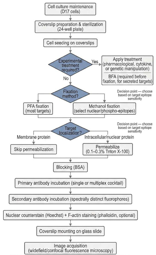
Figure 1: Overview flowchart of the mono- and multiplex immunofluorescence workflow. Principal steps from cell culture maintenance through image acquisition, including the two decision points requiring target-dependent selection: fixation method (PFA versus methanol) and permeabilization (based on target subcellular localization). Optional experimental treatment, including brefeldin A (BFA) pretreatment for rapidly secreted targets, is indicated as a branch prior to fixation. Optional steps not shown for clarity (e.g., aldehyde quenching) are described in the full Protocol. Please click here to view a larger version of this figure.
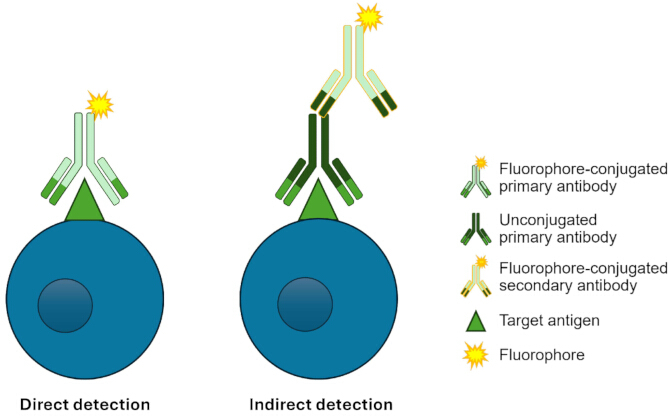
Figure 2: Schematic representation of antibody-based fluorescence detection strategies. Direct detection uses fluorophore-conjugated primary antibodies, while indirect detection uses unconjugated primary antibodies paired with fluorophore-conjugated secondary antibodies, the strategy adopted throughout this protocol. For multiplex applications, indirect detection requires unconjugated primary antibodies raised in different host species, allowing each secondary antibody to selectively recognize its corresponding primary without cross-reactivity. Please click here to view a larger version of this figure.
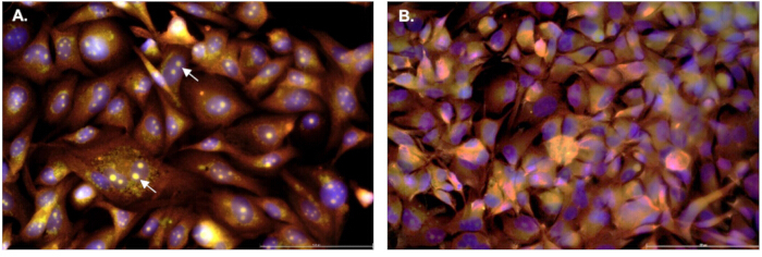
Figure 3: Multiplex immunofluorescence of D17 canine osteosarcoma cells. D17 cells (5 × 10⁴) were seeded on glass coverslips in 24-well plates and fixed with 4% paraformaldehyde (PFA) for 10 min at 25 °C. Cells were subjected to multiplex immunofluorescence using anti-p14ARF (1:200; green-emitting fluorophores), anti-actin (1:200; orange-emitting fluorophores), and anti-interferon-β (IFN-β) (1:200; far-red-emitting fluorophores) primary antibodies, with corresponding secondary antibodies at 1:500. Nuclei were counterstained. Prior to IFN-β detection, cells were treated with BFA (5 µg/mL, 4 h) to block protein secretion. (A) Cells were permeabilized with 0.3% non-ionic detergent for 10 min at 25 °C. (B) Cells processed without permeabilization (0% non-ionic detergent ). Representative images from 2 independent biological replicates, each performed in 2 technical replicates. Only merged channels are shown in this figure; individual channels are provided in Supplementary Figure 1. Differences in labeling are observable between conditions, particularly for p14ARF (A; arrows), an intranuclear target, whose signal was absent in the non-permeabilized group (B). Panels A and B were acquired and processed using identical settings (Supplementary Table 1). Negative and single-stain controls corresponding to this panel are shown in Supplementary Figure 2. Scale bar: 100 µm. Please click here to view a larger version of this figure.
Supplementary Figure 1: Individual fluorescence channels corresponding to Figure 3, organized by marker with permeabilized (left column) and non-permeabilized (right column). Conditions shown side by side: Hoechst 33342 (A, B), p14ARF (C, D; green-emitting fluorophores), actin (E, F; orange-emitting fluorophore), and IFN-β (G, H; far-red-emitting fluorophores), alongside the corresponding merged panels (I, J), matching Figure 3A and 3B, respectively. Arrows indicate representative p14ARF nuclear staining; ellipses indicate representative actin (E) and IFN-β (G) staining, respectively. Loss of p14ARF nuclear signal, along with sub-optimal labeling of IFN-β and actin, is evident under non-permeabilized conditions. Residual signal observed in (D; asterisk) reflects spectral crosstalk from co-present fluorophores, confirmed by its absence in the corresponding single-stain and secondary-only controls (Supplementary Figure 2). Scale bar: 100 µm. Please click here to download this file.
Supplementary Figure 2: Combined immunofluorescence controls corresponding to the multiplex panel in Figure 3. For each control group, individual channels (Hoechst 33342, green-emitting fluorophores, orange-emitting fluorophore, and far-red-emitting fluorophores) are shown alongside the merge (fifth column). (A–E) No-antibody control: cells processed without primary or secondary antibodies. (F–J) Secondary-only control: cells incubated with the secondary antibody cocktail in the absence of primary antibodies. (K–O) p14ARF single-stain control (green-emitting fluorophores). (P–T) Actin single-stain control (orange-emitting fluorophore). (U–Y) IFN-β single-stain control (far-red-emitting fluorophores). Acquisition and processing settings were identical to those used in Figure 3 (Supplementary Table 1). Scale bar: 100 µm. Please click here to download this file.
Supplementary Table 1: Fluorescence filter cube specifications and acquisition order. Images were acquired sequentially on an inverted widefield fluorescence microscope (40×/0.80 objective) using the filter cubes listed below, in the order shown, for every field imaged in Figure 3, Supplementary Figure 1, and Supplementary Figure 2. Images were acquired as single optical planes (no z-stacking).Excitation intensity was set using the fluorescence-intensity control at the indicated levels, with the incident-light field diaphragm held at position 6 for all channels. Camera settings (8-bit digitization, 3072×2048 format, gamma 1.0) and a fixed digital sharpening setting were identical for all channels and panels, including the control images in Supplementary Figure 2. A single linear brightness/contrast adjustment was applied identically to all channels and panels for display. Images were exported as TIFF. Please click here to download this file.
Supplementary Table 2: Troubleshooting guide for common technical issues in the immunofluorescence workflow. Common problems, likely causes, and corrective actions across the protocol.Please click here to download this file.
Discussion
Several steps are critical for the consistent performance of this coverslip-based immunofluorescence workflow. All procedures are performed within the confined environment of a 24-well culture well, which simplifies handling, limits sample manipulation, and enables complete coverslip coverage using low reagent volumes, thereby reducing antibody consumption per experiment. Because these low-volume solutions are prone to evaporation, incubations are best performed in a humidified chamber, and samples must never be allowed to dry: even brief drying increases background fluorescence and degrades cellular morphology. When aspirating, a thin film of buffer is left on the coverslip, and the next solution is added immediately.
Proper cell seeding density and controlled confluency at the time of fixation are essential to limit background and preserve morphology, as overgrowth increases background and compromises cellular architecture. Precise control of incubation time and temperature is likewise required throughout, since both influence fixation efficiency, antibody binding, and background signal5,13. When ambient conditions fluctuate, a temperature-controlled incubator or dry oven set to 25 °C, combined with consistent incubation timing across replicates, improves reproducibility.
Fixation is the first decisive step, as it determines antigen preservation and cellular architecture5, and the fixative is selected according to target localization and epitope sensitivity. PFA is recommended for most membrane-associated, cytoplasmic, nuclear, and cytoskeletal targets, as it preserves architecture, maintains antigen distribution, and is compatible with phalloidin-based F-actin staining. Methanol may be preferred for certain nuclear antigens poorly detected after cross-linking fixation, particularly phospho-epitopes or aldehyde-sensitive epitopes, but it disrupts membrane integrity, extracts soluble proteins, and impairs phalloidin/F-actin staining; compatibility should be validated for each target 13,14,15,16. For soluble cytokines and other rapidly secreted proteins, PFA fixation combined with BFA pretreatment retains intracellular signal prior to fixation: BFA blocks protein transport from the endoplasmic reticulum to the Golgi apparatus, promoting intracellular accumulation of secreted proteins and enabling their detection by immunofluorescence. When targets with conflicting fixation requirements must be detected simultaneously, empirical optimization is required. Glyoxal, a small dialdehyde, has been reported as an alternative to PFA, with faster cross-linking, improved antigen preservation, and compatibility with immunofluorescence and super-resolution microscopy; we have no direct experience with glyoxal fixation and present it as a literature-supported option17,18.
Following fixation, appropriate permeabilization enables antibody access to intracellular and nuclear epitopes while maintaining structural integrity5. Insufficient permeabilization typically yields weak or absent signal despite intact morphology, indicating restricted antibody access, whereas excessive permeabilization extracts soluble proteins, disrupts membranes, causes loss of cytoskeletal staining, and increases background. Detergent type, concentration, and incubation time are therefore optimized for the target. Adequate permeabilization can be judged by four criteria: (i) specific signal in the expected subcellular compartment; (ii) preserved morphology, with intact nuclear and cytoplasmic boundaries resolved by Hoechst 33342 and phalloidin, respectively; (iii) well-defined F-actin staining, since diffuse or lost cytoskeletal signal indicates over-permeabilization; and (iv) low background in negative controls. If these criteria are not met, octylphenol ethoxylate nonionic detergent concentration and/or incubation time are adjusted empirically. When membrane-associated and intracellular antigens are combined in a single panel, milder conditions (lower detergent concentration, shorter incubation) preserve membrane epitopes while allowing intracellular access, and a time course starting at 5 min and increasing incrementally is useful. If saponin is used instead, it must be maintained in all subsequent antibody and wash buffers, as its permeabilizing effect is reversible.
Antibody concentrations are established empirically by titration rather than adopted from generic recommendations. A practical approach is a serial dilution series spanning at least 3–4 concentrations above and below the manufacturer's suggested dilution (e.g., 1:50, 1:100, 1:200, and 1:400 for a recommended 1:200), applied to replicate coverslips under identical conditions. Each dilution is evaluated for specific signal intensity in the expected compartment; signal-to-background ratio, comparing stained samples with the corresponding no-primary control and secondary background with the no-antibody control; and preservation of morphology. The lowest concentration giving a strong, specific signal with minimal background is selected. Persistent high background is commonly mitigated by further titration, extended blocking, or increased wash stringency, whereas weak signal may reflect excessive fixation, suboptimal antibody concentration, failure of the primary antibody to recognize its target epitope, a mismatch between the secondary antibody and the host species of the primary (e.g., an anti-rabbit secondary applied to a mouse-host primary), or loss of fluorophore signal through secondary antibody degradation caused by expiry or improper storage, particularly repeated light exposure. If signal is absent across all dilutions, it is worth extending primary incubation or switching to overnight at 4 °C, confirming antigen expression in the cell line, verifying antibody compatibility with the fixation and permeabilization conditions, checking the lot, storage, and expiry of the primary and secondary antibodies, confirming that the microscope carries appropriate excitation and emission filters for the fluorophore, and, if the problem persists, testing an alternative clone. When preparing working solutions, the required cocktail volume is the number of wells multiplied by 150 µL plus roughly 10% excess to offset pipetting loss; the stock volume of each antibody equals this total divided by its dilution factor (e.g., 825 µL ÷ 200 = 4.13 µL for a 1:200 dilution), with the balance made up in TBST–BSA 1%. The same calculation applies to the secondary cocktail, with the corresponding dilution factor substituted.
Multiplexing performance ultimately depends on fluorophore panel design. Fluorophores assigned to the primary targets, the nuclear counterstain, and the cytoskeletal marker must be coordinated to avoid spectral overlap or signal duplication, and the imaging system must support excitation and detection of every selected fluorophore9,10; excitation and emission ranges for each filter cube are provided in Supplementary Table 1, allowing direct comparison of spectral compatibility across the panel. Users adapting this panel to other fluorophores are advised to select combinations with minimal adjacent-channel overlap where possible, and to verify the absence of crosstalk in single-stain controls before combining targets, as done here. Residual signal attributable to co-present fluorophores was observed across all acquired channels in the multiplex panel (Supplementary Figure 1) but was absent in the corresponding single-stain and secondary-only controls (Supplementary Figure 2), indicating spectral crosstalk from the broadband filter sets used in widefield acquisition (potentially reducible with confocal, laser-based excitation). Given the spatial separation between the nucleolar localization of p14ARF and the filamentous/cytoplasmic distribution of actin and IFN-β, this crosstalk does not compromise the interpretation of p14ARF localization in the multiplex panel. This workflow relies on conventional fluorophore-based multiplexing, in which the number of simultaneously resolvable targets is constrained by the available antibody host species and spectrally distinct fluorophores. Higher-order multiplexing can be achieved through cyclic immunofluorescence or spectral imaging, which permit iterative staining/bleaching or linear unmixing of overlapping fluorophores, respectively, but require specialized equipment and software not addressed here19,20.
Mounting medium affects both image quality and signal longevity. A commercial self-curing antifade medium or, as a homemade alternative, 80% glycerol in PBS (no antifade, no self-curing) may be used; homemade glycerol-based mounts are sealed with clear nail polish to prevent drying and coverslip movement, and media without nuclear stains are chosen once nuclear counterstaining has been performed. Images are best acquired promptly after curing. When immediate imaging is not possible, cured slides can be stored in a dark box at 4 °C for 2–4 weeks or at −20 °C for up to 6 months, bearing in mind that highly photolabile fluorophores (e.g., green-emitting fluorophores) lose signal over time even under optimal storage conditions. Commercial antifade media substantially extend fluorophore stability relative to homemade glycerol-based media; when the latter are used, acquisition within 24–48 h is strongly recommended. Clear nail polish is compatible with most fluorophores and does not appreciably affect signal preservation, provided that at least 30 min of dark curing is allowed before storage.
From the fixation step onward, sterile technique is no longer required, and staining can proceed on the bench, reserving a certified chemical fume hood for any reagent labeled CAUTION. D17 and other tumor-derived cell lines are handled in accordance with institutional biosafety guidelines for risk group 1 or 2 materials, as applicable. All materials that contact cultured cells, coverslips, tips, medium, and liquid waste are decontaminated before disposal: liquid waste is inactivated with bleach to a final concentration of 10% for at least 30 min, and solid waste is discarded in appropriate biological waste containers. Chemical waste containing PFA, methanol, or octylphenol ethoxylate nonionic detergent is segregated and disposed of in accordance with institutional chemical safety regulations.
Although designed to be broadly useful, this workflow is best regarded as amenable to target-dependent adaptation rather than a rigid, universal procedure; successful application depends on the investigator's understanding of each antigen's localization, solubility, epitope accessibility, and sensitivity to fixation and permeabilization. Two limitations should be emphasized. First, the staining assessment here is qualitative, based on comparison with negative controls and expected subcellular localization, rather than formal signal-to-background quantification; accordingly, the consistency reported here reflects reproducible qualitative staining across replicate coverslips and control conditions, rather than quantitatively validated performance. A consolidated troubleshooting guide summarizing common problems, likely causes, and corrective actions is provided in Supplementary Table 2. Laboratories requiring quantitative readouts can use open-source image-analysis software for mean-intensity measurement, background subtraction, Region of Interest (ROI)-based quantification, and intensity profiling21. Second, the representative data derive from a single adherent canine tumor line (D17), so extension to other cell types, species, or antigen combinations will require target-specific re-optimization of fixation, permeabilization, and antibody conditions.
In summary, this protocol provides a flexible, cost-effective workflow for mono- and multiplex immunofluorescence in adherent 2D cultures, combining low reagent consumption with consistent staining performance on standard laboratory equipment while accommodating informed, target-dependent adaptation.
Disclosures
The authors declare no competing financial interests.
Acknowledgements
This research was supported by Fundação de Amparo à Pesquisa do Estado de São Paulo (FAPESP) grant 2023/01697-7 (JL) and 2022/15913-0 (BES). Support was also received from the Conselho Nacional de Desenvolvimento Científico e Tecnológico (CNPq), including grant 401811/2024-7 (BES) and fellowship 310497/2021-3 (BES).
Materials
| Name | Company | Catalog Number | Comments |
|---|---|---|---|
| 24-well tissue culture plates | Corning | 3524 | Coverslip seeding and staining |
| Antibiotic-Antimycotic | Gibco | 15240062 | Used to prevent bacterial and fungal contamination in cell culture. |
| Antibody diluent solution (TBST-BSA 1%) | Prepared in-house | — | For 10 mL: 2 mL of the 5% BSA blocking solution + 5 µL Tween-20 + 7.995 mL TBS 1x (working solution) to a final volume of 10 mL (final: 1% BSA, 0.05% Tween-20 in TBS; pH 7.6). Prepare fresh; if stored, keep at 4 °C protected from contamination and use within 1 week. |
| Blocking solution | Prepared in-house | — | For 10 mL: 0.5 g BSA dissolved in 10 mL TBS 1x (1 mL TBS 10x stock + 9 mL distilled water); pH 7.6. 30 min incubation at 25 °C; prepare fresh. |
| (5% BSA in TBS) | |||
| Brefeldin A | Sigma-Aldrich | B7651 | 5 µg/mL; 4 h pretreatment to block protein secretion prior to cytokine detection. |
| BSA (bovine serum albumin) | Sigma-Aldrich | A7906 | 5% in TBS for blocking; 1% in TBS + 0.05% Tween-20 for antibody diluent. |
| Collagen based reagent (coverslip coating, optional) | Corning | 354231 | Bovine dermis-derived type I collagen; diluted to 50 µg/mL in 0.01 N HCl; applied at 5–10 µg/cm² (≈100 µL/well); incubate 1 h at room temperature (≈25°C); aspirate and rinse with PBS before cell seeding. |
| Coverslips, glass, 12 mm round | Knittel Glass | 100013 | Substrate for adherent cell culture and staining. |
| D17 canine osteosarcoma cell line | Banco de células do Rio de Janeiro | 276 | Representative cell model; maintained in DMEM supplemented with 10% FBS and antibiotic-antimycotic; maintain at 37 °C, 5% CO2. |
| (BCRJ) | |||
| Deionized/distilled water | Prepared in-house | — | Buffer preparation. |
| DMEM (Dulbecco's Modified Eagle Medium) | Gibco | 12100-046 | Complete medium for cell culture. |
| Ethanol | Supelco | 1.00983 | Coverslip/forceps decontamination. |
| Fetal Bovine Serum | Sigma-Aldrich | F7524 | Supplement in cell culture medium. |
| (FBS) | |||
| Fibronectin | Gibco | 33016015 | Extracellular matrix glycoprotein; diluted to 0.0125 mg/mL in cell culture medium; applied at 100 µL/well and incubated for 1 h at room temperature (≈25°C) before cell seeding. |
| Fine-tip metal forceps | Various | — | Coverslip handling. |
| Fluorescence microscope | Leica Microsystems | DMi8 | DMi8 widefield fluorescence microscope (Leica Microsystems), HC PL FLUOTAR 40×/0.80 DRY objective; compatible with fluorophores in the panel (AF488, AF555, AF647, Hoechst). |
| Glass microscope slides | Bioslide | — | Mounting stained coverslips for imaging. |
| Glass Petri dish or coverslip holder | Various | — | Coverslip autoclaving/storage. |
| Glycine | Sigma-Aldrich | 8.16E+09 | For 10 mL of 50 mM solution: 37.5 mg glycine dissolved in 10 mL TBS 1x; pH 7.6. 5 min at 25 °C; optional aldehyde-quenching step after fixation, before permeabilization; prepare fresh. |
| Hoechst 33342 | Invitrogen | H3570 | For 10 mL working solution: dilute stock 1: 5000 in TBS. Nuclear counterstain; incubate protected from light; prepare fresh. |
| Humidified chamber | Various/Prepared in-house | — | For antibody incubation; alternatively assembled from a sealed plastic box with moistened absorbent paper. |
| ImageJ | Open source (NIH) | — | Used for post-acquisition image analysis. |
| Imaging acquisition software | Leica Microsystems | LAS X 3.7.3 | Used for image acquisition on Leica DMi8. |
| Methanol | Merck | 1.06009 | Alternative fixation (nuclear antigens/phospho-epitopes); pre-chilled to −20 °C, 5 min incubation; can impair phalloidin/F-actin staining. |
| Mounting medium | Sigma-Aldrich | G2289 | For 10 mL: 8 mL glycerol + 2 mL PBS, pH 7.6, mix thoroughly. No antifade properties and does not self-cure. Seal coverslip edges with nail polish to prevent drying/drift, store slides at 4 °C protected from light, and image promptly to minimize photobleaching. |
| (Glycerol-based) | |||
| Mounting medium (ProLongTM Glass) | Invitrogen | P36984 | Contains antifade reagents and self-cures (hardens) within ~24 h at room temperature, protected from light, forming a permanent mount (no nail polish sealing required). Store slides at 2–8 °C, protected from light until imaging. |
| NaCl | Sigma-Aldrich | S9888 | Ionic component of TBS buffer; maintains physiological osmolarity to preserve cell and tissue morphology during washing and antibody incubation steps. |
| Needle (fine-gauge, e.g., 25–27G) | Various | — | Used as a lever to gently lift/detach the coverslip from the bottom of the well without damaging the cell monolayer. |
| Paraformaldehyde (PFA) | Sigma-Aldrich | 158127 | For 10 mL: 0.4 g paraformaldehyde powder dissolved in 8 mL PBS, heated to 60–65 °C with a few drops of 1N NaOH until clear, cooled, pH adjusted to 7.4, and brought to 10 mL with PBS; filter through a 0.2 µm filter before use. Prepare fresh or aliquot and store at −20 °C, avoiding repeated freeze-thaw cycles. |
| Phalloidin, fluorophore-conjugated. | Invitrogen | Depending on the chosen fluorescence. | For 10 mL working solution: dilute stock 1:400 in TBS, pH 7.6, according to the manufacturer. Alternative cytoskeletal/F-actin label; prepare fresh, protected from light. |
| Phosphate buffered saline | Sigma-Aldrich | P4474-1L | Isotonic phosphate buffer (pH 7.4); maintains physiological osmolarity to preserve cellular morphology during fixation and washing steps. |
| (PBS) | |||
| Poly-L-lysine (coverslip coating, optional) | Sigma-Aldrich | P8920 | Synthetic polycationic coating reagent that enhances electrostatic cell adhesion; used at 0.1 mg/mL in distilled water; sufficient volume to cover the coverslip surface; incubate 30 min at 25 °C before cell seeding. |
| Primary antibody, anti-actin (goat host) | Santa Cruz Biotechnology | SC-1615 | Anti-actin antibody; polyclonal, non-conjugated, detected with anti-goat secondary antibody. Used at 1:200 working dilution. |
| Primary antibody, anti-IFN-β (mouse host) | Aviva Systems Biology | OAPB00536 | Anti-IFN-β antibody; polyclonal, non-conjugated, detected with anti-mouse secondary antibody. Used at 1:200 working dilution. |
| Primary antibody, anti-p14ARF (rabbit host) | Calbiochem | PC409 | Anti-p14ARF antibody; polyclonal, non-conjugated, detected with anti-rabbit secondary antibody. This product has been discontinued, a new antibody will need to be validated if work on this target is planned. Used at 1:200 working dilution. |
| Secondary antibody, anti-rabbit, Alexa Fluor 488-conjugated | Thermo Fischer Scientific | A32731TR | Anti-rabbit AF488-conjugated antibody. Used at 1:500 working dilution. |
| Secondary antibody, anti-goat, Alexa Fluor 555-conjugated | Thermo Fischer Scientific | A-21432 | Anti-goat AF555-conjugated antibody. Used at 1:500 working dilution. |
| Secondary antibody, anti-mouse, Alexa Fluor 647-conjugated | Thermo Fischer Scientific | A-21235 | Anti-mouse AF647-conjugated antibody. Used at 1:500 working dilution. |
| Tris-buffered saline (TBS), 10x stock | Prepared in-house | — | For 100 mL: dissolve 6.06 g Tris base and 8.77 g NaCl in 80 mL distilled water; adjust pH to 7.6 with concentrated HCl; bring to final volume of 100 mL; autoclave. Dilute 1:10 in distilled water for working solution (TBS 1x) (final: 50 mM Tris, 150 mM NaCl, pH 7.6); store at RT or 4 °C for up to 3 months. |
| TBST (TBS + 0.05% Tween-20) | Prepared in-house | — | For 10 mL: 1 mL TBS 10x stock + 8.995 mL distilled water + 5 µL Tween-20 (0.05% v/v); pH 7.6. Wash buffer between staining steps; prepare fresh, use within ~1 week at 4 °C. |
| Tris Base | Sigma-Aldrich | 252859 | Buffering agent used to prepare TBS stock solution; maintains pH stability in the physiological range (pH 7.6) for immunofluorescence washing and dilution buffers. |
| Triton X-100 (octylphenol ethoxylate nonionic detergent) | Sigma-Aldrich | X100 | For 10 mL: 10 µL Triton X-100 in 9.99 mL TBS 1x for TX100 0.1%; 30 µL in 9.97 mL TBS 1x for TX100 0.3% (target-dependent; nuclear targets require higher concentrations). |
| Trypsin-EDTA | Gibco | 25200056 | Used for cell detachment. |
| Tween-20 | Sigma-Aldrich | P9416 | Used in TBS for wash buffer (TBST) and antibody diluent (TBST-BSA 1%). |
References
- Yoshida SR, Maity BK, Chong S. Visualizing protein localizations in fixed cells: caveats and the underlying mechanisms. J Phys Chem B. 2023;127:4165-73.
- Stadler C, et al. Immunofluorescence and fluorescent-protein tagging show high correlation for protein localization in mammalian cells. Nat Methods. 2013;10:315-23.
- Sule RO, Rivera G, Gomes AV. Western blotting (immunoblotting): history, theory, uses, protocol and problems. Biotechniques. 2023;75:99-114.
- Han Y, Gu Y, Zhang AC, Lo YH. Review: imaging technologies for flow cytometry. Lab Chip. 2016;16:4639-47.
- Im K, Mareninov S, Diaz MFP, Yong WH. An introduction to performing immunofluorescence staining. In: Yong WH, editor. Biobanking: methods and protocols. New York: Humana Press; 2019. p. 299-311.
- Francisco-Cruz A, Parra ER, Tetzlaff MT, Wistuba II. Multiplex immunofluorescence assays. In: Thurin M, Cesano A, Marincola FM, editors. Biomarkers for immunotherapy of cancer: methods and protocols. New York: Humana Press; 2020. p. 467-95.
- Harms PW, et al. Multiplex immunohistochemistry and immunofluorescence: a practical update for pathologists. Mod Pathol. 2023;36:100197. doi:10.1016/j.modpat.2023.100197.
- Paul I, White C, Turcinovic I, Emili A. Imaging the future: the emerging era of single-cell spatial proteomics. FEBS J. 2021;288:6990-7001.
- Radtke AJ, et al. IBEX: an iterative immunolabeling and chemical bleaching method for high-content imaging of diverse tissues. Nat Protoc. 2022;17:378-401.
- Gupta V, et al. Protocol for antibody optimization and panel design in high-dimensional multiplexed immunofluorescence imaging. STAR Protoc. 2025;6:104236. doi:10.1016/j.xpro.2025.104236.
- ibidi GmbH. µ-Slide 8 Well: instruction manual. Version 4.6. Gräfelfing: ibidi GmbH; 2026:https://ibidi.com/img/cms/downloads/in/IN_8082X_8well.pdf
- Thermo Fisher Scientific. Coating Nunc Lab-Tek and Lab-Tek II chamber slides and chambered coverglasses [protocol]. Waltham, MA: Thermo Fisher Scientific; 2017:https://documents.thermofisher.com/TFS-Assets/LCD/Instructions/ECM-Coating-Protocol-for-Lab-Tek-and-Lab-Tek-II.pdf
- Schnell U, Dijk F, Sjollema KA, Giepmans BNG. Immunolabeling artifacts and the need for live-cell imaging. Nat Methods. 2012;9:152-8.
- Piña R, et al. Ten approaches that improve immunostaining: a review of the latest advances for the optimization of immunofluorescence. Int J Mol Sci. 2022;23:1426. doi:10.3390/ijms23031426.
- Lopata A, et al. Affimer proteins for F-actin: novel affinity reagents that label F-actin in live and fixed cells. Sci Rep. 2018;8:6572. doi:10.1038/s41598-018-24953-4.
- Yamanushi TT, et al. Comparison of formaldehyde and methanol fixatives used in the detection of ion channel proteins in isolated rat ventricular myocytes by immunofluorescence labeling and confocal microscopy. Folia Morphol (Warsz). 2015;74:258-61.
- Richter KN, et al. Glyoxal as an alternative fixative to formaldehyde in immunostaining and super-resolution microscopy. EMBO J. 2018;37:139-59.
- Konno K, Yamasaki M, Miyazaki T, Watanabe M. Glyoxal fixation: an approach to solve immunohistochemical problems in neuroscience research. Sci Adv. 2023;9:eadf7084. doi:10.1126/sciadv.adf7084.
- Lin JR, et al. Highly multiplexed immunofluorescence imaging of human tissues and tumors using t-CyCIF and conventional optical microscopes. Elife. 2018;7:e31657. doi:10.7554/eLife.31657.
- Goltsev Y, et al. Deep profiling of mouse splenic architecture with CODEX multiplexed imaging. Cell. 2018;174:968–81.e15.
- Schindelin J, et al. Fiji: an open-source platform for biological-image analysis. Nat Methods. 2012;9:676-82.
Reprints and Permissions
This article has been published
Video Coming Soon