This article presents a standardized nursing protocol for assessing arteriovenous fistula maturation using bedside ultrasound in adults, detailing arterial and venous assessment parameters and nursing decision-making to support safe and reproducible clinical practice.
A subscription to JoVE is required to view this content. Sign in or start your free trial.
Method Article
Ultrasound-Based Assessment of Arteriovenous Fistula Maturation: A Reproducible Protocol for Nursing Practice
In This Article
Summary
Abstract
This article presents a standardized, evidence-informed nursing protocol for point-of-care ultrasound–guided assessment of arteriovenous fistula (AVF) maturation in adult patients undergoing hemodialysis. The protocol translates current best evidence into a reproducible, step-by-step method to support safe, accurate, and consistent bedside surveillance by trained nurses. It is structured using an operational adaptation of the Indication–Acquisition–Interpretation–Nursing Decision-Making framework to guide clinical reasoning and ultrasound-based evaluation. The protocol details patient positioning, infection prevention measures, systematic clinical examination of the AVF, ultrasound system preparation, and standardized ultrasound assessment of venous anatomy and hemodynamics, including vein diameter, depth, access flow, and resistance index. Key technical elements, such as transducer selection, image optimization, avoidance of vessel compression, and identification of stenosis or flow abnormalities, are explicitly described. Nursing decision-making related to fistula maturation status, suitability for cannulation, referral for further vascular evaluation, and documentation is also incorporated. The protocol assumes prior structured training and competency assessment in vascular point-of-care ultrasound for nurses performing the examination. By integrating point-of-care ultrasound as an extension of routine vascular access surveillance, this protocol aims to support early identification of non-maturing fistulas, guide timely clinical decision-making, and support the assessment of functional AVF use in clinical practice. This standardized approach is intended to promote consistency in maturation assessment and support the implementation of ultrasound-guided AVF surveillance as an advanced, evidence-informed nursing practice across diverse clinical settings.
Introduction
Arteriovenous fistulas (AVFs) are the preferred vascular access for hemodialysis because of their superior long-term patency and lower complication rates compared with grafts and central venous catheters. Despite these advantages, failure of AVFs maturation remains a frequent and clinically relevant problem, affecting a substantial proportion of newly created fistulas and leading to prolonged catheter dependence, increased infection risk, and higher healthcare costs1,2. Early recognition of fistulas unlikely to mature is therefore essential to enable timely intervention and optimize vascular access outcomes.
Point-of-care ultrasound (POCUS) has emerged as an effective method for assessing AVF maturation, enabling real-time evaluation of vascular morphology and hemodynamics at the bedside. Unlike conventional duplex ultrasound, which often requires referral to specialized imaging services, POCUS offers greater accessibility and flexibility, enabling repeated assessments during routine follow-up. The overall goal of this approach is to provide a practical, standardized, and clinically actionable method for predicting AVF maturation and supporting early decision-making in vascular access care3.
The rationale for implementing POCUS in AVFs maturation assessment is supported by evidence demonstrating that ultrasound-derived parameters—particularly blood flow volume and vein diameter—are associated with functional maturation4,5. Several studies suggest that evaluations performed between 4 and 6 weeks after fistula creation may provide useful diagnostic information during the period of hemodynamic stabilization and structural remodeling of the access6,7. Within this postoperative period, ultrasound assessment has demonstrated promising diagnostic performance for identifying fistulas at risk of non-maturation when used in conjunction with clinical evaluation8,9.
From a nursing perspective, the bedside and protocolized nature of POCUS is particularly relevant. Nurses play a central role in vascular access surveillance and ongoing patient monitoring in dialysis care, positioning them to potentially perform structured ultrasound assessments when appropriately trained. Evidence from studies involving trained non-specialist ultrasound operators suggests that bedside ultrasound assessment may achieve accuracy comparable to formal duplex sonography10. In addition, longitudinal Doppler surveillance studies—using ultrasound analysis of blood-flow velocity and waveform patterns to assess vascular hemodynamics—have shown that serial ultrasound assessments are effective for early identification of non-maturing fistulas, a process that aligns with routine vascular access monitoring workflows11.
Previous studies and clinical recommendations have increasingly recognized the role of nurses in vascular access surveillance and bedside ultrasound assessment in hemodialysis care. Nursing-led approaches incorporating point-of-care ultrasound have been described for vascular access monitoring, cannulation support, and early identification of AVF dysfunction, particularly within structured vascular access programs and multidisciplinary care models. Existing clinical frameworks also emphasize integrating physical examination findings with ultrasound parameters to support nursing decision-making during AVF surveillance. However, despite growing interest in nurse-performed vascular POCUS, standardized and reproducible protocols specifically detailing image acquisition, hemodynamic assessment, interpretation criteria, and nursing decision pathways for AVF maturation assessment remain limited in the literature.
Within the broader literature, several ultrasound-based diagnostic frameworks have been proposed to standardize AVF maturation assessment, including the “Rule of 6s” and more recent criteria such as the “Rule of 4,” which demonstrated improved clinical prediction at 6 weeks postoperatively in specific study populations12. More advanced predictive approaches, including machine learning models based on ultrasound parameters, have also been explored to improve risk stratification compared with traditional threshold-based criteria13,14. Nevertheless, translating these findings into clinical practice depends on the simplicity of the protocol, reproducibility, operator training, and consideration of patient-specific factors such as obesity and frailty15,16,17. Further validation studies are still needed to evaluate reproducibility, inter-operator reliability, and broader clinical implementation of standardized AVF POCUS protocols. In this context, the present work aims to describe a standardized, ultrasound-based protocol for AVF maturation assessment that is reproducible, objective, and feasible for implementation in nursing practice.
Access restricted. Please log in or start a trial to view this content.
Protocol
This study was approved by the Institutional Review Board of the Ribeirão Preto College of Nursing, University of São Paulo (CAAE No. 92291125.1.0000.5393). Written informed consent was obtained from all participants, and all images were collected and used with their explicit authorization.
1. Patient population and scope
- Apply this protocol to adult patients with newly created native AVF in the upper limbs who are undergoing maturation assessment between 4 and 6 weeks after surgical creation.
- Include radiocephalic, brachiocephalic, and brachiobasilic AVFs in patients on hemodialysis or preparing for hemodialysis therapy.
- Exclude patients with active local infection, severe upper-limb edema preventing adequate ultrasound visualization, hemodynamic instability, severe pain during examination, thrombosed AVFs, or inability to provide informed consent.
- Use this protocol primarily for maturation assessment of newly created AVFs.
- Perform additional vascular evaluation for chronically dysfunctional fistulas18,19,20,21,22,23,24,25,26.
2. Operator training and competency
- Ensure that operators performing this protocol have prior structured vascular POCUS training, including supervised acquisition and interpretation of AVF examinations.
- Perform at least 20 supervised AVF ultrasound assessments before independent clinical use.
- Review stored images and Doppler tracings with an experienced vascular-access ultrasound practitioner to confirm competency.
- Assess inter-operator reliability locally before routine implementation whenever possible18,19,20,21,26.
3. Pre-procedure patient assessment and preparation
- Explain the procedure to the patient and answer questions.
- Perform hand hygiene according to the institutional infection-control protocol.
- Clean and disinfect the ultrasound transducer according to manufacturer and institutional recommendations before and after each examination.
- Apply ultrasound gel to the area to be examined.
- Gather the required materials, including a stethoscope, ultrasound device, conductive gel, disposable towels, and documentation form (Figure 1).
- Position the patient in the supine position with the AVF's arm extended and supported on a pillow at heart level.
- Position the hand in a supinated orientation.
- Expose the entire AVF segment from the anastomosis to the proximal venous outflow tract.
- Clean the patient's skin with an institutionally approved antiseptic solution when clinically indicated18,19,20,21.
NOTE: Avoid excessive arm flexion or external compression during the examination to minimize alterations in venous diameter and flow measurements18,19,20,21.
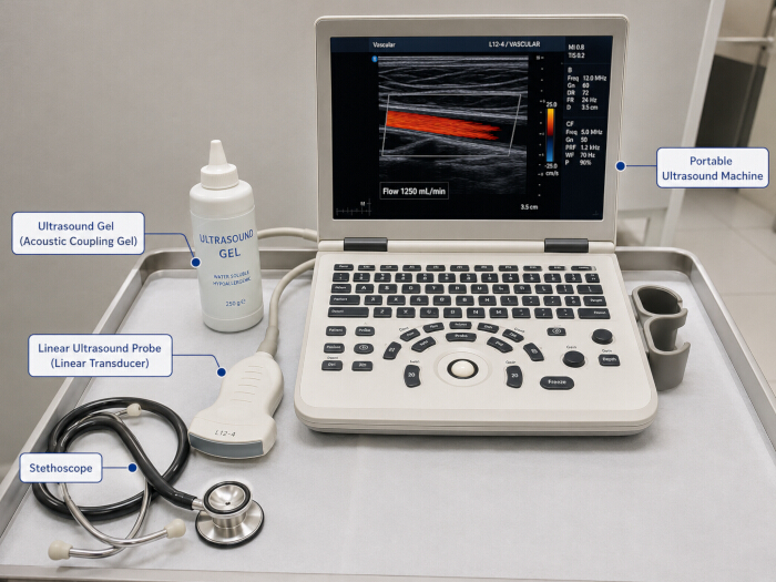
Figure 1: Materials for arteriovenous fistula assessment. Instrumental setup featuring a portable ultrasound system with a high-frequency linear transducer, conductive gel, and a stethoscope. This apparatus is used to evaluate Arteriovenous Fistula (AVF) maturation through physical examination (auscultation of bruit) and measurement of anatomical and hemodynamic parameters. Please click here to view a larger version of this figure.
4. Clinical examination of the arteriovenous fistula
- Inspection
- Inspect the AVF limb for pallor, cyanosis, erythema, edema, skin lesions, aneurysmal dilation, or signs of infection.
- Document collateral veins in the upper limb or thoracic region.
- Elevate the limb and observe venous collapse (arm lift test); if the vein does not collapse, identify probable outflow stenosis.
- Identify the presence of a developed, straight, and superficial vein18,19,20,21.
- Palpation
- Assess limb temperature and compare findings with the contralateral extremity.
- Palpate the AVFs along the entire draining vein and document the presence and extent (≥10 cm) of a thrill.
- Palpate the extent of the AVF and identify the reduction in the intensity of the thrill starting from the anastomosis (normal condition).
- Certify compressible veins and thickened walls through palpation.
- Manually occlude the arterialized vein and identify an increase in the pulse after anastomosis (Pulse augmentation test). If there is no increase in the pulse, inflow stenosis is likely. If, after occlusion of the AVF, it is still possible to palpate the thrill, the presence of accessory veins below the occlusion point is very likely.
- Evaluate the characteristics of the AVF thrill (continuous or discontinuous) and the pulse (hypopulsatile, hyperpulsatile, or soft).
- Assess the characteristic of the arterial pulse (present or absent)18,19,20,21.
- Auscultation
- Auscultate the AVFs along the entire vascular pathway using a stethoscope.
- Classify the bruit as continuous systolic-diastolic, discontinuous, high-pitched, or absent.
- Identify high-pitched systolic bruits or the absence of bruit as possible indicators of stenosis or thrombosis.
- Determine preliminary clinical suitability for cannulation and document the rationale18,19,20,21.
5. Preparation of the ultrasound system
- Power on the ultrasound device and select the vascular preset.
- Select a high-frequency linear transducer operating between 5 and 15 MHz (Figure 2).
- Ensure that the ultrasound system includes spectral Doppler functionality and supports hemodynamic measurements required for access flow and resistance index calculations.
- Apply the conductive gel on the transducer footprint over the skin in the AVF area.
- Use rotation, pressure, sliding, and tilting maneuvers to optimize image acquisition.
NOTE: Because some point-of-care ultrasound devices may have limited Doppler capabilities, equipment selection should be aligned with the intended scope of vascular access assessment. - Adjust imaging depth, gain, focal zone, pulse repetition frequency, and wall-filter settings before Doppler acquisition.
- Position the linear transducer over the AVF, with the index pointing to the patient's right18,19,20,21.
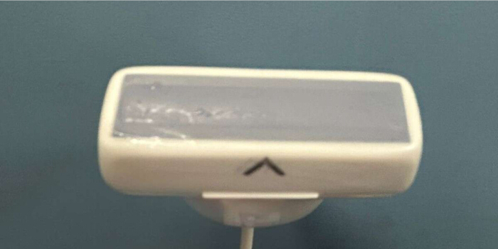
Figure 2: Linear probe used for vascular ultrasonography. High-frequency linear transducer used for ultrasound assessment of arteriovenous fistula morphology and hemodynamics, including measurements of vein diameter, depth, and blood flow. Please click here to view a larger version of this figure.
6. Ultrasound-based assessment of AVF maturation
- Identify the draining vein in longitudinal and transverse planes.
- Confirm venous characteristics using compression and color Doppler (Supplementary Files 1 and 2).
- Measure the venous diameter in a transverse view, about 5 centimeters distal to the arteriovenous anastomosis, ensuring the vessel is centered on the ultrasound screen. (Figure 3).
- Avoid aneurysmal segments, focal stenoses, or venous branches when selecting the measurement sites.
- Measure the venous diameter from the inner anterior wall to the inner posterior wall with the vessel centered on the screen and without probe-induced compression.
- Measure vein depth from the skin surface to the anterior vessel wall at the same site used for diameter assessment that best represents the intended cannulation segment. (Figure 4).
- Measure the usable cannulation segment length in longitudinal view.
- For the Doppler mode, adjust the Doppler scale according to the flow velocity to avoid aliasing.
- Use low wall-filter settings to preserve low-velocity flow signals.
- Evaluate the brachial artery using sliding and rocking movements and measure peak systolic velocity (PSV), resistance index (RI), and blood flow (ml/min).
- Measure peak systolic velocity and time-averaged mean velocity using an angle correction of 60°.
- Position the Doppler sample volume in the center of the vessel lumen during spectral Doppler acquisition.
- Calculate access flow using the following formula:
Access Flow (mL/min) = Time-Averaged Mean Velocity × Cross-Sectional Area × 60. - Calculate vessel cross-sectional area using the following formula:
Area = π × (vessel radius)2. - Calculate the resistance index (RI) using the following formula:
RI = (Peak Systolic Velocity − End Diastolic Velocity) / Peak Systolic Velocity.
NOTE: Interpret the resistance index together with physical examination findings, access flow, venous diameter, and other ultrasound parameters. Abnormal values may require further evaluation. - Record all flow measurements in mL/min and document representative Doppler waveforms.
- Assess focal stenosis by identifying luminal narrowing, focal flow acceleration, post-stenotic turbulence, or velocity increase greater than 2:1 compared with adjacent segments.
- Assess for thrombus by identifying echogenic intraluminal material, absent compressibility, or absent color Doppler flow.
- Document lesion location relative to the anastomosis and classify findings as juxta-anastomotic, cannulation-segment, or proximal outflow lesions18,19,20,21.
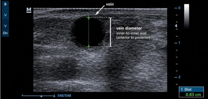
Figure 3: Ultrasound assessment of venous diameter. The B-mode image illustrates measurement of the drainage vein diameter in a cross-sectional (short-axis) view using electronic calipers, positioned from the inner anterior wall to the inner posterior wall of the vessel. The representative measurement shown is 0.83 cm. Please click here to view a larger version of this figure.
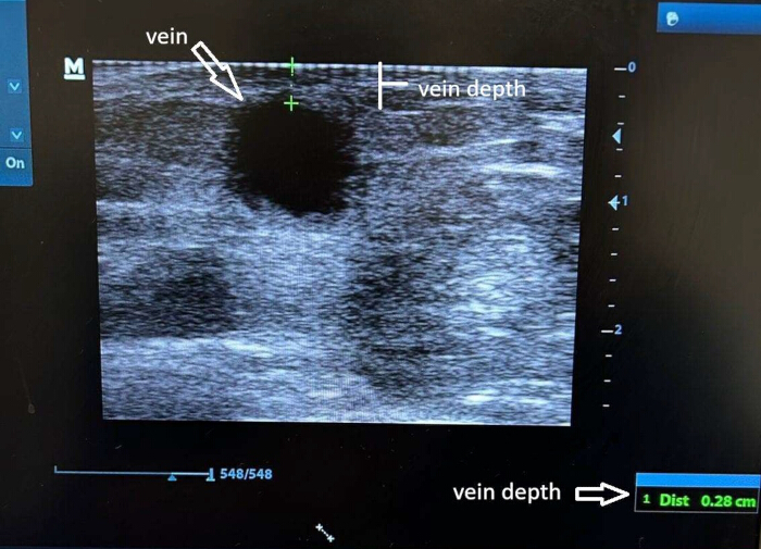
Figure 4: Ultrasound measurement of venous depth. The B-mode image illustrates the measurement of the drainage vein’s depth in a cross-sectional view (short axis). The measurement is taken using electronic calipers, with the distance from the skin surface to the anterior wall of the vessel recorded in centimeters. The measured value of 0.28 cm is highlighted in the lower right corner of the screen. This parameter is essential for assessing AVF maturation and ensuring that the vessel is accessible for safe cannulation, as clinical guidelines recommend an ideal depth of ≤ 0.6 cm from the skin surface. The result of 0.28 cm confirms that the vein is superficial, consistent with the Rule of 6 for AVF maturation. Please click here to view a larger version of this figure.
7. Interpretation and nursing decision-making
- Classify AVF maturation status using integrated ultrasound and clinical findings.
- Classify the AVF as adequately mature when the following criteria are met: diameter ≥6 mm, depth ≤6 mm, access flow ≥600 mL/min, and usable cannulation segment ≥6 cm (“Rule of 6s”)2,12.
- Identify AVFs with continuous thrill and continuous systolic-diastolic bruit associated with adequate flow and venous diameter as likely mature and potentially suitable for cannulation.
- Identify discontinuous or absent thrill, high-pitched systolic bruit, excessive pulsatility, collateral veins, marked edema, or abnormal pulse augmentation findings as possible indicators of stenosis, outflow obstruction, or non-maturation requiring additional ultrasound evaluation.
- Classify AVFs with adequate ultrasound maturation parameters but abnormal physical examination findings as requiring repeat assessment or vascular referral before cannulation.
- Classify AVFs with stenosis, thrombus, absent Doppler flow, absent thrill, or access flow <600 mL/min as unsuitable for cannulation and refer for vascular evaluation according to institutional protocol.
- Repeat the ultrasound assessment within 1–2 weeks when maturation criteria are partially met or when discordant physical-examination and ultrasound findings are identified.
- Interpretation of discordant findings
- When ultrasound maturation parameters and physical examination findings are discordant, determine AVF suitability for cannulation using an integrated assessment rather than isolated Rule-of-6 criteria.
- Classify AVFs with access flow ≥600 mL/min but vein depth >6 mm as physiologically mature but potentially unsuitable for routine cannulation because excessive depth may limit safe needle access. Repeat the assessment or refer the patient for vascular evaluation.
- Consider these AVFs suspicious for dysfunction and perform further ultrasound evaluation or refer for vascular evaluation before cannulation: AVFs with adequate vein diameter (≥6 mm) but abnormal physical examination findings (e.g., discontinuous thrill, absent thrill, high-pitched systolic bruit, excessive pulsatility, collateral veins, or abnormal pulse augmentation test).
- Do not classify these AVFs as fully mature if AVFs meet Rule-of-6 criteria but demonstrate ultrasound evidence of stenosis, thrombus, focal flow acceleration, or post-stenotic turbulence. Refer for vascular evaluation.
- Consider AVFs suitable for cannulation only when they demonstrate adequate ultrasound maturation parameters, normal physical examination findings, and no significant stenosis or thrombus.
- Identify optimal puncture sites within straight, superficial, compressible venous segments free of stenosis, thrombus, or aneurysmal dilation18,19,20,21. This protocol does not include real-time ultrasound-guided cannulation or skin-marking procedures for needle insertion.
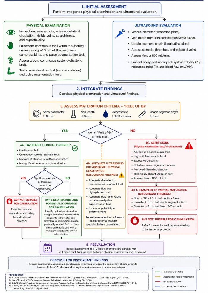
Figure 5: Flowchart for decision-making in the assessment of AVF maturation. Flowchart summarizing the integrated physical examination and ultrasound protocol used to assess AVF maturation and determine suitability for cannulation. Please click here to view a larger version of this figure.
8. Documentation
- Document AVF type and location, postoperative timing, venous diameter, depth, flow measurements, resistance index, stenosis findings, thrombus findings, and usable cannulation length.
- Document AVF maturation classification, cannulation suitability, referral decisions, and recommended timing for reassessment when applicable.
- Store representative grayscale and Doppler images or video clips according to institutional policy.
- Document interpretation summary, cannulation recommendation, referral decision, optimal puncture sites, examination date, and operator identity in the patient record18,19,20,21.
Access restricted. Please log in or start a trial to view this content.
Results
The protocol supports standardized assessment of AVF maturation by integrating physical examination findings with ultrasound-derived anatomical and hemodynamic measurements. Representative examples of mature, non-mature, and discordant AVFs are presented to illustrate protocol interpretation and clinical decision-making.
Representative case 1 – mature AVF
A brachiocephalic AVF was assessed 6 weeks after surgical creation. Physical examination demonstrated a continuous thri...
Access restricted. Please log in or start a trial to view this content.
Discussion
This protocol describes a standardized nurse-led approach to assessing arteriovenous fistula maturation using point-of-care ultrasound, addressing an important gap between available ultrasound evidence and its consistent application in clinical practice. Although preoperative venous mapping ultrasonography is widely recommended for vascular access planning, its ability to predict primary fistula maturation remains limited, particularly when vein diameter is used as an isolated predictor22. In cont...
Access restricted. Please log in or start a trial to view this content.
Disclosures
The authors have no conflicts of interest to disclose.
Materials
| Name | Company | Catalog Number | Comments |
|---|---|---|---|
| Disposable towels | N/A | N/A | Used for gel removal and patient preparation |
| Documentation form/data collection sheet | Institutional material | N/A | Recording of ultrasound findings and nursing assessment |
| High-frequency linear transducer (5–15 MHz) | Mindray | L12-4S | Linear vascular probe used for grayscale and Doppler imaging |
| Pillow/support cushion | N/A | N/A | Used to support the arm at heart level during examination |
| Portable ultrasound system | Mindray | UMT-150 | Ultrasound device used for bedside AVF maturation assessment |
| Spectral and Color Doppler software | Integrated with ultrasound system | N/A | Used for hemodynamic analysis and waveform acquisition |
| Stethoscope | BD | 7891463001146 | Used for AVF auscultation during physical examination |
| Ultrasound conductive gel | Multigel | 80316110001 | Sterile or single-use conductive gel for ultrasound examination |
Reprints and Permissions
This article has been published
Video Coming Soon