This paper outlines detailed protocols for maintaining a Carausius morosus laboratory colony, including key procedures for animal husbandry, egg handling, hatchling care, representative performance metrics, and an overview of the species lifespan and biological characteristics.
Method Article
A Standardized Protocol for Rearing Carausius morosus (Phasmatodea, Insecta) in Laboratory Settings
In This Article
Summary
Abstract
Carausius morosus, the Indian stick insect, is a slender twig-like insect endemic to India. Though widely introduced through captivity around the world and commonly used in laboratories or kept as a household pet, standardized animal husbandry laboratory protocols are lacking. Here we report detailed laboratory culture conditions for C. morosus. We maintain stocks at 23 °C, 70% relative humidity, and a 12:12 h light-dark photoperiod. This culture has been successfully sustained under these conditions for over two years, with standardized protocols in place for dietary and cage setup conditions. We also report methods for egg and hatchling care to support ongoing experiments with C. morosus. Under our culture conditions, 105 monitored batches of eggs laid in the same 24 hour period hatched within a consistent 12-day period, and 13.9% of hatchlings successfully reached adulthood. These standardized methods improve reproducibility and accessibility, enabling the broader use of C. morosus as a laboratory model system for a wide range of studies, including analyses of development, behavior, and physiology.
Introduction
As interest grows in alternative non-mammalian model systems, insects offer an opportunity to explore a wide range of biological diversity while also providing lower-cost research models, simpler care requirements, and fewer regulatory barriers than mammalian models1. Emerging insect model systems make it possible to investigate unique biological phenomena, including, for example, parthenogenesis in stick insects such as Carausius morosus (Sinéty, 1901)2. While the fruit fly Drosophila melanogaster has shaped our understanding of developmental and genetic processes since the early 20th century3, other insect model systems such as C. morosus present a unique opportunity not only for developmental biology, but also for investigating motor control, locomotion, and neurohormone-regulated systems4.
Additionally, within the field of evolutionary developmental biology, hemimetabolous (directly developing) insects remain underrepresented, with the holometabolous (indirectly developing or undergoing metamorphosis) D. melanogaster serving as the best-studied model insect. Studying directly developing insect species like C. morosus, which undergoes several nymphal instar stages before reaching sexual maturity5, allows us to fill key phylogenetic gaps in evolutionary developmental biology research. This enables the comparison between hemimetabolous and holometabolous insects, which diverged early in insect evolution but are not equally represented in current laboratory model systems. The broader use of C. morosus as a laboratory organism has been limited by inconsistent, unreproducible husbandry conditions and a lack of clear benchmarks for development and survival. Here, we outline a step-by-step protocol with defined culture conditions and expected outcomes that can be applied across labs, supporting multiple applications including embryogenesis tracking, longitudinal development, and reproductive output experiments.
C. morosus are easy to rear in captivity, making them well-suited for both laboratory research and teaching environments. Their ability to reproduce parthenogenetically6 eliminates the need for complex breeding setups, reducing maintenance demands and simplifying population management. Although successful rearing benefits from controlled environmental conditions, its husbandry remains straightforward and cost-effective compared to many other laboratory models. C. morosus feeds readily on common plant material such as ivy (Hedera helix), bramble (Rubus fruticosus), or rose leaves (Rosa spp.). Other advantages of this system include autotomy, the ability to regenerate legs7, large body size, and the ability to feed on a range of host plants. However, this species also has limitations, including the lack of a publicly available genome sequence, relatively long development time, and a hard embryonic exochorion that can make injections challenging8. Despite these limitations, C. morosus offers a practical and effective model for both research and teaching applications.
Standardized rearing protocols for C. morosus are critical for producing consistent and reproducible results in research studies. Although multiple laboratory studies of C. morosus have been reported in the literature6,9,10,11,12, to our knowledge, no in-depth protocols for rearing C. morosus in the laboratory have been published to date. In this study, we have successfully established a comprehensive set of standardized protocols for C. morosus laboratory cultures, including rearing of eggs, hatchlings, and reproductively mature adults. With these methods, we aim to make this insect more accessible to the broader biological research community.
Protocol
1. Environmental rearing conditions
- Maintain the laboratory culture in a climate-controlled room or incubator with environmental settings as follows: 23 °C, relative humidity 70%, light-dark photoperiod of 12:12 h.
NOTE: Under these conditions, C. morosus eggs are expected to hatch approximately 74-80 days after being laid.
2. Establishment and maintenance of mesh enclosures
NOTE: Mesh insect enclosures with fabric mesh ventilation on three sides are recommended for housing C. morosus. An enclosure measuring 61 x 61 x 91.5 cm is suitable for maintaining up to 50 adults or, when establishing a new colony, up to 500 hatchlings.
- Select a mesh enclosure of appropriate size. Ensure the container is clean and free of debris before introducing the animals.
- For establishing a new colony using mature adults, place 25–50 adult females into a clean 61 cm x 61 cm x 91.5 cm mesh enclosure.
NOTE: Mesh openings should be in the range of 0.2–1.0 mm to prevent escape of the small first instar hatchlings. - For establishing a new colony using hatchlings, transfer up to approximately 500 hatchlings into a clean 61 cm x 61 cm x 91.5 cm mesh enclosure.
3. Feeding and maintenance
- During cage cleanings, let the insects stay in the enclosure; gently take them off foliage and place them on the bottom of the mesh enclosure while cleaning around them.
NOTE: Expect cleaning to take roughly 10–20 min per cage. - Provide fresh foliage such as ivy, bramble, or rose once per week. Ensure all plant material is pesticide-free. Wash foliage with tap water prior to addition to cages to remove surface debris and contaminants.
- Trim stems to fit vessels (small jars or bottles) filled with tap water to maintain leaf hydration. Place the prepared foliage inside the enclosure, making sure leaves are elevated and accessible to the insects. Carefully close the enclosure after placement.
- During weekly cage cleanings, remove wilted or dried foliage and any accumulated excrement from the bottom of the enclosure.
NOTE: Replace foliage when leaves show signs of dehydration, such as dryness, brittleness, wilting, or discoloration. Maintaining a clean environment is essential for colony health and well-being. - Lightly mist each enclosure daily with 1–2 sprays of tap water from a 250 mL spray bottle to ensure proper hydration.
NOTE: This is especially crucial on the first day post-hatching. Reduce density if rapid food depletion occurs.
4. Collection and maintenance of Carausius morosus hatchlings
NOTE: In this study, we use “hatchling” to refer to individuals immediately after emerging from the egg (first instar), while “nymph” refers to juvenile stages following postembryonic molts.
- Gather 6 cm Petri dishes with newly emerged hatchlings.
- Using blunt plastic forceps, gently transfer no more than 500 hatchlings into a clean 61 cm x 61 cm x 91.5 cm mesh enclosure.
NOTE: Collect only fully emerged, stretched out nymphs that have completed exoskeleton expansion. Disturbing the nymphs before they have fully emerged from the egg may impair body extension and lead to premature death. For similar reasons, try to minimize direct handling of nymphs following transfer to the mesh enclosure. - Provide fresh foliage as described in section 3.
- Label new colony enclosures with the following information: egg batch collection date(s), date of hatching or range of hatching dates, and number of hatchlings added.
NOTE: This tracks enclosure age and predicts the start of reproductive maturity. - Lightly mist each enclosure immediately after hatchling transfer, and daily thereafter, with 1–2 sprays of tap water from a 250 mL spray bottle to ensure proper hydration.
NOTE: This is especially crucial on the first day post-hatching. Hatchlings may remain in the same enclosure throughout development until adulthood. Under the laboratory conditions described in section 1, approximately 13% of hatchlings survive to adulthood in 4–5 months (Table 1). - Remove any dead or damaged hatchlings during weekly cage cleaning to prevent contamination.
5. Collection and maintenance of C. morosus eggs
- Open an enclosure containing adult females.
- Using a gloved hand, gently sweep the substrates on the bottom of the cage, which contain both eggs and fecal matter, towards the front opening.
- Carefully transfer the material using a gloved hand to a 15 cm Petri dish for egg retrieval.
6. Termination of insects and disposal of enclosure waste
NOTE: When C. morosus are no longer needed for experiments, have died, or eggs are not required:
- Euthanasia/Disposal of insects: Transfer live or dead insects no longer needed for culture or experiments into an autoclavable waste bag, and store in a -20 °C freezer for at least 72 h.
- Egg destruction: To prevent overpopulation, remove any eggs not used for experiments from cages or Petri dishes, place them in an autoclavable waste bag, and store at -20 °C for at least 72 h.
- Waste handling: Place any contaminated waste from enclosures or materials used for egg or insect collection in an autoclavable waste bag, and store at -20 °C for at least 72 h.
- After freezing materials, transfer the autoclavable waste bag to a biohazard bin for proper disposal.
NOTE: As C. morosus is an invasive species outside of India, it is crucial that all materials are properly disposed of as described above to prevent accidental release into the environment.
7. Egg retrieval
- Using blunt plastic forceps, carefully pick up each egg and place it into a clean 6 cm Petri dish.
NOTE: Mature eggs are protected by a hardened exochorion and are dark brown, resembling plant seeds. Using a white background can help differentiate droppings from eggs when sorting. - Label the dish with the date and time of collection for tracking purposes. Keep the lid off during incubation to allow ventilation.
NOTE: A 6 cm Petri dish is sufficient for storing up to 200 eggs. - Approximately 1 week before the expected hatching window (74–80 days after egg laying; Kovacikova & Extavour, unpublished data), replace the Petri dish cover.
- Begin daily monitoring of egg containers before the expected hatching date to observe emerging hatchlings.
8. Life expectancy
- After hatching, monitor individuals as they undergo five to six molts before reaching reproductive maturity, which under the laboratory conditions described in Section 1, happens on average 139 days after hatching (Table 1).
- To follow this protocol, define developmental timing under these culture conditions as follows: embryogenesis duration (egg laying to hatching) is 74–80 days; time from hatching to sexual maturity (onset of oviposition) is approximately 135 days, corresponding to approximately 200 days from egg laying to sexual maturity, and total lifespan from egg laying to death is approximately 9.5 months.
Results
We collected data documenting the successful laboratory rearing of C. morosus from eggs to reproductively mature adults between September 2024 and November 2025. Over this 14-month period, we established, maintained, and monitored five cages for one or more of the following: adult mortality, eggs laid, timing of egg hatching, and hatchling survival to adulthood. Our method supports long-term colony maintenance without obvious signs of pathogen-related disease or reproductive decline, all of which would constitute suboptimal outcomes. Over more than two years of continuous culture, we observed no obvious pathogen-related mass deaths, and egg and hatchling production remained stable across generations.
Data shown in Figure 1 and Figure 2 present observations from the five monitored cages from setup to adulthood of hatchlings, including the eggs laid and adult mortality for each cage. After the founder individuals in each cage reached sexual maturity and began laying eggs, we recorded the number of adults present in each cage to establish the starting population at the onset of oviposition. The number of adults in each cage over time is shown in Figure 1 and Table 1. We interpret the steady decline in survival as age-related mortality.
We monitored egg production in parallel in the same five cages monitored for adult survival (Figure 1 and Figure 2). Daily egg counts per cage were highest during the first 30–50 days of egg-laying and decreased gradually thereafter. Although total egg production varied among cages (Table 2), this general pattern was consistent across all five replicate cages. We monitored egg hatching dynamics over a 50-day period for 105 egg batches overall, where each egg batch is a group of eggs oviposited in the same cage within a 24 h period. For the batch-size analyses shown in Figure 3, 75 batches were grouped into small, medium, and large categories. We found that once the first egg in a batch hatched, 90% hatch completion was reached within 12 days of the first hatchling's emergence (Figure 3D), independent of batch size (Figure 3B). Using the term “hatch completion” to describe the proportion of eggs in a batch that have hatched by a given time point following the emergence of the first hatchling from a batch, we note that the distribution of egg batches by the day of 90% hatch completion indicates that most batches reached this threshold between hatching days 10 and 14 (Figure 3C) after emergence of the first hatchling.
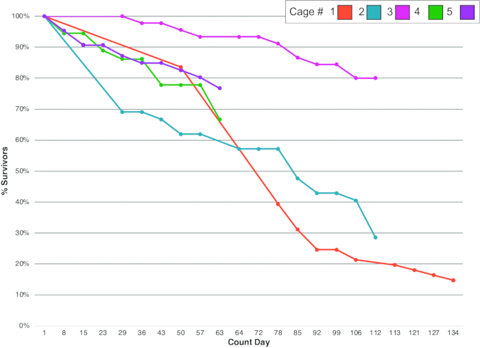
Figure 1. Adult mortality dynamics. Survival trajectories across five replicate C. morosus experimental cages over time. Percent survival is plotted over time for individuals in each monitored cage. See summary metrics in Table 1. Please click here to view a larger version of this figure.
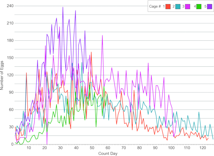
Figure 2. Egg production dynamics. The number of eggs collected from the same five cages monitored in Figure 1. Cages 1 through 5 were monitored daily for 47, 58, 106, 129, and 132 days, respectively. The plot shows data from the first 126 days of oviposition. See summary metrics in Table 2. Please click here to view a larger version of this figure.
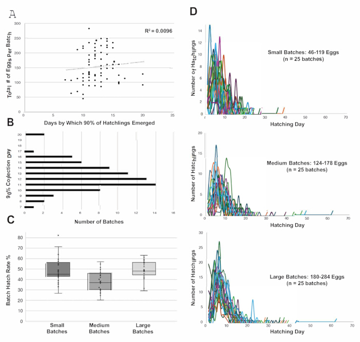
Figure 3. Egg hatching dynamics. (A) Relationship between total eggs per batch and the number of days required to reach 90% hatch completion. (B) Distribution of egg batches by the day on which they reached 90% hatch completion across hatching days 7–20. (C) Distribution of batch hatch rate across small, medium, and large egg batches. n = 25 batches per batch size category. (D) Hatching trajectories over a 70-day collection period for the 75 egg batches used in the batch-size analysis. A total of 105 egg batches were monitored; for the analyses shown here, 75 batches were grouped by total egg count using tercile cutoffs. Please click here to view a larger version of this figure.
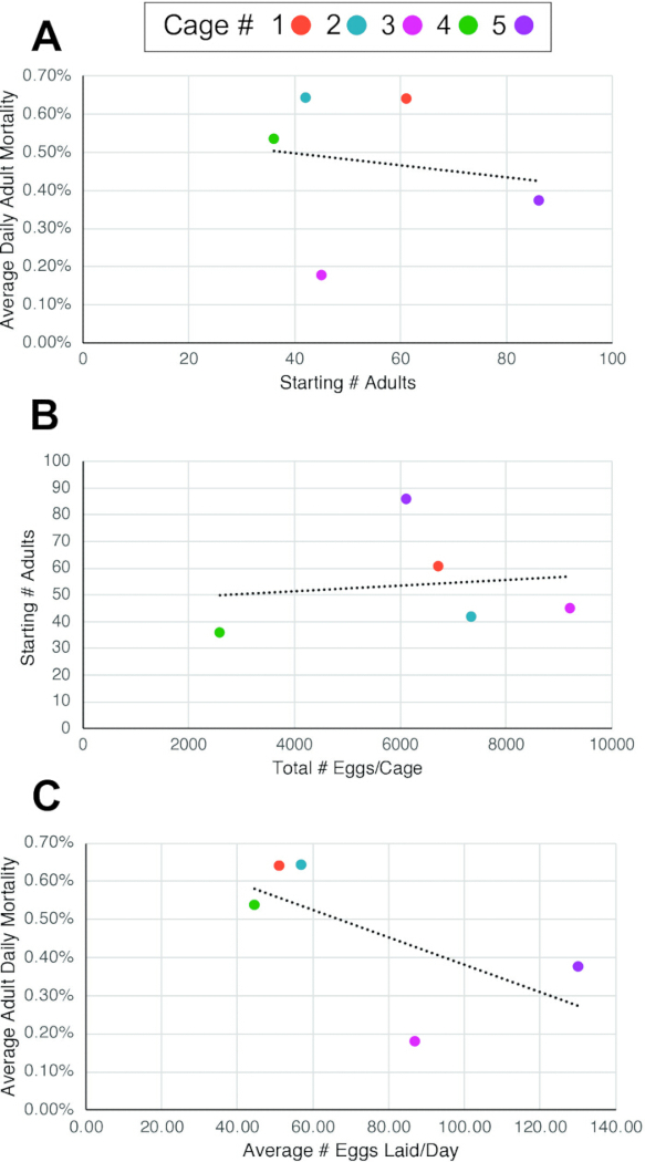
Figure 4. Relationships between adult numbers, adult mortality, and egg production per cage. (A) There is no significant relationship (R2 = 0.025) between average adult daily mortality and the number of founding adults in a cage. (B) There is no significant relationship (R2 = 0.017) between total egg production and the number of founding adults in a cage. (C) There is a moderate negative correlation (R2 = 0.411) between average daily egg production and average adult daily mortality. Average adult daily mortality = (% total mortality) / (total number of days monitored). Please click here to view a larger version of this figure.
| Cage # | old cage # | First day hatch lings added to founder cage | Last day hatch lings added to founder cage | # Hatch lings added to cage | Range of days hatch lings were added | Average Time Between Cage Start and Adult # Counting | First Day of Counting Adults | First Day Adults Started Laying Eggs | Time Elapsed Between Hatching and Adulthood | Time Elapsed Between Hatching and Egg Laying | Starting # of Adults | Hatchling Survival to Adult hood | Last Day of Counting Adults | Ending # of Adults | Length of Time Adult # Monitored [days] | Average Adult Mortality Over Monitored Time [%] | Average Daily Adult Mortality [%] |
| 1 | 7 | 12/7/ 2024 | 12/21/ 2024 | ND | 14.00 | 166.00 | 5/29/ 2025 | 5/29/ 2025 | 173.00 | 173.00 | 61.00 | ND | 10/9/ 2025 | 9.00 | 133.00 | 85.25% | 0.64% |
| 2 | 8 | 2/10/ 2025 | 3/9/ 2025 | ND | 27.00 | 130.00 | 7/17 /2025 | 7/17/ 2025 | 157.00 | 157.00 | 42.00 | ND | 11/5/ 2025 | 12.00 | 111.00 | 71.43% | 0.64% |
| 3 | 9 | 4/5/ 2025 | 4/12/ 2025 | ND | 7.00 | 110.00 | 7/17/ 2025 | 7/27/ 2025 | 103.00 | 113.00 | 45.00 | ND | 11/5/ 2025 | 36.00 | 111.00 | 20.00% | 0.18% |
| 4 | 10 | 5/8/ 2025 | 5/22/ 2025 | 350 | 14.00 | 121.00 | 9/4/ 2025 | 9/13/2 025 | 119.00 | 128.00 | 36.00 | 10.29% | 11/5/ 2025 | 24.00 | 62.00 | 33.33% | 0.54% |
| 5 | 11 | 5/23/ 2025 | 6/6/ 2025 | 536 | 14.00 | 117.00 | 9/4/ 2025 | 9/24/ 2025 | 104.00 | 124.00 | 86.00 | 16.04% | 11/5/ 2025 | 66.00 | 62.00 | 23.26% | 0.38% |
| Averages | 15.20 | 128.80 | 131.20 | 139.00 | 54.00 | 13.17% | 29.40 | 95.80 | 46.65% | 0.48% |
Table 1: Adult survival and mortality dynamics across experimental cages. Adult survival outcomes for five replicate experimental cages. For each cage, we report the date range of cage establishment, the number of hatchlings used to establish each cage (for 2 of the 5 cages), hatchling survival to adulthood (for 2 of the 5 cages), time between cage establishment, initiation of adult counts, and initiation of egg-laying, the duration of adult monitoring, starting and ending adult population sizes, adult mortality over the monitored period, and average daily mortality. Hatchling survival to adulthood was calculated only for cages with known starting hatchling numbers.
| Cage # | Date Egg Collection Start | Date Egg Collection End | Duration of Egg Collection [days] | Total # of Eggs/Cage | Average # Eggs Laid/Day | Starting # of Adult Individuals | Total # Eggs/Starting Adults | Average # Eggs/Starting Adult/Day |
| 1 | 5/29/2025 | 10/8/ 2025 | 132.00 | 6721.00 | 50.92 | 61.00 | 110.18 | 0.83 |
| 2 | 7/4/2025 | 11/10/2025 | 129.00 | 7333.00 | 56.84 | 42.00 | 174.60 | 1.35 |
| 3 | 7/27/2025 | 11/10/2025 | 106.00 | 9198.00 | 86.77 | 45.00 | 204.40 | 1.93 |
| 4 | 9/13/2025 | 11/10/2025 | 58.00 | 2583.00 | 44.53 | 36.00 | 71.75 | 1.24 |
| 5 | 9/24/2025 | 11/10/2025 | 47.00 | 6104.00 | 129.87 | 86.00 | 70.98 | 1.51 |
| Averages | 94.40 | 6387.80 | 73.79 | 54.00 | 126.38 | 1.37 |
Table 2: Egg production across experimental cages. Egg-laying output of five replicate experimental cages containing mature adults. For each cage, we report the start and end dates of egg collection, the total duration of egg collection, cumulative egg production per cage, and the average number of eggs laid per day.
Discussion
The husbandry methods detailed here provide a straightforward, reproducible framework for maintaining laboratory populations of C. morosus. Although C. morosus has long been kept in laboratories or as domestic pets, laboratory research husbandry protocols have varied across studies6,9,10,11,12. Ensuring consistent environmental conditions and feeding practices is important for making C. morosus a reliable laboratory model organism for developmental, physiological, and evolutionary research.
Developmental timing in C. morosus relative to other model insects
Compared to holometabolous insects such as the fruit fly Drosophila melanogaster, C. morosus exhibits slower developmental rates and a more prolonged juvenile phase. Whereas D. melanogaster completes embryogenesis in ~24 h and reaches reproductive maturity within 10–12 days at 25 °C13, under our rearing conditions, C. morosus requires 74–80 days for embryogenesis and approximately 135–139 days after hatching to reach reproductive maturity, corresponding to approximately 200 days from egg laying to reproductive maturity at 23 °C (data not shown). This extended timeline reflects fundamental physiological and evolutionary differences between hemimetabolous and holometabolous insects14,15 and is repeatedly found across the Phasmatodea16,17,18. While other hemimetabolous insects, such as the cricket Gryllus bimaculatus, complete embryogenesis and reach sexual maturity in a shorter timeframe (ten days and six weeks, respectively, at 29 °C19), the slower development of C. morosus provides a unique opportunity for reproducible research and detailed, longitudinal studies of embryogenesis and juvenile development.
Foliage source and feeding practices
In this protocol, we report using ivy (Hedera helix) as the primary food source for our C. morosus colony, with foliage collected from a consistent on-campus location year-round. While ivy may not be accessible to all laboratories, several alternative plant food sources have been reported to be suitable to maintain C. morosus colonies, including bramble (Rubus spp.), privet (Ligustrum spp.), firethorn (Pyracantha spp.), rose (Rosa spp.), hawthorn (Crataegus monogyna), oak (Quercus spp.), and hazel (Corylus spp.)20. One important consideration when using foliage collected from outside sources is that nutritional content is less controlled than in laboratory systems that rely on standardized diets (e.g., Drosophila melanogaster), potentially introducing nutritional variability. To help limit this, we used the same plant species from the same location across all seasons and provided foliage to all cages once per week during routine cage maintenance. Branches of ivy used for feeding typically had a mix of sprouting and mature leaves; we did not select branches for a specific leaf development stage. All leaves were washed prior to being added to cages. Our weekly replacement schedule, which removed all foliage and replaced it on the same day, was implemented for consistency and to manage the scale of the colony. Cut ivy foliage was kept in water-filled vessels within the cages to maintain hydration over the week. In most cases, the leaves remained in good condition for the full week, although occasional drying or depletion could occur, in which case we would replace the foliage earlier than 1 week.
Colony size, survival rates, and adult reproduction
Proper colony setup and careful management of population density were key to keeping the stocks healthy. Maintaining a maximum of approximately 500 hatchlings or 50 adults per 61 cm x 61 cm x 91.5 cm enclosure supported stable conditions, colony maintenance, and reproducible developmental observations.
Comparing average daily mortality with the number of founding adults in a cage (Figure 4A and Table 1) revealed no predictable relationship between these two parameters. Similarly, there was not a strong predictive relationship between the total number of founding adults and total egg production (Figure 4B, Table 1, and Table 2). Instead, cages with lower average daily adult mortality showed a mild tendency to have higher average daily egg output (Figure 4C). However, differences among cages suggest that egg production varied among adults (Table 1 and Table 2), which could mean that factors beyond adult survival contribute to fecundity patterns, and that individual females may also vary in how many eggs they lay each day.
Egg production in all monitored cages generally declined over the course of the experiment (Figure 2). Most cages had the highest daily egg-laying rates early in the reproductive period (first 50 days), after which production gradually tapered off (Figure 2). This pattern is consistent with our observation that individuals typically reach sexual maturity and begin laying eggs at approximately 135 days post-hatching, and that mortality begins approximately 90 days later (~225 days post hatching; data not shown).
Timing and dynamics of hatching
Across the monitored egg batches, most hatching occurred within a consistent 12-day period following the onset of emergence (Figure 3D). Similarly, there was no correlation between batch size and the timing of 90% hatch completion (Figure 3A), and most batches reached 90% hatch completion between days 10 and 14 after the first hatchling emerged (Figure 3B). This predictable hatching period is a useful feature of C. morosus colony management because it allows hatchlings to be grouped into cohorts of similar age, improving developmental staging and reducing experimental variability. Knowing when bulk hatching will occur also allows for better planning of enclosure turnover and resource allocation, including the timely preparation of enclosures, foliage, and tracking materials, which is especially important for large-scale or long-term studies. Although both sexual and parthenogenetic Phasmatodea species have been studied, we know of no evidence that reproductive mode alone drives differences in embryogenesis timing. Instead, variation in development has been reported as being species-specific and influenced by environmental conditions2.
Egg handling and substrate considerations
Our egg-handling method involves collecting eggs directly from the enclosure floor and incubating them in ventilated Petri dishes. This approach makes eggs easily observable, unlike cricket systems that collect eggs in substrates such as coconut fiber or cotton wicks21. The hard exochorion of C. morosus eggs provides protection during handling and against desiccation, enabling safe incubation in Petri dishes without additional substrate under our culture conditions. Taken together, these standardized procedures provide a structured approach for maintaining healthy C. morosus populations for reproducible developmental, behavioral, genetic, or physiological research. Consistent environmental controls (23 °C, 70% RH, 12:12 LD), regular enclosure maintenance, and predictable developmental timelines make C. morosus an accessible and low-cost model system. Importantly, unlike some cricket systems where crowding, cannibalism, or pathogen load can rapidly destabilize colonies22, C. morosus are easy to maintain, making them well-suited for large-scale experiments and studies spanning multiple generations. As C. morosus becomes more widely adopted as a hemimetabolous model species, the use of standardized methods should promote reproducibility and comparability across studies in multiple areas of insect biology.
Survival from hatching to adulthood
Survival from hatchling to adulthood in our study (~13%) is substantially lower than that reported by another quantitative study of C. morosus nymph survival under controlled laboratory conditions, in which 68% of individuals survived to adulthood under caloric restriction and up to 87% under ab libitum feeding23. We speculate that the lower survival we observed in our colony (~13%) relative to the previous report is due to differences in how the animals were reared, which in turn reflect the different goals of our culture and that of the previous study. Our protocol was designed to maintain large, continuously reproducing colonies, rather than to maximize survival of individual groups. Thus, we kept animals at relatively high densities, in contrast to the previous study, which raised isolated animals from egg to adulthood in individual cages23. We speculate that the high-density culture method we describe here may increase mortality compared with rearing in isolation, especially during early nymphal stages, when individuals may be more sensitive to crowding or competition. In our larger cages, variation in plant quality may also have played a role, compared with the individual rearing approach, which fed individuals discs of controlled size cut from plant leaves23. However, despite its comparatively lower survival rate, our colony consistently produced large numbers of eggs and remained stable over time (Figure 1, Figure 3, Table 1, and Table 2). This suggests that the approach we describe here is effective for maintaining a long-term laboratory culture.
Factors influencing survival and protocol limitations
Several factors in this protocol are likely to influence survival and should be carefully controlled. Population density is an important parameter, as overcrowding may reduce survival, particularly during early instar stages. Another parameter that influences survival is adequate hydration, especially during early instar stages. All procedures and timing described in the protocol are based on the specific environmental conditions used in this study. However, changes in humidity, temperature, airflow, or plant type may affect the frequency of water misting, how often foliage needs to be replaced, egg hatching time, and overall survival. For example, in drier culture conditions, more frequent water misting and plant replacement may be needed to maintain hydration. Different plant species may also influence feeding and development. These factors should be adjusted depending on the rearing conditions and experimental goals. For applications requiring high juvenile survival, individual tracking, or behavioral assays, lower starting densities may be preferable.
Disclosures
The authors have no conflicts of interest to declare.
Acknowledgements
This project was supported by National Science Foundation Award IOS 22207477 to CGE, who is an investigator of the Howard Hughes Medical Institute. We acknowledge that Carausius morosus is believed to originate from Tamil Nadu, India, and that laboratory cultures of this species have been maintained outside of India since at least 1908. We sincerely thank Stanislav Gorb and Thies Büscher (University of Kiel) for providing the initial group of adults used to establish our culture, and all members of the Extavour lab for their support, including former lab member Dr. Upendra Bhattarai (current affiliation: Brown University), who set up the initial colony.
Materials
| Name | Company | Catalog Number | Comments |
|---|---|---|---|
| Autoclavable polypropylene bags, 30.5 cm × 61 cm | VWR | 95042-554 | For insect, egg, and enclosure waste disposal |
| Biohazard bin | N/A | N/A | For disposal after freezing |
| Blunt plastic forceps | Amazon | B07YZ5FB27 | For collecting and handling eggs and hatchlings |
| Climate-controlled room or incubator | N/A | N/A | For maintaining 23 °C, 70% RH, and a 12:12 h light-dark photoperiod |
| Disposable gloves | N/A | N/A | For egg collection and waste handling |
| Disposable Petri dishes, 150 × 15 mm | VWR | 25384-326 | For egg collection |
| Disposable Petri dishes, 60 × 15 mm | VWR | 25384-092 | For egg incubation |
| Fresh foliage (ivy, bramble, or rose leaves) | N/A | N/A | Pesticide-free food source |
| Freezer, -20 °C | N/A | N/A | For euthanasia/disposal and freezing eggs or waste before disposal |
| Mesh insect enclosures | Amazon | B09GS2CH1G | Used to house insects |
| Spray bottle, 250 mL | Amazon | B000H88PCU | For misting mesh enclosures |
| Tall glass container, 18–23 cm height | N/A | N/A | Used to hold water and plant stems |
| Tap water | N/A | N/A | For misting and maintaining foliage hydration |
| White sorting surface or tray | N/A | N/A | For differentiating eggs from droppings when sorting |
References
- Adamski Z, et al. Beetles as model organisms in physiological, biomedical and environmental studies: a review. Front Physiol. 2019;10:319.
- Schwander T, et al. On the repeated evolution of parthenogenesis in stick insects. Evolution. 2026;80(3):513-24.
- Arias AM. Drosophila melanogaster and the development of biology in the 20th century. Methods Mol Biol. 2008;420:1-25.
- Dürr V, König Y, Kittmann R. The antennal motor system of the stick insect Carausius morosus: anatomy and antennal movement pattern during walking. J Comp Physiol A Neuroethol Sens Neural Behav Physiol. 2001;187(2):131-44.
- Roth HL. XIX. Observations on the growth and habits of the stick insect, Carausius morosus, Br.; intended as a contribution towards a knowledge of variation in an organism which reproduces itself by the parthenogenetic method. Trans R Entomol Soc Lond. 1917;64(3-4):345-86.
- Pijnacker LP. The maturation divisions of the parthenogenetic stick insect Carausius morosus Br. (Orthoptera, Phasmidae). Chromosoma. 1966;19(1):99-112.
- Browaeys-Poly E. Extracellular matrix of the regeneration chamber and plasma membranes of the epidermis during leg regeneration in an insect Carausius morosus. Tissue Cell. 1991;23(1):41-55.
- Di Cristina G, et al. Pioneering genome editing in parthenogenetic stick insects: CRISPR/Cas9-mediated gene knockout in Medauroidea extradentata. Sci Rep. 2025;15(1):2584.
- Cavallin M. [“Substitutive polyembryony” and the problem of germinal line origin in the phasma Carausius morosus Br]. C R Acad Hebd Seances Acad Sci D. 1971;272(3):462-5.
- Giorgi F, Cecchettini A, Masetti M. Changes in the patterns of proteins stored and synthesized by developing embryos of the stick insect Carausius morosus Br. Comp Biochem Physiol B. 1990;95(1):107-13.
- Pijnacker LP, Godeke J. Development of ovarian follicle cells of the stick insect, Carausius morosus Br. (Phasmatodea), in relation to their function. Int J Insect Morphol Embryol. 1984;13(1):21-8.
- Pijnacker LP. Effect of centrifugation of the eggs on the sex of Carausius morosus Br. Nature. 1966;210(5041):1184-5.
- Hales KG, Korey CA, Larracuente AM, Roberts DM. Genetics on the fly: a primer on the Drosophila model system. Genetics. 2015;201(3):815-42.
- Truman JW, Riddiford LM. The evolution of insect metamorphosis: a developmental and endocrine view. Philos Trans R Soc Lond B Biol Sci. 2019;374(1783):20190070.
- Truman JW, Riddiford LM. The origins of insect metamorphosis. Nature. 1999;401(6752):447-52.
- Bedford GO. Description and development of the eggs of two stick insects (Phasmatodea: Phasmatidae) from New Britain. Aust J Entomol. 1977;15(4):389-93.
- Kim MJ, et al. Development of a phenology model for egg hatching of walking-stick insect, Ramulus mikado (Phasmatodea: Phasmatidae) in Korea. Forests. 2023;14(9):1710.
- Lee J, et al. Temperature-dependent development and oviposition models of Ramulus irregulariterdentatus (Phasmida: Phasmatidae). J Asia Pac Entomol. 2018;21(3):903-13.
- Donoughe S, Extavour CG. Embryonic development of the cricket Gryllus bimaculatus. Dev Biol. 2016;411(1):140-56.
- Conle O, Hennemann F, Riquelme PV, Kneubühler B. Phasmatodea [Internet]. 2000. Available from: https://www.phasmatodea.com/
- Ventura MK, Stull VJ, Paskewitz SM. Maintaining laboratory cultures of Gryllus bimaculatus, a versatile orthopteran model for insect agriculture and invertebrate physiology. J Vis Exp. 2022; (184):e63277.
- de Miranda JR, et al. Virus diversity and loads in crickets reared for feed: implications for husbandry. Front Vet Sci. 2021;8:642085.
- Roark AM, Bjorndal KA. Bridging developmental boundaries: lifelong dietary patterns modulate life histories in a parthenogenetic insect. PLoS One. 2014;9(11):e111654.
Reprints and Permissions
