A rat skin wound model that enables the evaluation of various therapeutic agents under physiological or pathological conditions across the distinct stages of the healing process is described. Furthermore, a protocol for performing manual planimetry to monitor the wound contraction rate throughout tissue repair is detailed.
A subscription to JoVE is required to view this content. Sign in or start your free trial.
Method Article
A Rat Surgical Skin Wound Model: An Approach for Wound Healing Studies and Biomaterial Evaluation
In This Article
Summary
Abstract
Skin wound healing is a complex biological process involving coordinated phases of inflammation, proliferation, and remodeling. Given the need to better understand the underlying mechanisms of wound healing, excisional animal wound models are widely used to deepen the knowledge of this process and facilitate the development of novel therapeutic approaches. In such models, the induction of skin wounds using punch biopsies is frequently employed as it yields standardized lesions. However, this technique restricts the lesion to a circular shape, which may not be suitable for some experimental objectives. In this context, a simple, reproducible surgical protocol for inducing full-thickness excisional skin wounds in rats, using a standardized square model on the flank, is presented. This methodology allows for the creation of lesions of varying sizes and geometries, providing greater experimental flexibility tailored to the specific objectives of each study and enabling the monitoring of the healing process under physiological or pathological conditions. Additionally, a method for monitoring the wound contraction rate through manual planimetry is described. Although the execution of this protocol requires attention to detail and technical proficiency, advanced surgical skills are not required.
Introduction
Cutaneous wound healing is a complex and orchestrated biological process that comprises multiple coordinated events aimed at restoring tissue integrity. This phenomenon progresses through distinct yet overlapping phases, which include hemostasis and inflammation, proliferation, and remodeling1. The inflammatory phase begins minutes after wound formation, peaks between 24 and 48 h post-injury, and involves the recruitment of immune cells and the release of inflammatory mediators. The proliferative phase is marked by re-epithelialization and granulation tissue formation, starting 4 to 5 days post-injury and potentially lasting for weeks. Finally, the wound enters the remodeling phase, which begins in the third week post-injury and can extend for months or years, being characterized by tissue reorganization2. When healing fails to progress normally, it can result in a chronic wound, which poses a significant burden on both the patient and the healthcare system3, underscoring the need to develop novel therapeutic approaches.
The creation of an excisional wound via the surgical removal of all skin layers (epidermis, dermis, and subcutaneous tissue) from the animal allows for the investigation of inflammation, granulation tissue formation, re-epithelialization, angiogenesis, and the remodeling process4. Over recent decades, the adoption of wound-healing models has enabled numerous discoveries, expanding the knowledge and understanding of the molecular and cellular events that facilitate this process5. In vivo models remain the most predictive tools for studying human wound repair, as they more accurately represent the complete healing microenvironment due to the presence of multiple cell types, environmental cues, and paracrine interactions6. In this context, rats and mice are widely utilized due to their ease of handling and maintenance4.
In these models, punch biopsies are frequently used to induce surgical wounds because they provide standardized excisions. Consequently, most experimental models described in the literature are based on the establishment of circular lesions7—12. Although this approach is widely accepted, the use of alternative geometries can diversify the methodological approaches available for wound healing studies. In this regard, creating wounds with a scalpel enables the generation of lesions of varying sizes and in defined shapes outside the standard circular pattern used in punch biopsies13,14,15,16,17,18. Furthermore, comparative studies in animal models have demonstrated that scalpel incisions inflict less initial tissue damage compared to other techniques19.
This protocol describes an alternative surgical methodology for inducing full-thickness excisional skin wounds in rats, using a square lesion model on the flank. The presented approach facilitates the standardization of the lesion area and the monitoring of the tissue repair process. Furthermore, it is applicable to studies aiming to evaluate the effects of different therapeutic strategies and biomaterials throughout the distinct stages of cutaneous healing under physiological or pathological conditions.
Access restricted. Please log in or start a trial to view this content.
Protocol
All experimental procedures were approved by the Ethics Committee on Animal Use (CEUA) of the Universidade Estadual de Santa Cruz (UESC). Wistar rats (Rattus norvegicus) were obtained from the Animal Breeding, Maintenance, and Experimentation Laboratory (LaBIO)/Animal Facility at UESC, under CEUA/UESC protocol No. 010/25. All animal procedures were conducted in accordance with the NIH Guide for the Care and Use of Laboratory Animals and complied with institutional guidelines for animal welfare and ethical use. The overall design of the experimental protocol is shown in Figure 1, and the materials and equipment used in this protocol are detailed in the Table of Materials available as supplementary material.
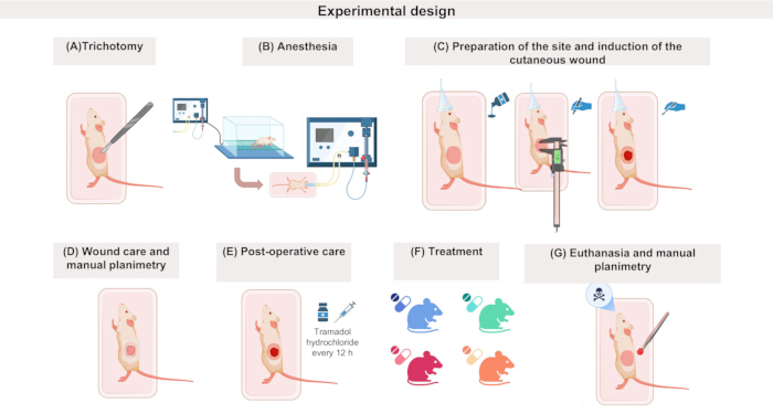
Figure 1: Experimental design of the cutaneous wound induction protocol in rat models. (A) Following animal sedation, shave the experimental region; (B) induce anesthesia in a chamber with isoflurane and, upon loss of reflexes, maintain an adequate anesthetic plane via a facial mask; (C) perform antisepsis of the shaved area, demarcate the lesion dimensions with a caliper, and create the surgical wound using a sterile scalpel; (D) protect the lesion with a transparent film dressing and perform manual planimetry using the adhesive backing paper; (E) administer postoperative analgesia with tramadol hydrochloride every 12 h for 3 days; (F) apply the experimental treatment 24 h after wound induction; (G) perform euthanasia at the previously established experimental time point and proceed with the collection of samples and planimetry tracings for subsequent analyses. This figure was created by the authors with BioRender. Galvão, M. (2026) https://BioRender.com/jsiygmf. Publication license is attached. Please click here to view a larger version of this figure.
1. Animals and handling
- Use adult male Wistar rats aged 2 to 3 months.
- On the day before the surgical procedure, ensure that the animal cages are clean and sanitized, and label them according to the experimental groups.
- On the day of the surgery, weigh each animal using a digital scale and record the values.
- Randomly distribute the animals into the previously sanitized and labeled cages, respecting housing capacity to ensure animal welfare conditions.
- Using a fine-tip permanent marker, identify each animal on the tail according to its experimental group and number, ensuring traceability throughout the entire experiment.
NOTE: Female animals may also be utilized in this protocol, depending on animal availability at the institutional animal facility and the specific objectives of the study. Alphanumeric identification of animals is recommended, with the letter indicating the experimental group and the number indicating the animal. e.g., A1 refers to group A, animal 1.
2. Pre-surgical procedure
- Sedation
- Prepare in advance: cotton, 70% ethanol, one 1 mL hypodermic syringe with a needle per animal, morphine sulfate (10 mg/mL), and prepare the CO₂ chamber.
- Perform antisepsis on the animal's abdominal region using 70% ethanol and cotton. As pre-anesthetic medication, administer morphine sulfate at a dose of 5 mg/kg (10 mg/mL) intraperitoneally in the lower right quadrant of the abdomen.
- Ten min after pre-anesthetic medication administration, place the animal in the CO₂ chamber.
CAUTION: Morphine sulfate, needles, CO₂ exposure systems, and 70% ethanol must be handled exclusively by trained personnel using appropriate personal protective equipment (PPE), including gloves, laboratory coat, and protective eyewear. Needles and syringes must be discarded in approved sharps containers, while chemical and biological waste must be disposed of in accordance with institutional biosafety and hazardous waste regulations.
NOTE: During the 10 min waiting period, perform the anesthetic procedure in the CO₂ chamber and prepare the wound area.
- Anesthetic procedure
- Prepare in advance: isoflurane inhalation anesthetic 4 V% (volume percent), CO₂ chamber, and 1 thermal blanket.
- Induce anesthesia with isoflurane at a concentration of 4 V% in a CO₂ chamber.
- After the animal’s loss of reflexes, transfer the animal from the CO₂ chamber to the facial mask.
- Maintain animal anesthesia with isoflurane at 2 V% or the concentration required to keep it in an adequate surgical anesthetic plane, with a fresh gas flow of 50 mL/min.
- Throughout the procedure, maintain the animal's body temperature using a thermal blanket.
NOTE: An adequate surgical anesthetic plane is considered achieved when the animal shows the absence of reflex responses, including no withdrawal reaction to interdigital mechanical stimulation, together with regular respiratory movements and absence of voluntary body movements throughout the procedure.
CAUTION: Isoflurane is a volatile anesthetic that poses a risk of inhalation exposure. Therefore, its administration using an anesthesia system with adequate exhaust ventilation is highly recommended. Additionally, operators must wear PPE, including gowns, gloves, masks, and caps. Continuous monitoring of the anesthetic plane and body temperature throughout the procedure is necessary. Residual anesthetic materials and contaminated disposables must be discarded in accordance with institutional chemical and biological waste regulations.
- Wound site preparation
- Prepare in advance: gauze sponges, stainless steel blade, 70% ethanol, and 10% povidone-iodine or 0.2% aqueous chlorhexidine.
- Apply a gauze sponge and soap to the animal's left flank region.
- On the surgical table, position the animal in right lateral decubitus.
- Shave the left flank using a stainless-steel blade or an electric shaver.
- Perform pre-surgical antisepsis of the shaved area with a gauze sponge and 70% ethanol (three times), followed by a gauze sponge and 10% povidone-iodine or 0.2% aqueous chlorhexidine (three times).
CAUTION: Sharps should be handled with care and disposed of in appropriate rigid containers after use. Furthermore, the chemicals described should be handled using recommended personal protective equipment and avoiding contact with the eyes and mucous membranes. Used blades must be discarded in approved sharps containers, while contaminated gauze, antiseptic residues, and biological materials must be disposed of in accordance with institutional biosafety and hazardous waste regulations.
3. Cutaneous wound induction
- Prepare in advance: 1 caliper, 1 fine-tip permanent marker, gauze, and sterile surgical instruments (No. 23 scalpel blade, Mayo scissors, and anatomic forceps)
- Using the caliper and permanent marker, demarcate four equidistant points on the skin, outlining a 1 x 1 cm square.
- Create the surgical wound by completely removing the skin within the demarcated area using a sterile No. 23 scalpel blade, Mayo scissors, and sterile anatomic forceps, exposing the subcutaneous tissue and the underlying musculature.
- Achieve hemostasis at the site using sterile gauze, applying gentle pressure directly to the wound until the bleeding ceases.
CAUTION: Blood and tissues must be considered potentially infectious biological waste. Following the procedure, ensure proper disposal in compliance with institutional biosafety standards.
NOTE: Ensure that appropriate aseptic techniques are applied throughout the wound induction procedure. Mandatory for staff are personal protective equipment (PPE), including a hairnet, gloves, a mask, and a lab coat, as well as the use of sterile surgical instruments.
4. Manual planimetry of the wound area
- Prepare in advance: transparent film dressing, white paper sheet (8.5 × 11 inches), fine-tip permanent marker, and transparent adhesive tape.
- Cut rectangles of the transparent film dressing with dimensions of 5 x 3.5 cm and cover the entire wound.
- On a white paper sheet, identify the group (e.g., Group A), the animal (e.g., A1, A2, A3, A4), and the day of wound planimetry collection. Assign D0 to the planimetry obtained on the day of wound induction and DX to the planimetries collected on the corresponding euthanasia day (e.g., D3, D7, D14, D21).
- Use the transparent film dressing to record the wound area and transfer it to the letter paper. To do this, position the film over the wound and, using a fine-tip permanent marker, perform manual planimetry by tracing the entire wound outline.
- Using transparent adhesive tape, attach the obtained planimetry to a letter paper according to its respective identification (group/animal/day). The planimetries will be used to monitor the wound area and determine the contraction rate throughout the experiment.
NOTE: The use of dressings is recommended to prevent fighting among animals, excessive licking of the injured area, and cannibalism, which could aggravate wounds or interfere with healing. D0 corresponds to the day of wound induction (baseline). The transparent film dressing was removed from its original packaging immediately before use and placed directly on the wound to obtain planimetry. A sterile film dressing may also be used according to institutional procedures and experimental requirements. Time points D3, D7, D14, and D21 indicate the post-induction days (3rd, 7th, 14th, and 21st) on which animal euthanasia and planimetry collection are performed, allowing the monitoring of the healing phases during the inflammatory, proliferative, and remodeling periods.
5. Immediate postoperative care
- Prepare tramadol hydrochloride in advance and set aside one sterile 1 mL hypodermic syringe with a needle for each animal, ensuring strict aseptic conditions. Also, prepare crepe bandages and medical tape for wound protection.
- Initiate analgesia with 5 mg/kg of tramadol hydrochloride (50 mg/mL) subcutaneously every 12 h and maintain for 3 days.
- Wrap the surgical wound region with a crepe bandage and secure it with medical tape to protect the wound site.
- Place the rat in an individual cage and monitor it to ensure recovery from anesthesia and surgery.
- After recovery, return the animal to its respective cage.
6. Treatment
- After 24 h of wound induction, initiate the desired experimental treatments, changing the dressings daily.
NOTE: The treatment duration will depend on each specific experimental protocol.
7. Euthanasia
- Perform euthanasia on the designated sample from each group according to the protocol approved by the animal ethics committee.
- Following the confirmation of death, perform manual planimetry of the wound as described in step 4.
NOTE: In this study, euthanasia was induced by decapitation using a guillotine device suitable for rodents, performed by trained personnel in strict accordance with institutional ethical and animal welfare guidelines. Death was confirmed by the absence of respiratory movements, heartbeat, and reflex responses following the procedure.
8. Measurement of the lesion area
- Image analysis software is required to assess the lesion area. Additionally, manual wound planimetry and a millimeter ruler are also required for wound measurement and monitoring.
- Download the chosen image analysis software and create an account, if necessary, to access the platform.
- Before starting the analysis, select two fixed points (e.g., A and B) on the planimetry image and, using the millimeter ruler, measure the distance between these points and record the obtained value separately (Figure 2, step A).
- Photograph the previously delimited planimetries with the aforementioned points. Save the files with their respective experimental group identification (Figure 2, step B).
- Open the image analysis platform and initiate a wound area assessment.
- With the image open in the software, use the cursor or the smartphone touchscreen to mark new points, superimposing them over the previously made ones, identifying them as regions A and B (Figure 2, step B).
- The program will automatically establish a line between points A and B and request the measurement, in millimeters or centimeters, obtained with the ruler. Enter this measurement into the software, and then click “measure”.
- The program will automatically detect the wound area and provide data on wound length, width, perimeter, area, and depth. In planimetry, the "depth" information will not be available (Figure 2, step C).
- Save the images with the generated analyses and export them to the desired location.
NOTE: The wound area assessment described in this protocol was performed using the commercially available image analysis software listed in the Table of Materials. However, equivalent image analysis platforms capable of wound measurement and digital planimetry may also be utilized according to user preference and institutional availability. If the software does not automatically detect the planimetry image, or if the outline differs from what is visually observed in the planimetry, it is recommended to reinforce the outline to facilitate the software's recognition of the area. This can be achieved by using a contrasting color (e.g., red) and applying texture to the external region over an approximate area of 4 cm2. Photographs of the planimetry tracings can be uploaded directly from the device's gallery or captured via camera for subsequent software analysis.
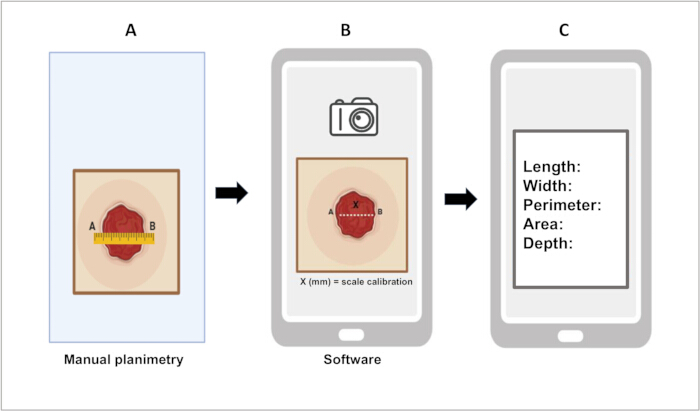
Figure 2: Measurement of the lesion area using ImitoWound. (A) On the manual planimetry, mark two points (A and B), and measure the distance (x) between them using a millimeter ruler; (B) upload the lesion image into the software, and demarcate the same previously defined points (A and B), superimposing them onto the originals; input the previously collected measurement (x) for scale calibration; (C) prompt the software to proceed with the lesion area measurement; wait as the area value is automatically calculated and provided by the system. This figure was created by the authors with BioRender. Lessa da Silva, M. (2026) https://BioRender.com/dzko62t. Publication license is attached. Please click here to view a larger version of this figure.
9. Determination of the wound contraction rate
- After determining the wound area at the evaluated time points, use the data to calculate the wound contraction rate20 using the following equation:
FF (%) = [(A0 - Ax) ÷ A0 ] × 100 (1)
Where A₀ = wound area on day 0 ; Ax = wound area on the day of euthanasia; FF = wound closure fraction
Access restricted. Please log in or start a trial to view this content.
Results
In this experiment, silver sulfadiazine was used as the reference treatment. The product was applied topically to the wound bed 24 h after lesion induction. The choice of silver sulfadiazine was based on its well-established clinical use as an antimicrobial agent and in the management of cutaneous wounds. Data are presented as the median and interquartile range, with n = 5 animals per experimental time point. Figure 3A illustrates the macroscopic evolution of the cutaneous wound on the day o...
Access restricted. Please log in or start a trial to view this content.
Discussion
Although knowledge regarding human wound healing has advanced considerably, inherent difficulties and limitations make the use of experimental models indispensable for research in this field4. In this context, the present protocol aimed to describe a full-thickness excisional wound model induced with a scalpel on the flank of rats as an alternative methodology to conventional techniques. The dorsal region associated with the use of circular punch biopsies is the most widely employed method in cuta...
Access restricted. Please log in or start a trial to view this content.
Disclosures
The authors declare no competing interests.
Acknowledgements
We gratefully acknowledge the Fundação de Amparo à Pesquisa do Estado da Bahia (FAPESB), and the Coordenação de Aperfeiçoamento de Pessoal de Nível Superior (CAPES), for the fellowship support; the Pró-Reitoria de Pesquisa e Pós-Graduação (PROPP/UESC), the Postgraduate program in Biology and Biotechnology of Microorganisms (PPGBBM/UESC), for the financial support. We also thank the Veterinary Medicine Hospital (HVET/UESC), for making the operating room and equipment available.
Access restricted. Please log in or start a trial to view this content.
Materials
| Name | Company | Catalog Number | Comments |
|---|---|---|---|
| Alcohol (ethanol 70%) | Dinâmica | P.01.0024.006.06.81 | Antisepsis |
| Anatomical forceps | Sigma-Aldrich | F3892 | Stainless steel |
| Bandage | Any | Not applicable | wound protection |
| Caliper | Mitutoyo | 530-104 | Wound delimitation |
| Chlorhexidine 0.2% aqueous antiseptic solution | Riohex | Not applicable | Antisepsis |
| CO2 chamber | Bonther | D149F1LYSZ | Anesthetic induction |
| Crepe bandage | Cremer | 818765 | 15 cm x 1,25 m |
| Disposable latex gloves | Descarpak | e0530101 | Non-sterile |
| Gauze pad | Cremer | 819342 | 7,5 x 7,5 cm |
| Hypodermic syringe | Dercarpack | e0324201 | 1 mL with needle |
| ImageJ | Image J | https://imagej.net/ij/ | Planimetric measurement |
| ImitoWound | ImitoWound | https://imito.io/pt/imitowound | Planimetric measurement |
| Interfolded paper towel | Scott Smart | 48260 | General cleaning |
| Isoflurane | Cristália | 7.89668E+12 | Inhalation anesthetic |
| Latex gloves for procedure | Latex gloves for procedure Latex gloves for procedure | Personal protective equipment | |
| Liquid soap | Any | Not applicable | Wound area cleaning |
| Mayo scissors, straight, sharp/sharp | Fine Science Tools | 14001-12 | Stainless steel |
| Millimeter ruler | Any | Not applicable | |
| Morphine sulfate | Sigma-Aldrich | M8777 | Injectable solution 1 mg/mL |
| Paper sheet | Any | Not applicable | |
| Permanent marker | Any | Not applicable | Skin marking |
| Povidone-iodine | Rioquimica | ZK48BN72E | Antiseptic |
| Scalpel blade no. 23 | Descarpak | 740801 | Stainless steel |
| Scalpel handle no. 4 | Sigma-Aldrich | Not applicable | Stainless steel |
| Stainless steel razor blade | Wilkinson | N/A | Hair removal |
| Sterile surgical glove | Descarpak | e0212101 | Sterile |
| Surgical mask | Any | Not applicable | Non-sterile |
| Tramadol hydrochloride | UniãoQuímica | 7.89601E+12 | Solução injetável 50 mg/mL |
| Transparent film dressing | Vital derme | 1503 | 15 cm x 10 m |
| Transparent polypropylene adhesive tape | Transparent polypropylene adhesive tape Transparent polypropylene adhesive tape | wound protection | |
| Veterinary thermal blanket with PVC cover | Prevtech | MSO2VT001 | Maintenance of body temperature |
| Waterproof adhesive tape | Cremer | 819113 | 10 cm x 4,5 m |
References
- Lin X, Lai Y. Skin scarring: mechanisms and therapies. Int J Mol Sci. 2024;25(3):1458.
- Desjardins-Park HE, Mascharak S, Longaker MT, Wan DC. Endogenous mechanisms of craniomaxillofacial repair: toward novel regenerative therapies. Front Oral Health. 2021;2:676258.
- Han G, Ceilley R. Chronic wound healing: a review of current management and treatments. Adv Ther. 2017;34(3):599-610.
- Benito-Martínez S, Pérez-Köhler B, Rodríguez M, Izco JM, Recalde JI, Pascual G. Wound healing modulation through the local application of powder collagen-derived treatments in an excisional cutaneous murine model. Biomedicines. 2022;10(5):960.
- Elliott S, Wikramanayake TC, Jozic I, Tomic-Canic M. A modeling conundrum: murine models for cutaneous wound healing. J Invest Dermatol. 2018;138(4):736-740.
- Chen JS, Longaker MT, Gurtner GC. Murine models of human wound healing. Methods Mol Biol. 2013;1037:265-274.
- Rowland MB, Moore PE, Bui C, Correll RN. Assessing wound closure in mice using skin punch biopsy. STAR Protoc. 2023;4(1):101989.
- De Moura Estevão LR, et al. Morphological evaluation of wound healing events in the excisional wound healing model in rats. Bio-protocol. 2019;9(13):e3285.
- Ansell DM, Campbell L, Thomason HA, Brass A, Hardman MJ. A statistical analysis of murine incisional and excisional acute wound models. Wound Repair Regen. 2014;22(2):281-287.
- Masson-Meyers DS, Andrade TAM, Caetano GF, Guimaraes FR, Leite MN, Leite SN, et al. Experimental models and methods for cutaneous wound healing assessment. Int J Exp Pathol. 2020;101(1-2):21-37.
- Chen Y, Chen X, Zhou Q. Different effects of a perioperative single dose of dexamethasone on wound healing in mice with or without sepsis. Front Surg. 2023;10:927168.
- Zhao J, Zhang S, Gong Z, Mao W, Bao W, Li Q, et al. NLRP3: a key regulator of skin wound healing and macrophage-fibroblast interactions in mice. Cell Commun Signal. 2025;23:55.
- Liu T, Qiu C, Ben C, Li H, Zhu S. One-step approach for full-thickness skin defect reconstruction in rats using minced split-thickness skin grafts with Pelnac overlay. Burns Trauma. 2019;7:19.
- Nayak BS, Maiya A, Kumar P. Influence of Helium-Neon laser photostimulation on excision wound healing in Wistar rats. Online J Biol Sci. 2007;7:89-92.
- Adhirajan N, Shanmugasundaram N, Shanmuganathan S, Babu M. Collagen-based wound dressing for doxycycline delivery: in vivo. evaluation in an infected excisional wound model in rats. J Pharm Pharmacol. 2009;61(12):1617-1623.
- Goorani S, Zangeneh MM, Koohi MK, Seydi N, Zangeneh A, Souri N, Hosseini MS. Assessment of antioxidant and cutaneous wound healing effects of Falcaria vulgaris aqueous extract in Wistar male rats. Comp Clin Pathol. 2019;28(2):435-445.
- Yassine KA et al. A topical ointment formulation containing powder of Lawsonia inermis leaves accelerates excision wound healing in Wistar rats. Vet World. 2020;13(7):1280-1287.
- Kuma DN, et al. Wound healing properties and antimicrobial effects of parkia clappertoniana keay fruit husk extract in a rat excisional wound Model. Biomed Res Int. 2022;2022:9709365.
- Wu AY, et al. Comparison of skin incision healing in swine made with a cold steel scalpel, a standard electrosurgical blade, and a novel tissue dissector. Med Res Innov. 2017;1(5):1000124.
- Ahmadi H, et al. Photobiomodulation-preconditioned diabetic stem cell transplantation accelerates ischemic wound healing in diabetic rats. Stem Cell Res Ther. 2020;11(1):494.
- Zhou Q, et al. Porous sodium carboxymethylamide microspheres for hemostasis and skin wound healing. ACS Appl Bio Mater. 2025;8(4):3076-3085.
- Corrêa JMX, et al. Treatment of surgical wounds in rats with copaiba oil-resin (Copaifera reticulata). Rev Bras Ciênc Vet. 2021;28(4):184-189.
- Huang L, et al. Topical administration of acyl homoserine lactone improves epithelialization of cutaneous wounds in hyperglycemic rats. PLoS One. 2016;11(7):e0158647.
- Dunn L, Prosser HC, Tan JT, Vanags LZ, Ng MK, Bursill CA. Murine model of wound healing. J Vis Exp. 2013;(75):e50265.
- Davidson JM, Yu F, Opalenik SR. Immobilization strategies to overcome wound contraction in experimental animal models. Adv Wound Care. 2013;2(4):142-148.
- Zhang Y, et al. Establishment and translational evaluation of animal models for skin wound healing: a systematic review. Front Physiol. 2026;17:1800001.
- Rizzo AE, Beckett LA, Baier BS, Isseroff RR. The linear excisional wound: an improved model for human ex vivo wound epithelialization studies. Skin Res Technol. 2012;18(1):125-132.
- Can H, et al. Clinical, histopathological, and immunohistochemical investigations of the effects of Aloe vera (Aloe barbadensis Miller) on open wound healing in rats. Pak Vet J. 2025;45(3):1135-1145.
- Shehasina KA, et al. Comparison of manual and digital planimetry techniques in full-thickness cutaneous wounds in dogs. Int J Vet Sci Anim Husb. 2024;9(2):603-607.
- Gill R, Poojar B, Bairy LK, Praveen KSE. Comparative evaluation of wound healing potential of Manuka and Acacia honey in diabetic and non-diabetic rats. J Pharm Bioallied Sci. 2019;11(2):116-126.
Access restricted. Please log in or start a trial to view this content.
Reprints and Permissions
