Here, we present a standardized diagnostic protocol that integrates urine cytology, histopathology, and immunohistochemistry for diagnosing primary bladder large-cell neuroendocrine carcinoma in elderly patients.
Case Report
Bladder Large Cell Neuroendocrine Carcinoma(LCNEC): A Case Report of Exfoliated Cytology and Pathological Characteristics
* These authors contributed equally
In This Article
Summary
Abstract
Primary large cell neuroendocrine carcinoma (LCNEC) of the bladder is an extremely rare and highly aggressive malignancy with a poor prognosis, bringing major challenges to clinical diagnosis and treatment. We report the case of a 77-year-old man who presented with recurrent gross hematuria for more than 8 months, accompanied by urinary frequency, urinary urgency, and lumbosacral pain. Imaging revealed a large soft-tissue mass on the posterior bladder wall without pathognomonic imaging findings. Urine exfoliative cytology revealed atypical tumor cells with large cellular size, high nucleocytoplasmic ratio, scant cytoplasm, and marked nuclear atypia. Given the advanced age and multiple underlying comorbidities, the patient underwent transurethral resection of the bladder tumor. Histopathologically, the tumor was arranged in diffuse sheets with organoid architecture, composed of large polygonal cells with prominent atypia, active mitosis, focal necrosis, and intravascular tumor thrombi. Immunohistochemically, CK-Pan showed a paranuclear punctate expression pattern; neuroendocrine markers INSM1, Syn, and CD56 were diffusely positive; Ki-67 showed a high proliferation index; and P53 displayed diffuse strong expression. The final diagnosis of primary bladder LCNEC was confirmed by integrated morphological and immunohistochemical findings, with preoperative contrast-enhanced CT defining an advanced clinical stage of cT3N0M0 (American Joint Committee on Cancer, AJCC 8th edition). The patient received postoperative adjuvant chemotherapy, and tumor recurrence was identified at the 3-month follow-up with a stable general condition during the observational period. This case indicates that bladder LCNEC lacks specific clinical and imaging manifestations. Urine exfoliative cytology is a valuable non-invasive ancillary tool for detecting high-grade malignant cells in the urinary tract, whereas combining histopathology and immunohistochemistry remains the gold standard for definitive diagnosis. Surgical resection combined with individualized chemotherapy constitutes the main therapeutic regimen, and close long-term follow-up is indispensable for this highly aggressive tumor. Further clinical studies are required to optimize therapeutic strategies and improve patient prognosis.
Introduction
Bladder cancer includes a variety of histological subtypes, among which primary neuroendocrine carcinoma of the bladder accounts for less than 1% of all bladder cancers. According to the WHO 5th Classification of Tumors of the Urinary System and Male Genital Organs, bladder neuroendocrine carcinoma is divided into four subtypes: small cell neuroendocrine carcinoma (SCCB), large cell neuroendocrine carcinoma (LCNEC), well-differentiated neuroendocrine tumor, and paraganglioma. Most bladder neuroendocrine carcinomas are of the small cell type, while primary bladder LCNEC is an extremely rare, high-grade neuroendocrine carcinoma characterized by strong invasiveness, rapid progression, and high risk of metastasis. It is typically diagnosed at an advanced clinical stage and is associated with an extremely poor long-term prognosis1,2. Due to its low incidence, non-specific clinical manifestations and imaging features, and the scarcity of published clinical data, the diagnosis and management of bladder LCNEC remain challenging.
Compared with pelvic computed tomography, cystoscopy, and surgical biopsy, urine exfoliative cytology is non-invasive, inexpensive, and suitable for preliminary detection of malignant cells and postoperative follow-up, especially for elderly patients with multiple underlying diseases who cannot tolerate invasive examinations, although it cannot define tumor staging alone.
We admitted an elderly patient with bladder LCNEC recently and report it as follows, aiming to improve clinicians’ awareness of this rare disease and provide a reference for the standardized diagnostic workflow of this tumor.
Case presentation:
A 77-year-old male patient was admitted on November 16, 2024, presenting with recurrent gross hematuria lasting more than 8 months. He had moderate-volume gross hematuria with occasional blood clots, accompanied by urinary frequency, urinary urgency, and lumbosacral pain, but no dysuria, abdominal distension, chills, or fever. Symptoms relapsed after empirical anti-infection and hemostatic treatment. The patient had a history of chronic obstructive pulmonary disease and cerebral infarction with residual left limb motor dysfunction, without drug allergy or previous surgical history. No obvious body weight loss was noted during the disease course.
Physical examination on admission
Vital signs on admission were as follows: body temperature 36.6 °C, heart rate 82 beats/min, respiratory rate 20 breaths/min, and blood pressure 140/79 mmHg. Physical examination revealed a barrel chest without abnormal pulmonary rales. The abdomen was flat and soft, with no tenderness, rebound tenderness, palpable mass, or shifting dullness. No percussion tenderness was detected over the kidneys. There was no tenderness in the bladder and ureteral regions, and no bleeding was observed at the external urethral orifice. Digital rectal examination revealed grade II benign prostatic hyperplasia without palpable pelvic masses or abnormal rectal bleeding.
Preoperative chest and abdominopelvic contrast-enhanced CT demonstrated a bulky soft-tissue lesion originating from the posterior bladder wall, with suspected transmural bladder wall infiltration and imaging manifestations suggestive of possible extravesical tumor extension, accompanied by left ureteral ostial involvement. No pelvic lymphadenopathy or distant organ metastatic lesions were detected on full-thoracoabdominal imaging. Accordingly, the patient’s tumor was assigned a clinical TNM stage (cT3N0M0) based on the AJCC 8th edition bladder cancer staging criteria. Notably, only fragmented tissue specimens were obtained via transurethral resection of bladder tumor (TURBT), which lacked intact full-thickness bladder wall tissue; therefore, formal pathological pTNM staging could not be completed postoperatively.
Diagnosis, assessment, and plan:
Given the patient’s recurrent gross hematuria and clinical manifestations, imaging was initially arranged to identify intravesical lesions. Routine urine testing and exfoliative cytology of urinary sediment were also performed to screen for malignant cells. Given the patient’s advanced age and cardiopulmonary comorbidities, combined with poor surgical tolerance, transurethral resection of bladder tumor (TURBT) was scheduled to achieve hemostasis and clarify the pathological nature of the lesion. Postoperative histopathological and immunohistochemical examinations were arranged to confirm the final diagnosis and guide the formulation of the subsequent treatment strategy.
Protocol
This study was approved by the Ethics Committee of the affiliated hospital (Approval No. 2026-015) and supported by the 2025 Science and Technology Personnel Training Project of Fujian Provincial Department of Science and Technology (Grant No. 2025350709). Written informed consent was obtained from the patient and his family prior to all clinical examinations and surgical interventions.
All reagents, instruments, and consumables used throughout this protocol are listed in the separately uploaded Table of Materials.
1. Imaging examination
- Chest and whole abdominal computed tomography (CT) was performed using a 64-slice spiral CT scanner. The scanning parameters were set as follows: 120 kVp, field of view 37.5 × 37.5 cm, slice thickness 5.0 mm. Iterative reconstruction was applied, and multiplanar reconstruction (MPR) was performed in coronal and sagittal planes to evaluate bladder wall lesions, tumor scope, and potential distant organ metastasis.
- All imaging data were independently interpreted by two senior radiologists, and a unified diagnostic conclusion was reached through consensus discussion.
2. Urine examination and exfoliative cytology detection
- Routine urine examination was completed to detect the levels of urine bilirubinogen, ketone body, occult blood, protein, and other indicators, and observe the physical and chemical properties of urine.
- For urinary sediment exfoliative cytology examination (Wright-Giemsa staining), urine was centrifuged at a relative centrifugal force of 400 g for 5 min at room temperature, the supernatant was aspirated and discarded, and 0.2 mL of urinary sediment was retained for smear preparation.
- Smear preparation was performed according to the blood smear method, with reference to viscosity. After the smear was air-dried naturally, Wright-Giemsa staining was performed: Solution A was applied to the smear for 3 min of fixation, then Solution B buffer was added at a 1:2 ratio and incubated for 5 min.
- For washing, slow, parallel-running tap water was used for 2 min; avoid direct vertical flushing of the smear surface and excessive washing time to prevent cell shedding and staining fading. Do not pour off the staining solution before rinsing.
- The stained smears were observed under a light microscope by two senior cytologists, and the morphological characteristics of cells were recorded and analyzed.
3. Histopathological Examination
- The resected tumor tissue was fixed in 10% neutral buffered formalin, routinely processed, and embedded in paraffin. Serial sections were cut at a thickness of 3–5 µm and then baked at 60 °C for 120 min.
- After standard deparaffinization and rehydration with xylene and graded ethanol, sections were stained with hematoxylin-eosin (HE) and observed under a light microscope to evaluate histopathological morphological features.
4. Immunohistochemical Detection
Complete metadata for all primary antibodies (including clone number, manufacturer, catalog number, working dilution, detection system, and matched positive/negative tissue controls) is fully listed in the supplementary Table of Materials.
- Immunohistochemical (IHC) staining was performed on serial paraffin sections for the markers CK-Pan, CK8/18, INSM1, Synaptophysin (Syn), CD56, Ki-67, and P53. Antigen retrieval was carried out using EDTA buffer (pH 8.0) in a pressure cooker; heated at 1600 W/210 °C until steam escaped, then maintained at 800 W/130 °C for 260 min.
- After blocking endogenous peroxidase activity, all primary antibodies were incubated at room temperature for 30 min, followed by corresponding secondary antibody incubation. DAB chromogenic reaction was performed at room temperature for 1–10 min with continuous microscopic monitoring to control staining intensity.
- All stained sections were independently evaluated by two senior pathologists to determine the positive expression location and positive rate of each biomarker.
5. Biosafety and waste disposal
- All biospecimens, including patient urine samples and resected tumor tissues, were handled strictly in accordance with hospital biosafety management specifications. All experimental procedures were performed in a standardized pathological laboratory, with personal protective equipment worn throughout.
- Biological waste such as residual urine specimens, paraffin debris, stained slides, and contaminated consumables was classified, sealed, and disposed of as medical biohazard waste. Chemical waste, including formalin fixative, xylene, ethanol, EDTA buffer, Wright-Giemsa staining solution, and DAB chromogenic solution, was collected separately in dedicated chemical waste containers to avoid environmental pollution.
- All waste was uniformly recycled and processed by a qualified medical waste disposal institution, ensuring full compliance with biosafety and environmental protection regulations.
Results
Combined findings from imaging, exfoliative cytology of urine, histopathology, and immunohistochemistry enabled a definitive diagnosis of primary bladder large-cell neuroendocrine carcinoma (LCNEC). Urine cytology allowed noninvasive identification of malignant atypical cells, histopathology demonstrated characteristic neuroendocrine tumor morphology, and immunohistochemistry verified neuroendocrine differentiation and high proliferative activity. The diagnostic summary table integrates key results across all modalities to support accurate and reproducible diagnosis (Table 1).
Imaging examination results
Chest and whole abdominal CT examinations revealed the following findings: 1. Chronic bronchopulmonary disease accompanied by emphysema and mild inflammatory lesions in the left lower lung; 2. Multiple small, calcified foci in both lungs; 3. A large soft-tissue mass originating from the posterior bladder wall and protruding into the bladder lumen, measuring approximately 73 × 74 × 65 mm. The mass exhibited heterogeneous density with an average CT value of 38 HU, irregular margins, partial calcification, and involvement of the left ureteral orifice, with suspected transmural bladder wall infiltration and suspected involvement of perivesical adipose tissue. 4. Minor prostatic calcifications; 5. Multiple simple cystic lesions in the liver (Figure 1). No obvious regional lymph node enlargement or distant metastatic lesions were detected in the chest and abdomen. Imaging showed only a bladder space-occupying lesion, lacking pathognomonic radiological features to confirm full-thickness bladder wall penetration and perivesical fat invasion. The suspected clinical stage is cT3N0M0, based on imaging findings suggestive of transmural infiltration and potential perivesical fat involvement.
Urine examination results
Preoperative routine urine tests showed urobilinogen 3+, ketone bodies 3+, occult blood 3+, protein 3+, nitrite positive, and leukocyte esterase 3+, with a specific gravity of 1.015 and a pH of 5.0. The urine sample was grossly red and turbid, consistent with hematuria. Microscopic examination suggested the presence of abnormal urinary tract cells, and further exfoliative cytology of the urinary sediment was performed.
Light microscopic examination of urine exfoliative cytology revealed malignant atypical cells with variable aggregation patterns: a solitary atypical cell (Figure 2A); small clusters of three tumor cells (Figure 2B); and densely packed, massive tumor cell aggregates (Figure 2C). These cells displayed irregular morphology, scant basophilic cytoplasm with cytoplasmic processes and vacuoles, markedly enlarged irregular nuclei, coarse granular chromatin, and prominent nucleoli, consistent with high-grade malignant atypia.
Histopathological results
Specimens obtained from transurethral resection of bladder tumor (TURBT) were subjected to hematoxylin-eosin (HE) staining. The tumor tissues exhibited diffuse sheet-like and organoid arrangements (Figure 3A); intravascular tumor thrombi were visible within stromal vessels (Figure 3B), accompanied by abundant scattered stromal microvessels throughout the lesion. The tumor cells were large and polygonal with marked cellular atypia, abundant cytoplasm, and coarse granular chromatin (Figure 3C). At high magnification, the tumor cells displayed prominent nucleoli and frequent atypical mitotic figures, with focal vascular invasion noted (Figure 3D). The TURBT specimen demonstrated tumor invasion into the muscularis propria, corresponding to at least pathological T2 in the sampled material; higher pathological T staging could not be assessed because an intact full-thickness bladder wall and perivesical adipose tissue were not captured in fragmented resection specimens. Eight formalin-fixed paraffin-embedded (FFPE) tissue blocks obtained from TURBT resection were processed, and all serial histologic sections were independently reviewed by two senior pathologists. No morphologic foci of small cell neuroendocrine carcinoma, conventional urothelial carcinoma, or squamous cell carcinoma were detected within all sampled sections. The diagnosis, limited exclusively to the submitted TURBT specimens, is large cell neuroendocrine carcinoma of the urinary bladder; mixed divergent histological components located in unsampled regions of the primary tumor cannot be fully excluded.
Immunohistochemical results
Specimens from transurethral resection of bladder tumor (TURBT) were subjected to immunohistochemical staining to further clarify the tumor phenotype. Tumor cells showed punctate paranuclear expression of pan-cytokeratin (CK-Pan) (Figure 4A); low-molecular-weight cytokeratin CK8/18 showed partial cytoplasmic positivity. Among neuroendocrine markers, INSM1 showed nuclear expression (Figure 4B); Syn exhibited cytoplasmic positivity (Figure 4C); CD56 was mainly membrane-positive with focal cytoplasmic staining (Figure 4D). The Ki-67 proliferation index was approximately 80% in hot spots, indicating high tumor proliferative activity (Figure 4E). P53 displayed diffuse strong positivity in about 80% of tumor cells, suggesting mutant-type expression (Figure 4F).
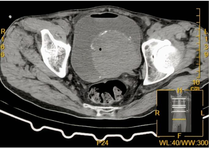
Figure 1: CT image of the bladder. A soft-tissue lesion is seen arising from the posterior bladder wall with intravesical protrusion, heterogeneous density, and focal calcification. No definite lymph node enlargement or distant metastatic lesion is identified on CT.
Please click here to view a larger version of this figure.
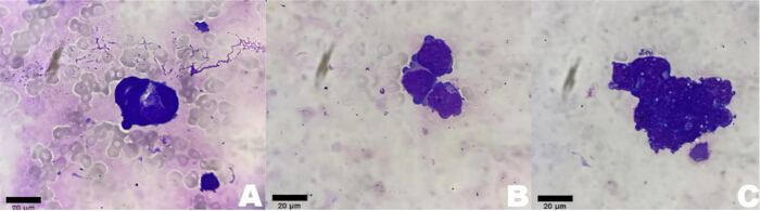
Figure 2: Preoperative urinary exfoliative cytology (Wright-Giemsa staining, 1000×). (A–C) Representative fields of preoperative urinary exfoliative cytological specimens under an oil immersion lens. Please click here to view a larger version of this figure.
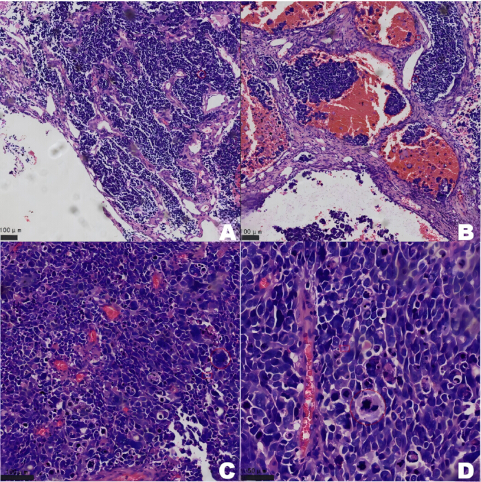
Figure 3: Histopathological microscopic morphology (HE staining). (A) Tumor characteristic growth pattern (100×). (B) Intravascular tumor thrombus within stromal vessels (100×). (C) Morphology of atypical tumor cells (200×). (D) Atypical mitotic figures and focal vascular invasion (400×). Please click here to view a larger version of this figure.
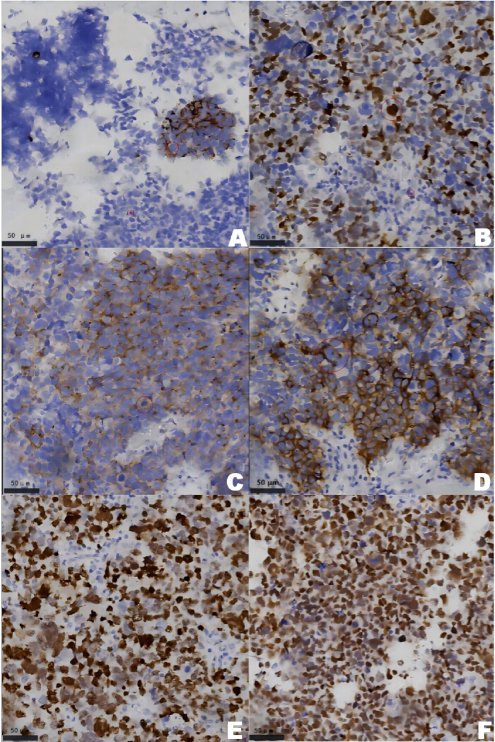
Figure 4: Immunohistochemical staining results. (A) CK-Pan immunohistochemical staining (400×); (B) INSM1 immunohistochemical staining (400×); (C) Syn immunohistochemical staining (400×); (D) CD56 immunohistochemical staining (400×); (E) Ki-67 immunohistochemical staining (400×); (F) P53 immunohistochemical staining (400×). Please click here to view a larger version of this figure.
| Diagnostic Modality | Key Findings | |||
| Computed Tomography (CT) | A 73 × 74 × 65 mm heterogeneous soft-tissue mass arises from the posterior bladder wall and protrudes into the bladder lumen, accompanied by focal intralesional calcification. No pelvic lymph node enlargement or distant visceral metastasis is identified in abdominal and thoracic scanning. | |||
| Urine Exfoliative Cytology | Malignant large atypical cells were observed in urine sediments, featuring elevated nucleocytoplasmic ratio, sparse basophilic cytoplasm and coarsely clumped chromatin with conspicuous nucleoli. Tumor cells exist as isolated single cells, small three-cell clusters and large compact cell aggregates. Cytology indicates high-grade malignant urothelial lesion without subtype-specific features for LCNEC. | |||
| Histopathology (HE staining) | Tumor tissues grow in diffuse sheet-like and typical neuroendocrine organoid patterns. Lesions consist of large polygonal neoplastic cells with severe nuclear atypia, frequent mitotic figures, focal tumoral necrosis and intraluminal vascular tumor thrombi, accompanied by rich interstitial microvascular proliferation; no mixed urothelial or squamous carcinoma components are found. | |||
Table 1: Summary of Diagnostic Findings Across Imaging, Cytology, Histopathology, and Immunohistochemistry. The table integrates key results across all modalities to support accurate and reproducible diagnosis.
Discussion
Bladder cancer is one of the most common malignant tumors of the genitourinary system and frequently occurs in elderly males. Over the past three decades, the incidence of bladder cancer has gradually increased in China, exceeding the global average, and the mortality risk rises significantly with advancing age3,4,5. Primary large cell neuroendocrine carcinoma (LCNEC) of the bladder is a rare high-grade subtype, first described in 1986, and is characterized by high aggressiveness and poor overall prognosis6. It predominantly affects males older than 50 years, with a male-to-female ratio of approximately 4:16. Approximately 50% of bladder LCNEC cases present as mixed tumors combined with urothelial carcinoma or squamous cell carcinoma, whereas pure LCNEC is extremely rare7,8. The cellular origin of bladder LCNEC remains controversial, with proposed origins including submucosal neuroendocrine cells, urothelial metaplasia, or urachal epithelial cells7,8.
Bladder LCNEC lacks specific clinical manifestations, with painless gross hematuria as the most common initial symptom. Some patients also present with urinary frequency, urinary urgency, pelvic discomfort, or paraneoplastic syndromes, which often delay early clinical recognition. Imaging can define tumor size and local scope, but cannot confirm pathological subtype due to non-specific radiological features. Most cases are diagnosed at an advanced stage owing to insidious early symptoms, and some present with local invasion or metastatic disease, leading to a generally poor prognosis9,10,11,12. Consistent with this clinical feature, our patient harbored a primary bladder LCNEC assigned as suspected clinical cT3N0M0, based on CT imaging manifestations suggestive of transmural bladder wall infiltration and potential perivesical adipose tissue involvement, with left ureteral ostial infiltration noted; no regional lymphadenopathy or distant metastatic lesions were identified on cross-sectional imaging, consistent with advanced clinical tumor staging; no regional lymphadenopathy or distant metastatic lesions were identified on cross-sectional imaging, consistent with advanced clinical tumor staging. The combined application of urine exfoliative cytology, histopathology, and immunohistochemistry facilitates the preliminary identification of high-grade malignant urothelial cells and the definitive pathological diagnosis.
Histologically, LCNEC exhibits typical neuroendocrine architecture, including organoid and rosette-like arrangements. Tumor cells are large, generally larger than three lymphocytes in diameter, with coarse chromatin, prominent nucleoli, frequent mitoses, and often focal hemorrhage and necrosis. LCNEC commonly expresses neuroendocrine markers, including Syn, CD56, CgA, and INSM113,14. Typical morphological features together with positivity for at least one neuroendocrine marker support the diagnosis. Epithelial markers, including CK-Pan, are also frequently expressed. Ultrastructurally, cytoplasmic neuroendocrine granules are identifiable.
In the present case, chest and abdominal CT revealed no lymphadenopathy or distant metastatic lesions, supporting a primary bladder origin without systemic spread. Fresh midstream urine was collected for exfoliative cytology, a simple, noninvasive test suitable for preliminary identification of malignant urothelial cells.
Bladder LCNEC readily sheds tumor cells into the urine, enabling detection of high-grade atypical cells by cytological examination. However, morphological overlap exists between LCNEC, high-grade urothelial carcinoma, and other bladder malignancies. Urine cytology alone cannot confirm the exact pathological subtype and serves only as an auxiliary diagnostic method; definitive diagnosis relies entirely on histopathology and immunohistochemistry. The cytological findings in this case reflected high-grade malignant atypia, consistent with a neuroendocrine tumor phenotype.
Postoperative histopathology showed diffuse sheet-like growth and organoid structures typical of neuroendocrine neoplasms. Tumor cells were large and polygonal with marked atypia, active mitoses, focal necrosis, and focal intravascular tumor thrombi. Immunohistochemically, tumor cells showed paranuclear punctate CK-Pan expression, positive neuroendocrine markers (INSM1, Syn, CD56), a high Ki-67 proliferation index, and mutant P53 expression. These immunohistochemical profiles do not support conventional urothelial or squamous epithelial differentiation within the sampled tumor cells, and negative melanoma, lymphoid, and mesenchymal markers rule out corresponding differential entities.
At present, there is no unified international guideline for the treatment of bladder LCNEC. Multidisciplinary therapy combining surgery and chemotherapy is the mainstream strategy. TURBT is a feasible option for elderly patients with multiple comorbidities who cannot tolerate radical cystectomy15,16,17. Notably, TURBT provides only partial tissue specimens and cannot support complete TNM staging. In the present case, only imaging-based clinical cT3N0M0 staging could be assigned. Complete pathological pTNM staging was not feasible because fragmented TURBT tissue specimens failed to capture the intact full-thickness bladder wall layers required for accurate assessment of depth of invasion, consistent with the routine clinical limitations of transurethral biopsy/resection for bladder malignancies. In this case, the patient underwent TURBT followed by adjuvant chemotherapy, and tumor recurrence was detected at the 3-month follow-up with a stable general condition.
Standardized specimen processing, smear preparation, staining quality control, and experienced pathological interpretation are critical to avoid misdiagnosis of urine cytology and immunohistochemical results. Strict procedural standardization helps improve diagnostic accuracy for rare bladder neuroendocrine tumors.
This study has inherent limitations as a single-case report. The sample size is small, and long-term survival and recurrence outcomes require further extended follow-up. Large-sample prospective studies are still needed to unify diagnostic criteria and optimize treatment regimens.
In conclusion, primary bladder LCNEC is a rare yet highly aggressive malignancy. Non-invasive urine exfoliative cytology is a valuable ancillary assay for identifying high-grade malignant urothelial cells, whereas histopathology combined with immunohistochemistry remains the gold standard for definitive diagnosis. For this elderly patient complicated with multiple underlying diseases, transurethral resection combined with intravesical therapeutic instillation and rigorous long-term surveillance was delivered. No universal standardized systemic chemotherapy regimen can be generalized from this single case; large-scale prospective clinical trials are required to optimize treatment strategies for bladder LCNEC. Further clinical studies are required to establish standardized management strategies and improve patient prognosis.
Disclosures
The authors declare no conflicts of interest.
Acknowledgements
This study was supported by the 2025 Science and Technology Personnel Training Project of the Science and Technology Department of Fujian Province (No.2025350709). We would like to thank the medical staff who participated in the patient's diagnosis and treatment, as well as the patient and his family for their cooperation.
Materials
| Name | Company | Catalog Number | Comments |
|---|---|---|---|
| 64-slice spiral CT scanner (LightSpeed VCT) | GE Medical Systems, LLC | SFDA(I)20113300100; Serial No.: VCTC1001CT | |
| Anti-CD56 antibody | AnBiPing | 1216241224 | |
| Anti-CK-Pan antibody | AnBiPing | 0714251118 | |
| Anti-CK8/18 antibody | AnBiPing | 0827240911 | |
| Anti-INSM1 antibody | AnBiPing | 1122241126 | |
| Anti-Ki-67 antibody | AnBiPing | 0209260320 | |
| Anti-P53 antibody | AnBiPing | 0105260121 | |
| Anti-Synaptophysin (Syn) antibody | AnBiPing | 0819250901 | |
| DAB chromogenic kit | AnBiPing | T2603001 | |
| EDTA buffer | AnBiPing | 1224251225 | |
| Flexible cystoscope | RUIDE | 069772520300066 | |
| Hematoxylin-eosin (HE) staining kit | JinShengLai | 2025102178, 2025061277 | |
| Neutral buffered formalin fixative | TongHe Bio | 20260301 | |
| Resectoscope system | Zhuhai Simai Technology Co., Ltd. | 1550085C | |
| Wright-Giemsa staining solution | BASO Diagnostics Inc., Zhuhai | C240807 | |
| Xylene | FuYu Chemical | 20260201F |
References
- Moch H, et al. The 2022 World Health Organization Classification of Tumors of the Urinary System and Male Genital Organs-Part A. Eur Urol. 2022;82(5):458–468.
- Raspollini MR, et al. News in the classification of WHO 2022 bladder tumors. Pathologica. 2023;115(1):32–40.
- Wei YG, et al. Analysis of the incidence and mortality of bladder cancer in urban and rural residents of Shandong Cancer Registry in 2018. Chin J Cancer Prev Treat. 2024;31(20):1240–1244.
- Yu L, et al. Incidence and mortality trends of bladder cancer in China based on age-period-cohort model. Mod Prev Med. 2022;49(23):4253–4258.
- Ji XC, Guo HY, Wang W. Incidence trend of bladder cancer in China from 1990 to 2019: an age-period-cohort analysis. J Jining Med Univ. 2023;46(2):90–95.
- Pusol T, Zorzi MG, Morichetti D. "Pure" primary large cell neuroendocrine carcinoma of the urinary bladder. Turk Patoloji Derg. 2013;29:241–245.
- Gupta S, et al. High grade neuroendocrine carcinoma of the urinary bladder treated by radical cystectomy: a series of small cell, mixed neuroendocrine and large cell neuroendocrine carcinoma. Pathology. 2015;47(6):533–542.
- Hata S, Tasaki Y. A case of the large cell neuroendocrine carcinoma of the urinary bladder. Case Rep Med. 2013;2013:804136.
- Li HM, Ni JY, Zheng ZG. Clinical and pathological analysis of 6 cases of neuroendocrine carcinoma of the bladder. Zhejiang Pract Med. 2019;24(4):266–270.
- Gong ZX, Xu QG. A case report of primary large cell neuroendocrine carcinoma of the bladder. J Mod Urol. 2022;27(4):59–60.
- Zeng Z, Zhou XJ. Meta-analysis of the diagnostic value of fluorescence in situ hybridization in bladder cancer. Chin J Pathol. 2010;39(2):75–78.
- Yang B, et al. A case report of large cell neuroendocrine carcinoma of the ureter and bladder with literature review. Chin J Urol. 2018;39(2):95–98.
- Colarossi C, Pino P, Giuffrida D. Large cell neuroendocrine carcinoma (LCNEC) of the urinary bladder: a case report. Diagn Pathol. 2013;8:19.
- Xia K, et al. Clinical characteristics, treatment strategy, and outcomes of primary large cell neuroendocrine carcinoma of the bladder: a case report and systematic review of the literature. Front Oncol. 2020;10:1291.
- He ZL, Liu LL, Chen SL. A case report of primary large cell neuroendocrine carcinoma of the bladder accompanied by urothelial carcinoma. Chin J Urol. 2024;45(1):59–60.
- Martín IJ, Vilar DG, Aguado JM. Large cell neuroendocrine carcinoma of the urinary bladder. Arch Esp Urol. 2011;64(2):105–113.
- Zare A, Aminirad O, Binesh F. A case of large-cell undifferentiated carcinoma of the bladder. J Surg Case Rep. 2023;2023(12).