This protocol demonstrates the modified Wiltse Transforaminal Lumbar Interbody Fusion (W-TLIF), a muscle-sparing paraspinal approach that minimizes soft tissue disruption while achieving lumbar decompression and fusion.
Method Article
Muscle-Sparing Wiltse Transforaminal Lumbar Interbody Fusion: A Technical Guide for the Modified Paraspinal Approach
In This Article
Summary
Abstract
The Wiltse transforaminal lumbar interbody fusion (W-TLIF) modifies the traditional open posterior lumbar fusion to treat the same degenerative pathologies through a smaller, muscle-sparing incision that accesses the lumbar spine through the paramedian corridor between the multifidus and longissimus muscles, preserving the posterior musculature and limiting iatrogenic soft tissue trauma. This protocol provides a reproducible, step-by-step guide to performing the muscle-sparing W-TLIF for lumbar decompression and interbody fusion. After prone positioning and fluoroscopic level localization, a paramedian incision is made, and the lumbodorsal fascia is divided to develop the natural interval between the multifidus and longissimus, and sequential tubular dilation is used to dock a tubular retractor on the facet complex. An ipsilateral hemilaminectomy and medial facetectomy are performed to decompress the neural elements, followed by discectomy and endplate preparation. An interbody cage packed with autograft and allograft is placed, and the construct is completed with percutaneous pedicle screws under fluoroscopic guidance. The technique yields low intraoperative blood loss and short hospital stays, with same-day discharge feasible in selected patients and improvement in patient-reported outcomes. The muscle-sparing W-TLIF is a safe, reproducible, and teachable evolution of the traditional posterior lumbar fusion that emphasizes anatomical preservation and improved recovery.
Introduction
Dr. Wiltse first described the paraspinal approach in 1968 as a muscle-splitting technique that accesses the lumbar spine through a natural intermuscular plane, avoiding midline stripping of the paraspinal musculature1. Originally developed to reduce muscle injury during lumbar exposure, this approach laid the groundwork for modern minimally invasive spinal surgery2,3. The muscle-sparing Wiltse Transforaminal Lumbar Interbody Fusion (W-TLIF) modifies the traditional posterior TLIF approach by exploiting the intermuscular plane between the multifidus and longissimus to minimize iatrogenic soft tissue trauma and preserve the multifidus muscle, a critical stabilizer of the lumbar spine4,5.
Compared with the midline open TLIF, the W-TLIF provides equivalent decompression and fusion outcomes with significantly less postoperative pain, reduced blood loss, and shorter hospital stays6,7,8. Biomechanically, preservation of the multifidus reduces adjacent segment disease and postoperative instability by maintaining paraspinal integrity9,10.
The W-TLIF is indicated for single- or two-level degenerative lumbar pathology requiring both decompression and interbody fusion, including low-grade (Meyerding grade I to II) degenerative or isthmic spondylolisthesis11, recurrent disc herniation, symptomatic foraminal or lateral recess stenosis with segmental instability, and degenerative disc disease with mechanical instability12,13. Optimal candidates have focal, unilateral, or predominantly unilateral pathology at one or two levels, as the paramedian corridor provides direct ipsilateral access to the transforaminal space. Patient selection accounts for body habitus and operative level, as increasing body mass index (BMI) is associated with poorer operative corridors and more difficult access to anatomical landmarks14, with corridor depth further increasing at more cephalad lumbar levels in the authors’ experience. While no large consensus guidelines exist, in the authors’ experience, some relative contraindications for a muscle-sparing approach include high-grade spondylolisthesis, severe central canal stenosis requiring wide bilateral decompression, multilevel disease exceeding two to three levels, significant coronal or sagittal deformity requiring open correction, and tumor or infection requiring wide exposure.
Conventional open midline TLIF remains preferable when the pathology demands wide bilateral central decompression, multilevel exposure, or deformity correction, settings in which the transforaminal corridor limits access and coronal or lordotic correction13, and in revision cases where scarring obscures the multifidus-longissimus plane, in the authors’ experience. This protocol outlines a reproducible, step-by-step guide to the modified Wiltse incision, exposure, decompression, and interbody fusion technique.
Protocol
No new subject data were generated for this study protocol and manuscript. The institutional outcome data reported is cited with an associated study. The accompanying technique video and images were obtained with written informed consent for filming and publication.
1. Preoperative preparation
- Review preoperative magnetic resonance imaging (MRI), computed tomography (CT), and standing anteroposterior (AP) and lateral radiographs to define the pathology and confirm the operative level(s) and decompression side.
- Administer general anesthesia.
- Position the patient prone on a radiolucent operating table with padding under the chest, pelvis, and bilateral lower extremities. Ensure the abdomen hangs somewhat free of compression, and maintain the lumbar spine in a neutral to slightly flexed alignment on the frame.
- Position the arms at 90° of shoulder abduction and 90° of elbow flexion (the “90/90” position) (Figure 1).
- Establish somatosensory evoked potential (SSEP) neuromonitoring. Obtain baseline neuromonitoring signals prior to skin incision to serve as an intraoperative reference.
NOTE: Maintain the head, cervical spine, and arms in neutral alignment to prevent perioperative neuropraxia.
2. Level localization
- Obtain a true AP fluoroscopic image of the target level using a C-arm, confirming symmetric pedicles and a midline spinous process.
- Mark the midline overlying the spinous processes of the target level(s) with a surgical marking pen (Figure 2).
- Mark a paramedian line just lateral to the pedicles of the vertebral bodies above and below the target disc. Determine this line fluoroscopically rather than from surface landmarks, projecting it through the lateral pedicle borders on the true AP image so that it approximates a line connecting the lateral pedicle margins of the levels above and below the target disc.
- Insert a spinal needle at the marked level and confirm the target disc space on a true lateral fluoroscopic image (Figure 3).
NOTE: Confirm true AP and lateral fluoroscopic images, with well-defined endplates and symmetric pedicles, before skin marking, as inadequate imaging is a common source of wrong-level exposure.
3. Skin incision and fascial exposure
- Select the approach side ipsilateral to the symptomatic pathology.
- Create a longitudinal paramedian skin incision approximately 2–3 cm in length, positioned 2 cm lateral to the midline and centered over the target disc space under lateral fluoroscopy. Carry the incision sequentially through the skin, subcutaneous fat, and the superficial fascial layer down to the thoracolumbar (lumbodorsal) fascia.
NOTE: Lengthen the incision by approximately 1 cm for patients with higher BMI or when multilevel decompression is performed through a single incision, to allow cranial-caudal angulation of the working corridor and tubular retractor as needed. - Divide the subcutaneous tissue with a mixture of sharp and electrocautery dissection and place a self-retaining retractor to expose the lumbodorsal fascia. Obtain hemostasis with bipolar forceps or pressure.
- Incise the lumbodorsal fascia in line with the skin incision to expose the underlying paraspinal muscles.
NOTE: Paraspinal muscle fibers interdigitate and run in multiple directions at this level and may not split cleanly, so minimize sharp dissection.
4. Muscle-sparing dissection
- Identify the natural interval between the multifidus (medial) and longissimus (lateral) muscles using a blunt finger sweep, feeling for the intermuscular septum while advancing the finger obliquely in a main cranial-to-caudal and subtle medial-to-lateral direction toward the transverse process.
NOTE: The interval may be recognized visually by a thin fatty septum separating the more vertically oriented multifidus fibers medially from the obliquely oriented longissimus fibers laterally (however, this is not always readily appreciable). When performing blunt dissection between these muscles, a natural split may be palpated, indicating that the surgeon has entered the intermuscular plane. - Palpate the transverse process, then direct dissection to the junction of the transverse process and superior articular process (SAP), and then over the ipsilateral lamina.
NOTE: Confirmation of the correct plane is tactile. The intermuscular septum and the bony landmarks (transverse process, TP-SAP junction, and lamina) are palpable, with minimal muscle bleeding. - Insert the smallest dilator through the developed interval and dock it onto the facet complex. Separate the muscle fibers sufficiently to admit approximately 2 fingerbreadths and seat the smallest dilator. Separate additional fibers as needed to accommodate a larger working portal during sequential dilation.
5. Tubular retractor exposure
- Confirm correct docking of the initial dilator on a lateral fluoroscopic image.
- Perform sequential dilation over the initial dilator and place a tubular retractor sized to the procedure (18–22 mm for decompression, 26 mm for interbody fusion) (Figure 4). Advance each dilator along the trajectory established by the initial dilator, maintaining a slight medial-to-lateral angulation directed at the facet complex, and confirm the position of the intermediate dilators on intermittent lateral fluoroscopy before seating the final retractor to a docking depth on the facet complex.
- Attach the tubular retractor to a table-mounted retractor arm. Dock the tubular retractor onto the facet complex and confirm the operative level on AP and lateral fluoroscopy (Figure 5).
- Adjust the operating light and loupe or operating microscope magnification to visualize the neural elements (Figure 6).
NOTE: Obtain high-quality AP and lateral fluoroscopic images to minimize pedicle screw malposition12.
6. Decompression
- Perform an ipsilateral hemilaminectomy and medial facetectomy using a high-speed burr and Kerrison rongeur as needed for decompression. Begin at the inferior edge of the cranial lamina and proceed caudally, removing the inferior portion of the cranial lamina and the medial third to half of the superior articular process until the lateral border of the traversing nerve root and the medial pedicle wall are exposed, preserving the pars interarticularis where possible.
NOTE: Preserve as much bone as possible for use as a local autograft (if fusion is intended). - Remove the ligamentum flavum and epidural fat, working in the direction of the traversing nerve root. Palpate the pedicle with a long ball-tip probe to confirm adequate skeletonization.
- Retract the traversing nerve root medially with a nerve retractor. Apply only gentle medial retraction sufficient to expose the disc space, release the retractor intermittently to allow neural reperfusion, and avoid sustained or forceful retraction to minimize the risk of neural or dural injury.
NOTE: Adequate decompression is confirmed when the traversing and exiting nerve roots are visually free, the thecal sac is decompressed and pulsatile, and a ball-tip probe may be passed freely into the foramen. - Place a 1/2 x 1/2 inch cottonoid cranial to the disc space to protect the exiting nerve root.
NOTE: Treat small cerebrospinal fluid leaks with a collagen sponge, fibrin glue, and flat bed rest12. If contralateral neural compression is present, decompress by repositioning (“wanding”) the ipsilateral tubular retractor or by placing a second tubular retractor through the contralateral incision.
7. Discectomy and endplate preparation (if fusion is desired)
- Incise the annulus over the exposed posterolateral disc space as a rectangular (box) annulotomy using a long-handled no. 15 blade, then perform a thorough discectomy with pituitary rongeurs and straight and angled curettes.
NOTE: Alternatively, create the rectangular annulotomy using an initial incision directed toward the surgeon followed by three additional incisions. Select the annulotomy technique according to surgeon preference. - Prepare the endplates using disc shavers, curettes, and rasps to remove the cartilaginous endplate while preserving the underlying bony endplate.
- Size the disc space using sequential trial implants. Select the trial that provides a snug fit with restoration of disc and foraminal height without over-distraction, and confirm trial size and position on AP and lateral fluoroscopy before choosing the final implant.
NOTE: Appropriate trial size may be determined by the surgeon based on factors such as desired sagittal and coronal correction, contact with the apophyseal ring, and amount of disc removal. - Avoid overzealous curettage to preserve endplate integrity and reduce the risk of cage subsidence.
NOTE: Endplate preparation is confirmed by the tactile feeling of removal of the cartilaginous tissue, with the goal of punctate bleeding of the subchondral bone and an intact bony endplate underneath.
8. Interbody fusion and instrumentation
- Pack the anterior disc space with a mixture of autograft and allograft to support fusion.
- Insert the interbody cage packed with autograft and allograft under fluoroscopic guidance. Insert the cage along an oblique transforaminal trajectory from lateral to medial while protecting the traversing and exiting nerve roots with a nerve-root protector, keeping the neural elements under direct vision throughout impaction.
- Position the cage within the disc space where desired to maximize coronal or sagittal plane correction. Confirm placement on AP and lateral fluoroscopy by visualizing the fluoroscopic markers on the cage.
NOTE: Positioning the cage within the anterior third of the disc space aids lordotic correction, and contact with the apophyseal ring is advisable to reduce the risk of subsidence. Cage type is at the surgeon's preference. An expandable cage may be used to achieve greater coronal or sagittal plane correction, with careful placement within the disc space. Titanium cages are used in the authors’ experience, although other cage materials have been reported. - Place percutaneous or mini-open pedicle screws under fluoroscopic guidance.
NOTE: This process may be chosen by the practicing surgeon based on their preferences. If navigation is used, the surgeon may elect to drill each screw hole directly. Next, a pedicle probe is used to feel for any cortical breaches. If no breach exists, an undersized tap (by approximately 1 mm) may be inserted. Once complete and the tap is removed, a pedicle probe is inserted again to confirm the absence of any breaches. Then, the final screw may be inserted along this path. If a Jamshidi-based approach is desired, this process differs slightly. Cannulate each pedicle with a Jamshidi needle at the junction of the transverse process and superior articular process, keeping the needle tip lateral to the medial pedicle wall on the AP view until the posterior vertebral body is reached on the lateral view, then pass a guidewire, tap over the wire, and place each cannulated screw over the guidewire, removing the wire before final seating. - Confirm construct alignment and hardware position on AP and lateral fluoroscopy before closure.
CAUTION: Radiation safety measures must be employed in every case15. These measures include, but are not limited to, lead aprons, thyroid shields, and radiation dosimeters. Personnel, when able, should maximize their distance from the radiation source and step back during image acquisition.
NOTE: Objective intraoperative endpoints include a cage well seated across the apophyseal ring without endplate violation, restored disc and foraminal height, a decompressed and pulsatile thecal sac with freely mobile traversing and exiting nerve roots, and pedicle screws with intact medial and inferior pedicle walls confirmed by palpation and, where available, triggered electromyography.
9. Wound closure and completion
- Remove the tubular retractor under direct visualization and confirm hemostasis of the muscle corridor. Irrigate the corridor copiously with sterile saline and achieve hemostasis with bipolar cautery. Supplement with a topical hemostatic agent if needed.
NOTE: A wound drain may be placed at the discretion of the surgeon. - Inspect the wound and confirm final hemostasis. Obtain final intraoperative AP and lateral fluoroscopic images to document construct position.
- Close the lumbodorsal fascia with interrupted absorbable sutures (e.g., 0 or 2-0 polyglactin).
- Close the subcutaneous layer with inverted absorbable sutures and approximate the skin with a running subcuticular absorbable suture or skin adhesive.
- Apply a sterile dressing.
10. Postoperative management
- Perform an immediate postoperative neurological examination in the recovery unit to document motor and sensory function and compare with the preoperative baseline.
- Obtain standing AP and lateral radiographs before discharge to confirm implant position and construct alignment.
- Begin early mobilization, typically on the day of surgery or postoperative day one, with physical therapy and progressive activity as tolerated; same-day discharge is appropriate in selected patients.
Results
The muscle-sparing W-TLIF has been used as the routine minimally invasive lumbar fusion technique at the authors’ institution and has been evaluated in two prior IRB-approved institutional analyses employing the technique described here.
In our W-TLIF experience, a prospectively maintained registry of 120 consecutive patients undergoing single-level non-tubular W-TLIF (2013–2023, minimum one-year follow-up) demonstrated intraoperative estimated blood loss ranging from 128.7 ± 112.3 mL in normal-BMI patients to 218.2 ± 203.2 mL in obese patients (p = 0.018) (Table 1). Oswestry Disability Index and Short Form-12 scores improved at one year, with mean improvements of approximately 5.9 points (ODI) (p = 0.029) and 3.3 points (SF-12 PCS) (p = 0.011) (Table 1)16.
When performed with a tubular retractor, a separate series of 87 consecutive W-TLIF procedures showed a mean estimated blood loss of 35 mL in the outpatient subgroup and 52 mL in the inpatient subgroup. Performed as an outpatient procedure, mean length of stay was 3.7 h for the tubular W-TLIF technique, with same-day discharge safely achieved17.
These findings are consistent with the literature, in which minimally invasive TLIF is associated with reduced blood loss, lower opioid consumption, and shorter hospitalization than open TLIF6,8,18, with same-day discharge feasible in appropriately selected patients19,20, fusion rates comparable to open TLIF6,7,21, and preservation of paraspinal musculature that may reduce adjacent segment degeneration9,10,22.
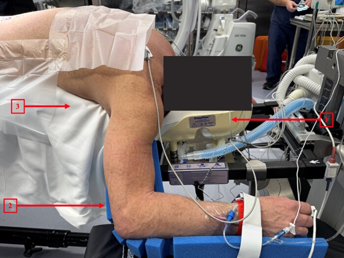
Figure 1: Patient positioning. The patient is positioned prone on a radiolucent operating table. 1 = Prone headrest; 2 = arm in the 90/90 position with the hand at or below the level of the elbow; 3 = radiolucent operating table. Please click here to view a larger version of this figure.
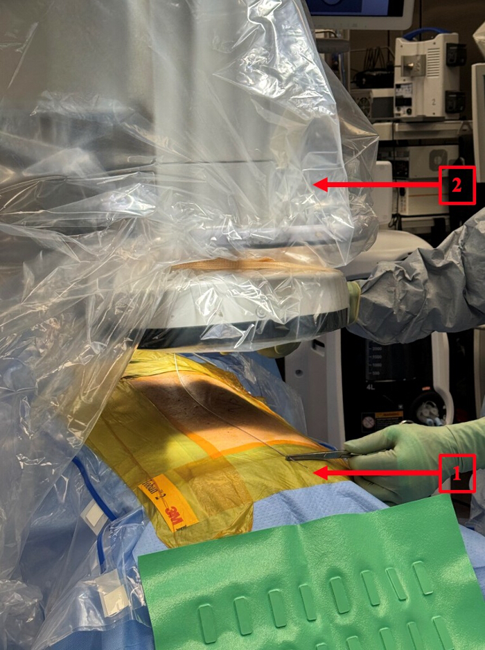
Figure 2: Midline identification and fluoroscopic setup. 1 = marking of the midline using a radiopaque flexible rod; 2 = draped mobile C-arm for intraoperative fluoroscopy. Please click here to view a larger version of this figure.
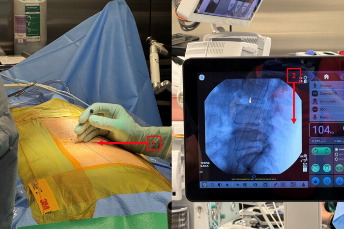
Figure 3: Level localization and targeting for the initial incision. 1 = spinal needle inserted through the skin to target the disc space and operative level; 2 = confirmation of needle position at the target level on a lateral fluoroscopic image. Please click here to view a larger version of this figure.
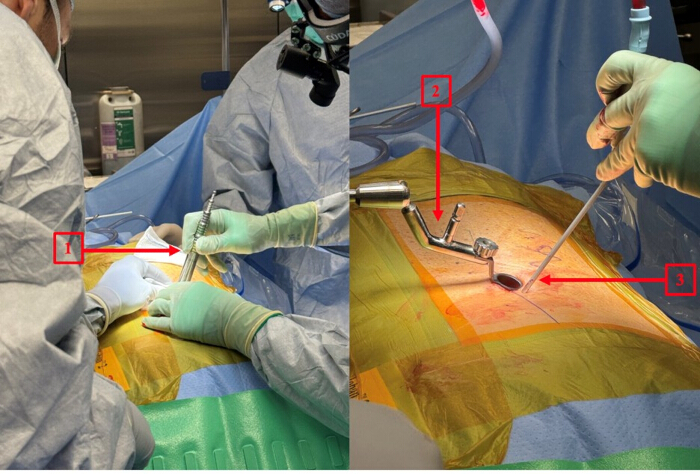
Figure 4: Tubular dilation and retractor docking. 1 = initial docking of the tubular dilator followed by sequential dilation; 2 = table-mounted flexible arm holding the tubular retractor; 3 = working corridor. Please click here to view a larger version of this figure.
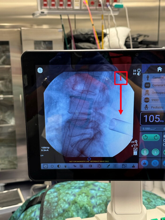
Figure 5: Fluoroscopic confirmation of portal placement. 1 = tubular retractor docked at the operative level on a lateral fluoroscopic image. Please click here to view a larger version of this figure.
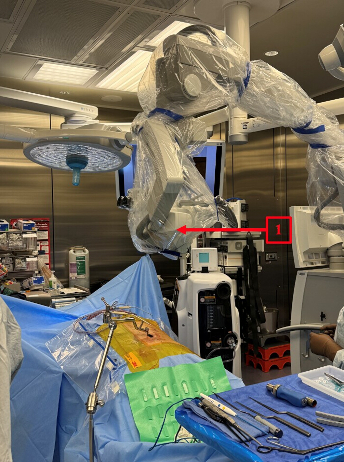
Figure 6: Operative visualization. 1 = draped operative microscope positioned for magnified visualization of the neural elements. Please click here to view a larger version of this figure.
| Cohort (single institution) | n | Approach | EBL (mL) | Length of stay | Follow-up | Mean PRO improvement at 1 yr |
| Comorbidity registry | 120 | Non-tubular W-TLIF | 128.7 ± 112.3 (normal BMI) to 218.2 ± 203.2 (obese); p = 0.018 | Not reported | ≥1 yr | ODI 5.9 (p = 0.029), SF-12 PCS 3.3 (p = 0.011), SF-12 MCS 2.6 (p = 0.089) |
| Outpatient series | 87 | Tubular W-TLIF | 35 (outpatient) to 52 (inpatient) | 3.7 h (outpatient) | ≥3 mo | Not reported |
| PRO improvement is the mean within-cohort change from preoperative to one year by paired t-test (two-sided p). Paired n = 82 for ODI and 112 for SF-12 PCS and MCS. Continuous data are presented as mean ± standard deviation (SD). | ||||||
Table 1: Representative institutional outcomes of the muscle-sparing W-TLIF.
Discussion
The muscle-sparing W-TLIF achieves lumbar decompression and interbody fusion through the natural multifidus-longissimus plane, minimizing iatrogenic paraspinal injury while providing direct access to the transforaminal corridor3,23.
Critical steps
Technical success depends on precise identification of the multifidus-longissimus interval by blunt finger dissection, docking on the transverse process-SAP junction, adequate transforaminal decompression confirmed by a free and pulsatile thecal sac, meticulous endplate preparation without violating the bony endplate, and cage placement to optimize any coronal or sagittal deformity.
Troubleshooting
Wrong-level exposure is avoided by confirming true AP and lateral fluoroscopic images before incision. Incidental durotomy is managed with a collagen sponge, fibrin glue, and flat bed rest12. Contralateral neural compression is addressed by “wanding” the tubular retractor or by a contralateral docking2. In higher-BMI patients or at cephalad levels, lengthening the incision and adjusting corridor angulation improves access.
Learning curve, patient selection, and radiation
A learning curve exists, most relevant for surgeons without prior minimally invasive or tubular experience, and is mitigated by prior open TLIF and tubular familiarity17,24. Appropriate patient selection, focal one- or two-level pathology, remains central to reproducible outcomes. As with all fluoroscopically guided minimally invasive fusions, radiation exposure is greater than in open surgery, particularly during the learning phase, and warrants disciplined radiation-safety practice15.
Comparison with other approaches
Compared with open TLIF, W-TLIF offers equivalent fusion with reduced blood loss, less postoperative pain, and faster recovery6,7,21, with lower surgical-site infection rates than open surgery25. Relative to tubular MIS-TLIF and endoscopic or lateral interbody techniques, the Wiltse corridor requires no specialized endoscopic equipment and preserves the midline while achieving comparable muscle-sparing benefit.
Limitations
The supporting data are retrospective, single-institution, and were not collected as a prospective outcomes study. Fusion rates and visual analog scale (VAS) pain scores were not formally assessed in these analyses. The technique is also limited in settings requiring wide bilateral decompression, multilevel exposure, or deformity correction. A prospective comparative study is warranted.
Conclusion
The muscle-sparing W-TLIF is a reproducible, teachable technique that preserves paraspinal integrity while achieving effective decompression and fusion in appropriately selected patients.
Disclosures
The authors have no conflicts of interest to declare.
Materials
| Name | Company | Catalog Number | Comments |
|---|---|---|---|
| Ball-tip probe / nerve hook | Standard surgical instrument | N/A | Pedicle palpation |
| Bipolar forceps | Standard surgical instrument | N/A | Bipolar cautery for hemostasis |
| C-arm fluoroscopy unit | GE Healthcare | OEC Elite | Intraoperative AP/lateral fluoroscopy |
| Collagen sponge | Standard surgical supply | N/A | Dural repair |
| Cottonoid patties, 1/2 x 1/2 in | Standard surgical supply | N/A | Nerve-root protection |
| Curettes | Standard surgical instrument | N/A | Endplate preparation |
| Disc shavers | Standard surgical instrument | N/A | Discectomy |
| Double-action rongeur | Standard surgical instrument | N/A | Bone removal |
| Fibrin glue | Standard surgical supply | N/A | Dural repair |
| Interbody (TLIF) cage | Varies (surgeon preference) | N/A – varies by surgeon preference | Titanium or other material, static or expandable |
| Kerrison rongeur | Standard surgical instrument | N/A | Bone removal |
| Local autograft / allograft | Standard surgical supply | N/A | Interbody fusion |
| METRx flexible arm | Medtronic | 9560524 | Table-mounted retractor arm |
| METRx instrument set | Medtronic | https://www.medtronic.com/in-en/index.html | MIS instrument set |
| METRx sequential dilators | Medtronic | https://www.medtronic.com/in-en/index.html | Sequential soft-tissue dilation |
| METRx tubular retractor (tubes) | Medtronic | https://www.medtronic.com/in-en/index.html | Straight, 18 / 22 / 26 mm |
| Midas Rex MR8 high-speed drill | Medtronic | https://www.medtronic.com/in-en/index.html | Hemilaminectomy / facetectomy |
| Nerve root retractor | Standard surgical instrument | N/A | Neural retraction |
| Operating microscope / surgical loupes | Equipment varies by institution | N/A | Magnification |
| Pedicle screw system | Orthofix | https://orthofix.com/ | Firebird Spinal Fixation System, percutaneous / mini-open |
| ProneView headrest | Mizuho OSI | https://www.mizuhosi.com/ | Prone head positioning |
| Radiolucent operating table | Mizuho OSI | https://www.mizuhosi.com/ | Jackson table |
| Rasps | Standard surgical instrument | N/A | Endplate preparation |
| Self-retaining (Weitlaner) retractor | Standard surgical instrument | N/A | Fascial exposure |
| Spinal needle | Standard surgical supply | N/A | Level localization |
| SSEP neuromonitoring system | System varies by institution | N/A | Somatosensory evoked potentials |
| Sterile dressing | Standard surgical supply | N/A | Wound dressing |
| Surgical marking pen | Standard surgical supply | N/A | Skin marking |
| Sutures | Standard surgical supply | N/A | Fascial and wound closure |
| Trial implants | Standard surgical instrument | N/A | Disc-space sizing |
References
- Wiltse LL, Bateman JG, Hutchinson RH, Nelson WE. The paraspinal sacrospinalis-splitting approach to the lumbar spine. J Bone Joint Surg Am. 1968;50(5):919-926.
- German JW, Foley KT. Minimal access surgical techniques in the management of the painful lumbar motion segment. Spine (Phila Pa 1976). 2005;30(16 Suppl):S52-S59.
- Jin YM, Chen Q, Chen CY, Lyu J, Shi B, Yang C, et al. Clinical research and technique note of TLIF by Wiltse approach for the treatment of degenerative lumbar. Orthop Surg. 2021;13(5):1628-1638.
- Wang K, Deng Z, Chen X, Shao J, Qiu L, Jiang C, Niu W. The role of multifidus in the biomechanics of lumbar spine: a musculoskeletal modeling study. Bioengineering (Basel). 2023;10(1):67.
- Freeman MD, Woodham MA, Woodham AW. The role of the lumbar multifidus in chronic low back pain: a review. PM R. 2010;2(2):142-146.
- Ge DH, Stekas ND, Varlotta CG, Fischer CR, Petrizzo A, Protopsaltis TS, et al. Comparative analysis of two transforaminal lumbar interbody fusion techniques: open TLIF versus Wiltse MIS TLIF. Spine (Phila Pa 1976). 2019;44(9):E555-E560.
- Miller LE, Bhattacharyya S, Pracyk J. Minimally invasive versus open transforaminal lumbar interbody fusion for single-level degenerative disease: a systematic review and meta-analysis of randomized controlled trials. World Neurosurg. 2020;133:358-365.e4.
- Singh K, Nandyala SV, Marquez-Lara A, Fineberg SJ, Oglesby M, Pelton MA, et al. A perioperative cost analysis comparing single-level minimally invasive and open transforaminal lumbar interbody fusion. Spine J. 2014;14(8):1694-1701.
- Malakoutian M, Street J, Wilke HJ, Stavness I, Dvorak M, Fels S, et al. Role of muscle damage on loading at the level adjacent to a lumbar spine fusion: a biomechanical analysis. Eur Spine J. 2016;25(9):2929-2937.
- Lee CW, Yoon KJ, Ha SS. Which approach is advantageous to preventing development of adjacent segment disease? Comparative analysis of 3 different lumbar interbody fusion techniques (ALIF, LLIF, and PLIF) in L4-5 spondylolisthesis. World Neurosurg. 2017;105:612-622.
- Wu AM, Hu ZC, Li XB, Feng ZH, Chen D, Xu H, et al. Comparison of minimally invasive and open transforaminal lumbar interbody fusion in the treatment of single segmental lumbar spondylolisthesis: minimum two-year follow up. Ann Transl Med. 2018;6(6):105.
- Holly LT, Schwender JD, Rouben DP, Foley KT. Minimally invasive transforaminal lumbar interbody fusion: indications, technique, and complications. Neurosurg Focus. 2006;20(3):E6.
- Mobbs RJ, Phan K, Malham G, Seex K, Rao PJ. Lumbar interbody fusion: techniques, indications and comparison of interbody fusion options including PLIF, TLIF, MI-TLIF, OLIF/ATP, LLIF and ALIF. J Spine Surg. 2015;1(1):2-18.
- Xie Q, Zhang J, Lu F, Wu H, Chen Z, Jian F. Minimally invasive versus open transforaminal lumbar interbody fusion in obese patients: a meta-analysis. BMC Musculoskelet Disord. 2018;19(1):15.
- Kumar A, Merrill RK, Overley SC, Leven DM, Meaike JJ, Vaishnav A, et al. Radiation exposure in minimally invasive transforaminal lumbar interbody fusion: the effect of the learning curve. Int J Spine Surg. 2019;13(1):39-45.
- Garg S, Muir M, Jamar K, Young P, Gill SS. Impact of medical comorbidities on outcomes following muscle-sparing Wiltse transforaminal lumbar interbody and fusion. Eur Spine J. (In Press).
- Garg S, Gill SS. Outpatient minimally invasive transforaminal lumbar interbody fusion surgery: assessing safety, efficacy, feasibility, and learning curve. Presented at: Society for Minimally Invasive Spine Surgery (SMISS) Annual Forum; 2022.
- Hockley A, Ge D, Vasquez-Montes D, Moawad MA, Passias PG, Errico TJ, et al. Minimally invasive versus open transforaminal lumbar interbody fusion surgery: an analysis of opioids, nonopioid analgesics, and perioperative characteristics. Global Spine J. 2019;9(6):624-629.
- Eckman WW, Hester L, McMillen M. Same-day discharge after minimally invasive transforaminal lumbar interbody fusion: a series of 808 cases. Clin Orthop Relat Res. 2014;472(6):1806-1812.
- Boissiere L, Haleem S, Liquois F, Vermorel PH, Le Huec JC, Kieser DC, et al. Prospective same day discharge instrumented lumbar spine surgery - a forty patient consecutive series. Eur Spine J. 2024;33(9):3452-3456.
- Hammad A, Wirries A, Ardeshiri A, Nikiforov O, Geiger F. Open versus minimally invasive TLIF: literature review and meta-analysis. J Orthop Surg Res. 2019;14(1):229.
- Street JT, Andrew Glennie R, Dea N, DiPaola C, Wang Z, Boyd M, et al. A comparison of the Wiltse versus midline approaches in degenerative conditions of the lumbar spine. J Neurosurg Spine. 2016;25(3):332-338.
- Wang S, Duan C, Yang H, Kang J, Wang Q. Wiltse approach versus conventional transforaminal interbody fusion for unstable thoracolumbar fracture with intervertebral disc lesions. Orthop Surg. 2022;14(4):694-703.
- Silva PS, Pereira P, Monteiro P, Silva PA, Vaz R. Learning curve and complications of minimally invasive transforaminal lumbar interbody fusion. Neurosurg Focus. 2013;35(2):E7.
- Sweitzer SF, Sickbert-Bennett EE, Seidelman J, Anderson DJ, Lim MR, Weber DJ. The impact of minimally invasive surgical approaches on surgical-site infections. Infect Control Hosp Epidemiol. 2024;45(5):557-561.
Reprints and Permissions
This article has been published
Video Coming Soon