Robot-assisted radical prostatectomy in a heart transplant recipient on tacrolimus was complicated by a tacrolimus-associated left ureteral leak and secondary anastomotic insufficiency, successfully managed with staged urinary diversion and multidisciplinary care.
Case Report
Robot-Assisted Radical Prostatectomy in a Heart Transplant Patient
In This Article
Summary
Abstract
Literature on major urological surgery in heart transplant recipients remains limited because these patients historically had shorter life expectancies, limiting long-term surgical follow-up. However, advances in medical care, immunosuppressive therapy, and patient management have prolonged survival, increasing the incidence of age-related diseases, including prostate cancer. This intersection of cardiac transplantation and urological disease presents unique clinical challenges requiring further study of optimal surgical and postoperative management. Heart transplant patients receiving lifelong tacrolimus-based immunosuppression are particularly prone to impaired wound healing and postoperative complications. The authors report a 71-year-old man who underwent heart transplantation in November 2015 for ischemic cardiomyopathy. His immunosuppressive regimen included tacrolimus (target trough 5–7 µg/L), mycophenolate mofetil, and low-dose steroids. The patient underwent robot-assisted radical prostatectomy (RARP) with bilateral pelvic lymphadenectomy for prostate adenocarcinoma (iPSA 9.3 ng/mL). Histopathology revealed pT3b pN1 (5/30) Gleason 7b (4 + 3, 80% + 20%) disease with negative margins. Recovery was initially uneventful, and he was discharged on postoperative day 5, catheter-free after spontaneous micturition. Fifteen days postoperatively, the patient was readmitted with acute flank pain. Imaging demonstrated left ureteral extravasation. Review of the surgical recording excluded thermal injury during lymphadenectomy, suggesting tacrolimus-associated impaired healing as a possible cause. Management included placement of a double-J stent, antibiotics, and cardiac monitoring. Because urinary drainage remained insufficient, urinary diversion with a Mono-J stent and percutaneous nephrostomy (PCN) was performed, considering the patient’s cardiologic risk profile. A cystogram demonstrated minimal secondary urethrovesical anastomotic insufficiency, possibly related to endourological manipulation, although tacrolimus-associated healing impairment could not be excluded. The Mono-J stent and the nephrostomy catheter were removed after 2 and 3 months, respectively, following complete ureteral healing. Secondary percutaneous radiotherapy with androgen deprivation therapy was recommended. This case demonstrates the feasibility of RARP in heart transplant recipients while highlighting the potential impact of immunosuppressive therapy on postoperative healing.
Introduction
The existing literature concerning major urological surgeries performed on heart transplant patients is quite limited. This scarcity of information is largely attributable to the fact that, in the past, these patients tended to have a shorter overall life expectancy, which limited the opportunities to document and study such surgeries1. However, advancements in medical care, immunosuppressive therapies, and overall patient management have significantly extended the lifespan of heart transplant recipients1. As a result, many of these patients are now living longer and are more likely to encounter new health challenges, including various forms of cancer. One such example is prostate cancer, which has become an increasingly common diagnosis among this patient population2,3.
Chronic immunosuppression with tacrolimus (often combined with mycophenolate mofetil and steroids) effectively prevents rejection but is associated with impaired tissue and wound healing, possibly through reduced nitric oxide synthesis and effects on fibroblast function and angiogenesis4,5. In the era of prostate-specific membrane antigen- positron emission tomography-computed tomography (PSMA-PET-CT) imaging, a novel approach to prostate cancer management is emerging. Patients who historically received lymph node dissection (LAD) based on nanogram assessments, even though they did not require the procedure, may be spared this additional intervention6. Conversely, in advanced disease, PSMA-PET-CT has revealed lymph node metastases that conventional staging methods, such as CT scans and skeletal scintigraphy, have missed7. Some of these metastases are located in regions typically not targeted during standard LAD procedures for prostate cancer. Removal of such Lymph nodes necessitates an extended or superextended lymphadenectomy, which comes with higher risks and morbidity8.
This case highlights the successful management of a 71-year-old heart transplant patient with locally advanced cancer, and PSMA-PET-CT suspected lymph node metastasis, including pre-sacral and bilateral common iliac nodes. The patient, maintained on ongoing tacrolimus therapy, remains in excellent general health with no significant medical issues, urinary obstructive symptoms, or erectile dysfunction. After thorough discussion in the multidisciplinary tumor board and considering the patient's preferences, a decision was made to proceed with robot-assisted radical prostatectomy (RARP) combined with super-extended lymphadenectomy with curative intent. While RARP provides reduced blood loss and faster recovery compared with open surgery9, an extended pelvic lymphadenectomy carries risks of ureteral injury that may be exacerbated by immunosuppression-related healing deficits10. Tacrolimus has been shown to impair wound healing in experimental models11. Although radical prostatectomy is feasible in selected transplant recipients12. Detailed reports of RARP in heart transplant patients with tacrolimus-associated complications remain scarce13. This case is relevant as it illustrates the need for meticulous intraoperative care and close monitoring in this high-risk population.
Case Presentation:
The patient is a 71-year-old male with heart transplantation in November 2015 for ischemic cardiomyopathy and explantation of the defibrillator system. Lifelong immunosuppression consisted of tacrolimus (target trough 5–7 µg/L), mycophenolate mofetil, and low-dose steroids. Comorbidities included arterial hypertension, chronic kidney disease stage G3a, left thoracic wall lymph node resection (2017), resection of M. Bowen on the back (2021), and left knee total endoprosthesis (2021). He presented for RARP after histological confirmation of prostate adenocarcinoma (iPSA 9.3 ng/mL). Physical examination showed a suspect digital rectal finding; the patient was in good general condition. The patient had no relevant lower urinary tract symptoms (LUTS) with an international prostate symptom score of 3/35, post-void residual urine of 50 mL.
Diagnosis, Assessment, and Plan:
Diagnosis was confirmed histologically. 18F-PSMA-1007 PET/CT showed uptake in the right prostate, and suspicious presacral/left iliac lymph nodes (Figure 1). Prostate volume was 36 mL on ultrasound. Robot-assisted radical prostatectomy with bilateral pelvic lymphadenectomy was planned.
Protocol
The patient provided written informed consent for all aspects of the treatment plan, including each surgical intervention, and consented to the anonymous publication of his case. This study complies with the ethical standards outlined in the Declaration of Helsinki and is approved by the ethics committees of the Westfalen-Lippe Medical Association and the University of Muenster (2023–500-f-S) for the retrospective management of patient data processed within the department. All the materials used in the study are listed in the Table of Materials.
1. Preoperative preparation
- The patient underwent standard preoperative assessment. Cardiologic evaluation included electrocardiography (ECG) to assess cardiac rhythm and detect ischemic or conduction abnormalities, and transthoracic echocardiography to evaluate ventricular function and valvular status. In consultation with the treating cardiac team, tacrolimus therapy was adjusted, and the dose was subsequently reduced.
- Prophylactic intravenous antibiotic therapy with ampicillin/sulbactam was administered 30 min prior to skin incision in accordance with the institutional perioperative infection prevention guidelines.
- Perioperative thromboprophylaxis was initiated according to institutional protocol to reduce the risk of venous thromboembolism, taking into account the patient’s surgical risk profile.
2. Anesthesia and patient positioning
- General anesthesia was induced under comprehensive invasive hemodynamic monitoring. Continuous electrocardiography (ECG), pulse oximetry, and capnography were applied to monitor cardiac rhythm, oxygenation, and ventilation status throughout the procedure.
- Invasive arterial blood pressure monitoring via radial arterial catheter was established prior to induction to allow real-time beat-to-beat blood pressure assessment.
- Central venous access was inserted for fluid and vasoactive agent administration and for central venous pressure monitoring. Anesthetic induction was performed using carefully titrated intravenous agents to avoid hypotension.
- Maintenance of anesthesia focused on strict hemodynamic stability, avoidance of fluid overload, and preservation of cardiac allograft function through balanced fluid administration and vasopressor support as required.
- The patient was placed in a Trendelenburg position, with the legs in stirrups, to optimize pelvic exposure.
- All pressure points were carefully padded to prevent any neurovascular injury. Pneumoperitoneum was established using carbon dioxide insufflation, gradually increased to an intra-abdominal pressure of 8 mm Hg.
- Hemodynamic response to insufflation was closely monitored, and adjustments were made as needed to maintain stable cardiovascular parameters.
3. Port placement and robotic setup
- A standard four-port transperitoneal configuration for robot-assisted radical prostatectomy (RARP) was utilized.
- Trocar insertion was performed under direct vision. The robotic arms were then positioned, ensuring adequate spacing to prevent external arm collision during instrument manipulation.
- The robotic system was docked in a midline position, aligned with the patient’s pelvis to facilitate optimal instrument access.
4. The radical prostatectomy and lymphadenectomy
- Bladder neck dissection was performed with careful identification and preservation of anatomical landmarks. The neurovascular bundles were preserved bilaterally, where oncologically feasible, using meticulous nerve-sparing techniques.
- Apical dissection was carried out with attention to urethral length preservation to facilitate a tension-free anastomosis. Hemostasis was maintained throughout using bipolar cautery and Hem-o-loc clips.
- Bilateral extended pelvic lymph node dissection was performed, including the external iliac, internal iliac, and obturator lymph node regions.
- Due to suspected lymph nodes metastasis in PSMA-PET-CT, the lymph nodes around the common iliac vessels and the pre-sacral area were also dissected (Figure 1).
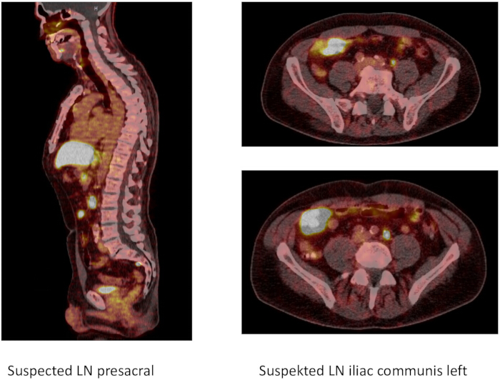
Figure 1: Suspected lymph nodes as seen on PSMA-PET-CT. On the left (sagittal), showing a presacral lymph node, on the right (axial), showing 2 left common iliac artery lymph nodes. Abbreviations; PSMA-PET-CT = prostate-specific membrane antigen- positron emission tomography-computed tomography; LN = lymph node. Please click here to view a larger version of this figure.
- The prostate and lymph node specimens were retrieved through the supraumbilical incision.
- A watertight urethrovesical anastomosis was performed using continuous resorbable sutures. Urinary diversion was achieved through a transurethral catheter. Additionally, a suprapubic catheter was placed in accordance with institutional standards.
- Hemostasis and wound closure: Final inspection of the operative field was performed under reduced pneumoperitoneum to identify and control any residual bleeding points.
- All trocar sites were removed under direct vision, and fascial closure was performed at the mini-laparotomy site and the assistant trocar (12 mm) site. Skin incisions were closed.
- The patient was extubated and transferred to a monitored unit with continuation of baseline immunosuppression and close tacrolimus level monitoring.
Results
Histopathology confirmed pT3b pN1 (5/30) Gleason 7b ( 4 + 3, 80% + 20% ) with negative margins. Initial recovery was uneventful, and the patient was discharged on postoperative day 5. The patient was readmitted 10 days after the initial discharge (15 days postoperatively) due to acute lower abdominal pain. The CT-scan revealed left ureteral extravasation with urinoma and hydronephrosis. A retrograde pyeloureterography confirmed the diagnosis (Figure 2).
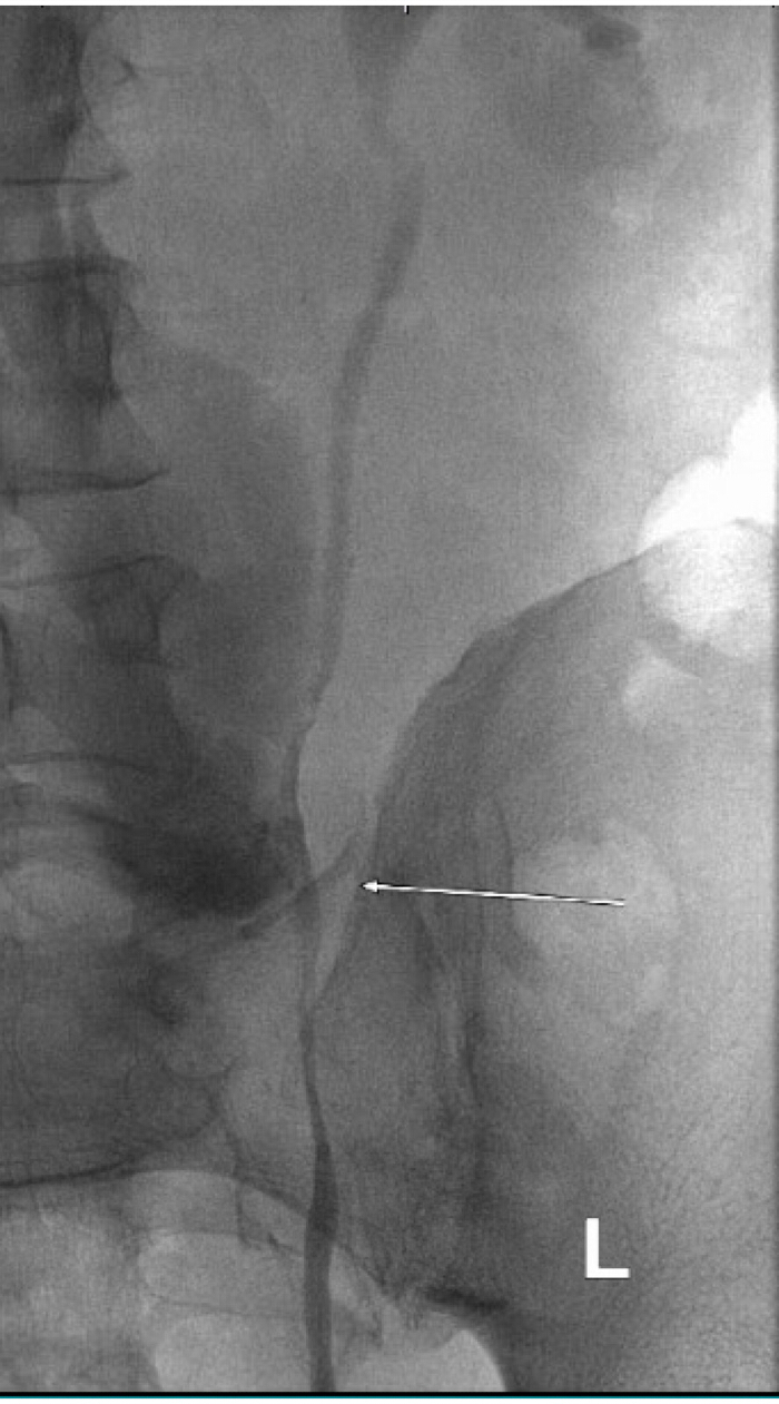
Figure 2: Uretal lesion. Contrast media leakage in the middle third of the left ureter was found. L shows lesion on the left ureter. Please click here to view a larger version of this figure.
Due to insufficient urinary drainage with the DJ-ureteral catheter, a urinary diversion with a Mono-J stent and percutaneous nephrostomy (PCN) was selected, given the cardiological risk profile. The cystogram indicated minimal secondary insufficiency at the urethrovesical anastomosis. A transurethral catheter was inserted and remained in place for two weeks. The catheter was subsequently removed following a cystography that showed no abnormalities, along with the Mono-J catheter. The nephrostomy catheter was removed three months post-radical prostatectomy, by which time complete healing of the ureteral injury had occurred (Figure 3).
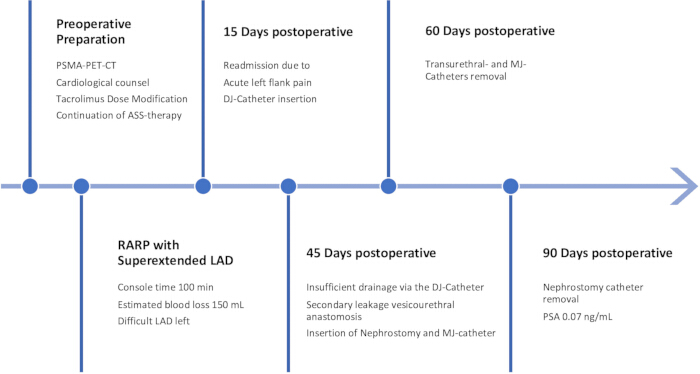
Figure 3: Timeline. Timeline outlining the first 90 days after surgery. Abbreviations; PSMA-PET-CT = prostate-specific membrane antigen- positron emission tomography-computed tomography; MJ = mono J; DJ = double J; LAD = lymph node dissection; ASS = acetyl salicylic acid; PSA = prostate-specific antigen; RARP = robot-assisted radical prostatectomy. Please click here to view a larger version of this figure.
This case was reviewed in the interdisciplinary post-therapeutic tumor board. Adjuvant percutaneous radiotherapy combined with androgen deprivation therapy was recommended with caution. The immunosuppressive therapy was continued as recommended by the cardiologic team. The repeated cariological checkups showed normal findings.
Discussion
This case demonstrates the technical feasibility of robot-assisted radical prostatectomy in a heart transplant recipient on tacrolimus, but also sheds light on possible involvement of the immunosuppressive therapy in the postoperative course. Principally, robot-assisted radical prostatectomy in heart transplant patients sounds safe and feasible. The patient had no major complications or cardiovascular events. The initial left ureteral leak is a very rare complication in experienced hands. In a series of more than 1500 radical prostatectomies performed by the primary surgeon (MF), there was not a single such injury to the ureters. Because of an extended lymphadenectomy, institutional standards require identifying and locating the ureters before removing lymph nodes. After carefully reviewing the surgical video, no ureteral injuries, thermal or cold, were detected. A microinjury or decreased blood flow to the ureter from the extended LAD might go unnoticed and remain subclinical in other patients. However, in patients on immunosuppressive therapy, such as tacrolimus, with impaired tissue repair, a leak may become clinically evident two weeks after the initial surgery5.
Immunosuppressive regimens containing tacrolimus effectively prevent rejection after heart transplantation, but can complicate urological procedures by impairing tissue repair4. In this patient, the combination of surgical trauma from pelvic lymphadenectomy and chronic tacrolimus exposure likely contributed to the prolonged need for urinary diversion until eventual resolution. Close cardiologic monitoring ensured stable allograft function without rejection. Literature reports variable wound-healing complications in transplant recipients on calcineurin inhibitors, with some studies showing no early effect, while others demonstrate clear impairment14. The interdisciplinary tumor board discussed and issued a very cautious recommendation concerning adjuvant radiotherapy combined with GnRH-antagonist-based androgen deprivation.
This report emphasizes the importance of meticulous ureteral protection, reduced energy use near critical structures, and vigilant postoperative monitoring in tacrolimus-treated heart transplant patients undergoing RARP. Long-term oncologic and functional outcomes in this population require further investigation. The literature on major urological surgeries in heart transplant patients is limited. This is partly because these patients historically had a shorter life expectancy. However, with improved medical care, many now receive new cancer diagnoses, such as prostate cancer. This case report aims to enhance understanding of this topic. It also covers the management of unexpected complications and their treatment.
Disclosures
The authors have nothing to disclose.
Acknowledgements
The authors thank the interdisciplinary teams in transplant cardiology, nuclear medicine, and pathology for excellent collaboration. No specific funding was received.
Materials
| Name | Company | Catalog Number | Comments |
|---|---|---|---|
| Double-J stent | Boston Scientific | https://www.bostonscientific.com/en-US/products/stents--ureteral.html | 6 Fr 28 cm |
| Guidewire | Terumo | https://www.terumois.com/products/product-type/guidewires.html | Hydrophilic |
| Monopolar shears | Intuitive Surgical | https://www.intuitive.com/en-in/products-and-services/da-vinci | Standard |
| Robotic system | Intuitive Surgical | https://www.intuitive.com/en-in/products-and-services/da-vinci | da Vinci X |
| Vicryl sutures (4-0) | Ethicon | J494G | Absorbable |
References
- Hajj S, et al. Imaging of de novo malignancy after solid organ transplant. Radiology. 2026;319(1):e251384.
- Ciancio G, et al. Prostate cancer after heart transplantation. J Urol. 1995;153(1):158-160.
- Mohammadi S, et al. Prostate cancer after heart transplantation: unicenter case-control study. J Heart Lung Transplant. 2005;24(8):995-997.
- Bootun R. Effects of immunosuppressive therapy on wound healing. Int Wound J. 2013;10(1):98-104.
- Schäffer MR, et al. Tacrolimus impairs wound healing: a possible role of decreased nitric oxide synthesis. Transplantation. 1998;65(6):813-818.
- Morgans AK, et al. Future opportunities and nuances with the use of PSMA PET in prostate cancer (MD PET 1). Theranostics. 2026;16(11):5816-5829.
- Bruins Slot AS, et al. Nodal maximum standardized uptake value improves PSMA-PET/CT-based nodal staging in intermediate- and high-risk prostate cancer. BJU Int. 2026.
- Wan QC, Min KY, Wei L, Xie LJ, Ji B. Can pelvic lymph node dissection be spared in intermediate-risk prostate cancer patients with negative PSMA PET scan? A systematic review and diagnostic meta-analysis. Prostate Cancer Prostatic Dis. 2026.
- Novara G, et al. Systematic review and meta-analysis of perioperative outcomes and complications after robot-assisted radical prostatectomy. Eur Urol. 2012;62(3):431-452.
- Cacciamani GE, et al. Impact of pelvic lymph node dissection and its extent on perioperative morbidity in patients undergoing radical prostatectomy for prostate cancer: a comprehensive systematic review and meta-analysis. Eur Urol Oncol. 2021;4(2):134-149.
- Stenman C, et al. Malignancies after heart transplantation. Transpl Int. 2024;37:12109.
- Piana A, et al. Robot-assisted radical prostatectomy in renal transplant recipients: a systematic review. J Clin Med. 2023;12(21):6754.
- Axcrona K, et al. Robot-assisted laparoscopic prostatectomy in a 68-year-old patient with previous heart transplantation and pelvic irradiation. J Robot Surg. 2012;6(1):81-83.
- Dean PG, et al. Wound-healing complications after kidney transplantation: a prospective, randomized comparison of sirolimus and tacrolimus. Transplantation. 2004;77(10):1555-1561.
Reprints and Permissions
