This protocol aims to demonstrate the feasibility and safety of a hybrid robotic-assisted thoracic and transcervical approach for resection of massive substernal thyroid goiters, highlighting its potential to avoid sternotomy, reduce surgical morbidity, and enable effective mobilization of mediastinal disease while preserving critical structures.
Method Article
Combined Robotic-Assisted Thoracic Surgery and Transcervical Approach for Substernal Thyroidectomy: A Sternotomy Sparing Technique
In This Article
Summary
Abstract
Significant extension of thyroid masses into the mediastinum has historically necessitated high-morbidity sternotomy or thoracotomy incisions during substernal thyroidectomy. This preliminary experience from a single-center, tertiary-care academic institution illustrates a combined minimally invasive robotic-assisted thoracic surgery (RATS) and transcervical approach for substernal thyroidectomy. Patients are considered for the minimally invasive approach for substantial goiter extension into the anterior and posterior mediastinum, in addition to risk factors for wound healing complications associated with sternotomy. Relative exclusion criteria include prior thoracic surgery or radiographic extrathyroidal invasion into mediastinal structures. Using the Intuitive Surgical da Vinci Xi platform, right-sided RATS mobilization of the mediastinal component is immediately followed by transcervical excision. The robotic approach enables precise identification and dissection of critical neurovascular and aerodigestive structures, thereby facilitating safe transcervical delivery. Two procedures utilizing this approach were completed without complications. By avoiding sternotomy, both patients were discharged home following abbreviated inpatient stays of just 2 days. Robotic-assisted thoracic surgery represents a promising alternative to traditional invasive approaches for substernal thyroidectomy, preliminarily showing safety and feasibility, and the potential to reduce morbidity and improve recovery times.
Introduction
While most substernal thyroid masses extending into the mediastinum can be excised via a transcervical incision, up to 5% require an additional thoracic approach1,2,3,4. Traditional incisions like sternotomy or thoracotomy disrupt the thoracic skeleton, leading to significant morbidity, particularly in obese patients5. Median sternotomy is associated with postoperative pain and prolonged hospital length of stay, with estimated rates of wound complications ranging from 1%–8%6,7,8. These potential morbidities provide a strong rationale for developing minimally invasive approaches to safely avoid sternotomy in massive substernal goiters.
For this purpose, video-assisted thoracoscopic surgery (VATS) combined with a transcervical approach has been reported8,9. However, posterior mediastinal extension and revision cases pose distinct challenges. Robotic-assisted thoracic surgery (RATS), a contemporary evolution of VATS, offers improved 3D visualization, instrument precision, and recovery times10,11,12,13,14,15,16. The hybrid approach described herein offers promise in these challenging situations. To date, published literature on both thoracoscopic and robotic-assisted approaches is sparse and only consists of a few isolated case reports13,14,15,16. These studies demonstrate the technical feasibility of thoracoscopic and robotic assistance and suggest potential advantages over sternotomy. However, there are currently no dedicated prospective series, comparative studies, or systematic reviews that objectively analyze these alternative approaches compared with conventional sternotomy. Moreover, widespread adoption remains limited due to increased procedural costs, prolonged operative times, specialized equipment and training requirements, and strict patient selection criteria.
Herein, we describe a combined unilateral RATS and transcervical technique for the treatment of massive substernal thyroidectomy to avoid a sternotomy. The overall goal of this approach is to reduce the morbidity associated with sternotomy for substernal thyroidectomy, particularly in at-risk patients, including those with obesity, large breasts, and diabetes. The study aimed to assess safety, feasibility, length of hospital stays, and complication rates in cases during which this approach was employed. This technical report aims to expand the previously described applications of this technique by demonstrating its feasibility for both posterior mediastinal extension and the revision surgical setting.
Protocol
This report follows standards and guidelines of the institution’s human research ethics committee and was deemed exempt by the Mount Sinai Health System IRB (no protocol exemption number assigned). Written informed consent was obtained for publication of the data and images.
1. Assess patient candidacy.
- Perform routine diagnostic workup, including ultrasound-guided fine needle aspiration of thyroid nodules (when appropriate), thyroid function tests, and serum calcium levels.
- Obtain contrast-enhanced computed tomography (CT) imaging of the neck and chest (1.5 mm slice thickness, standard protocols for contrast timing).
- Assess inclusion criteria.
- Identify patients with massive substernal goiters who are anticipated to require sternotomy for removal.
- Evaluate the size of the thyroid mass and craniocaudal extent. Identify mediastinal extension relative to key landmarks, including the aortic arch, carina, trachea, and esophagus.
- Assess for posterior mediastinal extension, extension to the level of the carina, or anterior mediastinal extension below the aortic arch.
- Assess for comorbidities that place the patient at an increased risk for complications of sternotomy, including obesity, large breasts, diabetes, and conditions that impair wound healing. These inclusion criteria support the use of the hybrid approach.
- Assess for exclusion criteria.
- Assess for prior thoracic surgery as scarring in the thoracic cavity or mediastinum may limit the feasibility of a minimally invasive approach.
- Assess for inflammatory conditions such as Graves’ or Hashimoto’s disease, often associated with neovascularization, which may hinder video-assisted visualization.
- Assess for extrathyroidal extension of malignant lesions that may necessitate greater access to mitigate procedural risks.
2. Anesthesia and airway management
- Induce general anesthesia using standard protocols. Avoid the use of long-acting muscle paralysis. Have standard preparations for a difficult airway.
- Insert an electromyographic endotracheal tube to enable recurrent laryngeal nerve monitoring.
- Use video-assisted laryngoscopy to ensure proper positioning of the nerve monitoring tube.
- Establish lung isolation.
- Perform bronchoscopy to confirm airway anatomy.
- Place a bronchial blocker to achieve single-lung ventilation on the operative side. Typically, use a 7 Fr bronchial blocker or a 9 Fr blocker for large adults.
- Employ lung-protective ventilation protocols, such as tidal volumes of 4–6 mL/kg predicted body weight and a positive end-expiratory pressure (PEEP) of 5 cm H₂O, and adjust the respiratory rate to maintain normocapnia.
- Titrate the fraction of inspired oxygen (FiO₂) to maintain arterial oxygen saturation >92%.
3. Patient positioning
- Position for the thoracic phase.
- Place the patient in the lateral decubitus position.
- Select laterality based on the side of greatest mediastinal extension.
- Place the dependent arm on a padded arm board with the shoulder abducted less than 90° and support the nondependent arm on a padded arm holder in slight flexion.
- Stabilize the patient using bariatric straps and adhesive tape across the torso to prevent movement during robotic docking.
- Apply mild table flexion to increase the intercostal spaces, with slight posterior tilt and reverse Trendelenburg as needed to optimize exposure of the superior mediastinum.
4. Thoracic access and robotic setup
- Establish thoracic access.
- Prepare and drape in standard fashion.
- Insert the optical trocar in the 8th intercostal space at the midaxillary line.
- Initiate carbon dioxide insufflation at 8–12 mmHg to facilitate ipsilateral lung collapse and enhance visualization of the mediastinum.
- At 12 mmHg, monitor for altered venous return, reduced cardiac output, hypercapnia, and increased airway pressures.
- Place an 8 mm robotic camera port at this location.
- Under thoracoscopic visualization, place additional 8 mm robotic working ports in the 6th intercostal space along the anterior axillary line, and the 9th intercostal space just posterior to the posterior axillary line.
- Place a 12 mm assistant port between the camera and anterior working port, typically in the 7th intercostal space, to facilitate suction, specimen retrieval, and introduction of clips or sutures.
- Robotic system setup
- Dock the robotic surgical platform from the patient's posterior aspect to optimize access to the superior mediastinum.
- Position a 30° robotic endoscope through the central camera port, oriented superiorly toward the thoracic inlet.
- Equip the anterior robotic arm with Maryland bipolar forceps. Equip the posterior robotic arm with monopolar curved scissors or a Tip-Up fenestrated grasper, based on the surgeon's preference. Fit the fourth arm with a Cadiere forceps (Figure 1).
- Use surgeon console settings that include high-definition three-dimensional visualization with 10× magnification and tremor filtration enabled.
5. Mediastinal dissection and mobilization
- Identify the thyroid mass.
- Identify the mediastinal component of the thyroid gland; look for fullness in the thoracic inlet and superior mediastinal aperture.
NOTE: The capsule may appear congested or darker due to venous stasis in the thoracic inlet. - Identify thyroid extension posterior to the sternum and clavicle.
- Identify the mediastinal component of the thyroid gland; look for fullness in the thoracic inlet and superior mediastinal aperture.
- Locate adjacent anatomical structures, including major vessels and airway structures. Use preoperative imaging to predict these locations.
- Perform true capsular dissection.
- Dissect along the capsular plane immediately adjacent to the capsule using blunt and sharp techniques.
NOTE: This is not an extracapsular dissection; this is an avascular plane directly on the surface of the thyroid capsule.- Use Maryland bipolar forceps to provide traction and facilitate precise tissue handling and dissection. Use monopolar curved scissors for sharp dissection in the capsular plane.
- Use Cadiere forceps to provide dynamic lung or mediastinal retraction and maintain exposure throughout the dissection.
- Mobilize the mass from adjacent vascular structures, including the azygous vein and innominate vessels.
- Use bipolar energy and oxidized cellulose hemostatic packing to achieve hemostasis as required.
- Dissect along the capsular plane immediately adjacent to the capsule using blunt and sharp techniques.
- Protect critical structures.
- Identify and preserve the trachea and esophagus.
- Identify and mobilize neural structures, including the vagus and the phrenic nerve.
- Identify the vagus nerve through gentle blunt dissection at the internal jugular and subclavian venous confluence region (left) or the superior vena cava interface (right).
- Protect the phrenic nerve during mediastinal fat dissection at the lateral pericardial border.
- Protect the recurrent laryngeal nerve in the retrovascular space, particularly if the innominate vein overlies the mass.
- Maintain readiness for conversion to open sternotomy or thoracotomy for emergency management.
- Promptly recognize critical complications of vascular injury that necessitate conversion to open sternotomy. These include severe bleeding preventing direct vascular injury, or indirect injury with hemorrhage that prevents safe visualization, or an inability to be controlled with robotic instrumentation, or hemodynamic instability.
- Maintain communication with anesthesiology and follow emergency resuscitation methods. Identify pulmonary injury to the ventilated lung.
- Pause after a potential nerve injury (recurrent laryngeal, vagus, phrenic) is either directly visualized or indirectly suspected as noted from changes in nerve monitoring signals. If a nerve injury is confirmed, resume the procedure only if the benefits of resection outweigh the risks. Consider conversion to open sternotomy to preserve the integrity of the contralateral nerve.
- Complete mediastinal mobilization.
- Continue dissection until the inferior portion of the gland is fully mobilized.
- Confirm that the mass is free from mediastinal attachments. Pack absorbable hemostatic sponges around the mass to help confirm adequate mobilization and aid identification during the cervical phase.
- Conclude the thoracic phase.
- Remove robotic instruments and undock the system.
- Insert a 28 Fr chest tube into the thoracic cavity, typically between the sixth or seventh intercostal space at the midaxillary line. Secure it using 2-0 silk sutures.
- Close all port sites.
- Place figure-of-eight absorbable 2-0 braided sutures through the intercostal muscle and fascia.
- Reapproximate the dermis with buried, interrupted 3-0 monofilament sutures.
- Apply adhesive skin-closure strips or skin glue.
6. Transcervical thyroidectomy phase
- Reposition and prepare.
- Place the patient in supine position.
- Prepare and drape the neck in a sterile fashion.
- Perform cervical exposure.
- Create a horizontal incision along a natural skin crease. Select the incision length according to surgeon preference; typically, use an incision no wider than the medial borders of the sternocleidomastoid muscles bilaterally.
- Elevate subplatysmal flaps to expose the thyroid gland.
- Place self-retaining retractors in the subplatysmal flaps.
- Identify the median raphe of the strap muscles and divide them longitudinally until the thyroid capsule is identified.
- Bluntly dissect along the thyroid capsule, separating the overlying strap muscle. Place retractors under the muscles, retracting them laterally away from the gland.
- Anticipate increased technical difficulty in revision cases due to fibrosis and scarring.
- Identify critical structures.
- Perform blunt dissection lateral to the gland to identify the carotid sheath.
- Identify and preserve the recurrent laryngeal nerve. Use the surgeon’s preferred method to identify the nerve inferiorly in the tracheoesophageal groove near the thyrothymic ligament and gently dissect it superiorly, separating the gland from the nerve.
- Preserve parathyroid glands and their vascular supply by maintaining the correct true capsular plane of dissection.
- Complete thyroid resection.
- Mobilize the superior pole of the thyroid gland. Ligate the superior thyroid vessels.
- Continue blunt dissection inferiorly toward the thoracic inlet. Ensure the recurrent laryngeal nerve is protected.
- Deliver the mediastinal component into the cervical field. Use a finger-sweeping motion to release any remaining attachments while providing adequate traction on the specimen for delivery. Consider the use of Babcock instruments to grab the mass if additional traction is needed.
- Remove the thyroid gland en bloc.
- Finalize the procedure.
- Confirm nerve integrity using intraoperative monitoring.
- Place a surgical drain in the neck: bring a 10 mm flat Jackson-Pratt (JP) drain out through a separate incision and secure it using 3-0 silk suture.
NOTE: Both neck and chest drains are used by surgeons to allow for independent monitoring. - Close the incision in layers.
- Close the superior portion of the strap muscles, leaving space inferiorly.
- Close the platysma using absorbable 3-0 braided suture and the subcuticular layer using absorbable 4-0 monofilament suture. Apply adhesive skin-closure strips or skin glue.
7. Postoperative management
- Immediate postoperative care
- Monitor airway patency and vocal cord function. Perform bedside laryngoscopy if there are signs of hoarseness or stridor.
- Frequently assess for neck swelling, crepitus, edema, or respiratory compromise.
- Cardiac monitoring may help identify early signs of mediastinitis (e.g., tachycardia).
- Monitor serum calcium levels every 8 h. Monitor parathyroid hormone levels once postoperatively, at least 30 min after surgery.
- Drain management
- Monitor output from chest and neck drains.
- Remove drains when output is less than 30 mL over a 24 h period.
- Recovery and discharge
- Advance diet as tolerated. Allow a regular diet immediately if no esophageal dissection was required.
- Administer analgesics per institutional protocols. Typically, use acetaminophen as the first-line agent, with oxycodone or morphine as needed for breakthrough pain.
- Discharge the patient once clinically stable.
8. Outcome assessment
- Define procedural success criteria. Track rates of complete resection, injury to critical neurovascular structures, and sternotomy avoidance.
Results
Using the hybrid robotic-assisted thoracic and transcervical approach, two patients with massive substernal goiters underwent successful en bloc resection without sternotomy or conversion to thoracotomy. In both cases, robotic mediastinal mobilization provided circumferential visualization of the substernal thyroid and surrounding neurovascular structures, facilitating subsequent transcervical delivery while avoiding excessive cervical traction. Estimated blood loss was limited to 50 mL and 100 mL, with operative times of 210 min and 298 min, respectively. No intraoperative or postoperative complications occurred. Both patients were started on a regular diet immediately after surgery, and had no reported changes in baseline voicing or acute dyspnea. PTH levels and calcium levels were normal. The second patient required supplemental oxygen via a 2 L nasal cannula for the first 24 h postoperatively. Exact pain scores and analgesic requirements were not collected. Chest drains were removed on post-operative day 1, and neck drains were removed on the second post-operative day. Both patients were discharged on postoperative day two. Both patients remain asymptomatic and disease-free at 12 and 18 months of follow-up, respectively.
Case 1 demonstrated the utility of this technique for a large posterior mediastinal goiter extending to the level of the carina. Preoperative computed tomography in Figure 2 illustrates the degree of posterior mediastinal extension, compression of the trachea and esophagus, and intimate relationship to the azygos vein, highlighting why a conventional transcervical approach would have required substantial blind inferior dissection or consideration of sternotomy.
During the robotic portion of the procedure, the mediastinal component was approached first. Figure 3 demonstrates the correct true capsular dissection plane along the thyroid capsule, separating from the azygos vein. Maintaining this avascular plane permitted progressive circumferential mobilization of the substernal component while minimizing manipulation of the adjacent mediastinal vessels. Continued dissection exposed the trachea, major vessels, esophagus, and right vagus nerve. Figure 4 demonstrates preservation of the vagus nerve after complete separation from the thyroid capsule, confirming adequate visualization of critical mediastinal neurovascular structures prior to cervical delivery. Once circumferential mobilization was completed, the remaining cervical attachments were released through the transcervical incision, allowing en bloc extraction without sternotomy. Figure 5 shows the intact specimen, whose final pathology demonstrated a 215 g multinodular goiter.
Case 2 illustrates the application of the technique in a technically challenging revision operation with dense cervical scarring and substernal extension below the aortic arch. Figure 6 shows pre-operative CT imaging demonstrating marked airway compression, extension into the anterior mediastinum, and compression of the innominate vein. Robotic dissection enabled direct visualization and preservation of the innominate vessels and right phrenic nerve, and the release of the mediastinal component from the anterior chest wall and internal mammary vessels before cervical dissection. Following complete mediastinal mobilization, the remaining thyroid was successfully delivered through the previous cervical incision despite significant pretracheal fibrosis. Final pathology demonstrated a 355 g multinodular goiter containing an incidental 0.6 cm papillary thyroid microcarcinoma without adverse pathological features. Surgical extent or adjuvant therapy was not adjusted.
These representative cases demonstrate several technical advantages of the hybrid approach. Robotic thoracic dissection provided enhanced visualization of mediastinal anatomy, enabling safe identification and preservation of major vascular structures and critical nerves, and establishing a circumferential capsular dissection plane prior to cervical mobilization. Complete mediastinal release reduced the need for blind traction through the thoracic inlet and enabled en bloc transcervical extraction without sternotomy in both patients. Although successful in these complex cases, the technique remains dependent on careful patient selection, multidisciplinary coordination, and experience with robotic mediastinal surgery. In patients with extensive vascular encasement, invasive malignancy, or inability to achieve a safe capsular dissection plane, conversion to sternotomy should remain a consideration.
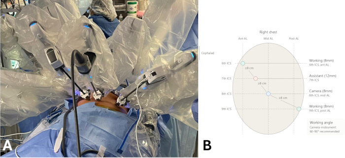
Figure 1: Intraoperative port positioning and docking of the Da Vinci Xi robot. (A) Patient in left lateral decubitus positioning, with the camera in the 8th intercostal port position, and corresponding ports positioned adjacently. Note that a working port is being used in the 7th intercostal position, which is typically designated for the assistant port. (B) Schema of right lateral chest silhouette depicting typical placement with anatomic landmarks, angulation, and spacing to avoid external robotic arm collision. Please click here to view a larger version of this figure.
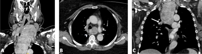
Figure 2: CT imaging demonstrating craniocaudal extension. (A) Mass effect on the laryngotracheal framework. (B) Posterior extension to the trachea abutting the esophagus (arrowhead). (C) Inferior extent at the carina abutting the azygous vein (arrow). Abbreviation: CT = computed tomography. Please click here to view a larger version of this figure.
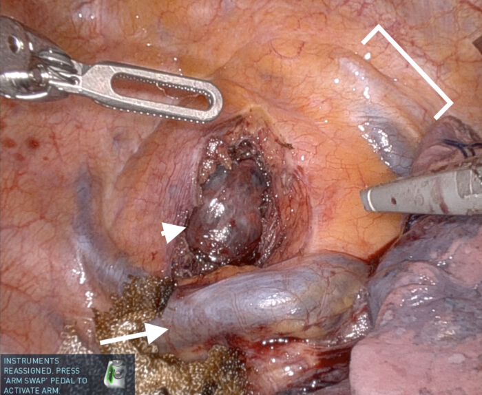
Figure 3: Robotic view of the mediastinal dissection. The image shows the anatomic relationship of the mass (arrowhead) to the azygous vein (arrow), the subclavian vessels (bracket), and the undissected trachea anteriorly. The right upper lung lobe is gently retracted (bottom right). Please click here to view a larger version of this figure.
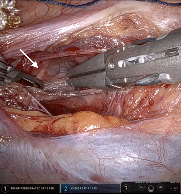
Figure 4: Preservation of the vagus nerve. Magnified robotic endoscopic view during the posterolateral dissection of the mass, mobilizing the lesion (retracted by the right-handed instrument) away from the vagus nerve (arrow). Please click here to view a larger version of this figure.
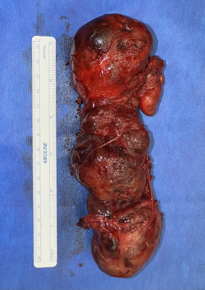
Figure 5: Resected hemithyroidectomy specimen. Hemithyroidectomy lesion excised en bloc, measuring roughly 16 cm in craniocaudal dimension and 215 g. Please click here to view a larger version of this figure.
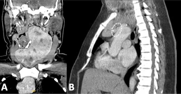
Figure 6: CT imaging with anterior mediastinal extension. (A) The multilobulated mass extends into the retropharynx superiorly and the anterior mediastinum inferiorly. (B) Sagittal view highlighting inferior extension at the aortic arch, compressing the innominate vein (dashed circle). Abbreviation: CT = computed tomography. Please click here to view a larger version of this figure.
Discussion
The hybrid robotic-assisted thoracoscopic and transcervical approach described here extends the application of minimally invasive surgery to selected patients with massive substernal goiters that would otherwise be considered for sternotomy. The principal objective of the thoracic robotic component is not thyroid resection itself but controlled circumferential mobilization of the mediastinal thyroid extension under direct visualization before transcervical delivery. This strategy allows critical mediastinal structures—including the innominate vessels, azygos vein, trachea, esophagus, vagus nerve, phrenic nerve, and recurrent laryngeal nerve—to be identified and preserved while minimizing blind traction through the thoracic inlet. Although the majority of substernal goiters can be excised via a transcervical incision, several factors predict the need for a transsternal or transthoracic approach, including radiographic extension below the aortic arch or posterior to the trachea, revision surgery, and unfavorable neck anatomy such as short length, limited extension, or elevated body mass index (BMI)3,20,21. This report supports the feasibility of this morbidity-sparing technique in such cases and its extension to both the anterior and posterior mediastinal spaces.
Several technical principles are critical to the successful execution of this method. Careful preoperative review of cross-sectional imaging is essential for operative planning, including assessment of the relationship between the thyroid and major mediastinal vessels, airway, and esophagus. During robotic dissection, maintenance of the true capsular avascular plane facilitates progressive circumferential mobilization while minimizing bleeding and protecting adjacent neurovascular structures. Early identification of the vagus and phrenic nerves, followed by meticulous preservation throughout dissection, provides additional anatomic orientation and reduces the risk of nerve injury. Equally important is avoiding excessive cervical traction before complete mediastinal mobilization, as blind inferior traction may increase the risk of vascular or recurrent laryngeal nerve injury. If the capsular plane cannot be safely developed, visualization becomes inadequate, or there is concern for vascular invasion or uncontrolled hemorrhage, prompt conversion to sternotomy or thoracotomy should be undertaken rather than persisting with minimally invasive dissection.
The reproducibility of this technique depends upon appropriate patient selection and multidisciplinary expertise. Candidates should undergo high-resolution contrast-enhanced CT to define the extent of mediastinal involvement and identify radiographic features suggestive of invasive disease. Prior cervical or thoracic surgery, dense mediastinal fibrosis, inflammatory thyroid disease, or extensive vascular encasement may significantly increase technical difficulty and should be considered relative contraindications. Likewise, successful implementation requires coordinated collaboration between surgeons familiar with robotic mediastinal anatomy, one-lung ventilation, and management of major thoracic vascular complications. Institutions without established robotic thoracic experience should exercise caution during the adoption of this technique.
The present report expands the limited literature describing combined video-assisted and robot-assisted thoracoscopic surgery in substernal thyroidectomy9,12,13,14,15,16,17,18,19. This technical report demonstrates successful application in both posterior mediastinal extension and revision thyroid surgery, two scenarios traditionally associated with increased operative complexity. Unlike previously described hybrid video-assisted approaches, high-definition three-dimensional visualization and superior mechanical control through articulated instrument wrists facilitate precise dissection around confined mediastinal neurovascular structures. Nevertheless, this study is limited by the small number of patients and the absence of comparative outcomes. Although both patients avoided sternotomy and experienced uncomplicated recoveries, larger prospective studies are needed to define patient selection criteria, perioperative outcomes, cost-effectiveness, learning curves, and long-term functional results compared with conventional transcervical surgery and open transthoracic approaches.
Future investigations should evaluate this hybrid technique in larger multicenter cohorts with standardized reporting of operative time, blood loss, conversion rates, complications, postoperative pain, length of stay, and patient-reported quality-of-life outcomes5,20,22. Comparative studies against conventional sternotomy and video-assisted thoracoscopic techniques will be particularly valuable in determining if this technique translates into measurable clinical benefit. There are several other limitations to this report, namely the small sample size, single-center experience, and retrospective design. Although robotic surgery requires specialized equipment and training, expertise in this technology is rapidly expanding in the United States23. Furthermore, robotic techniques have established yet controversial precedent in thyroid surgery, including transoral, transaxillary, and retroauricular approaches. This existing familiarity with robotic platforms should facilitate the adoption of this extended application. As robotic thoracic surgery continues to expand, this hybrid approach may provide a reproducible minimally invasive alternative for carefully selected patients with complex substernal thyroid disease while avoiding the morbidity associated with thoracic skeletal division.
Disclosures
The authors have no conflicts of interest to declare.
Materials
| Name | Company | Catalog Number | Comments |
|---|---|---|---|
| 28 Fr chest tube | Teleflex / Pleur-evac | DSTC-28S | 28-Fr straight soft thoracic catheter |
| Teleflex / Pleur-evac | DRAC-28S | 28-Fr right-angle soft thoracic catheter | |
| Teleflex / Pleur-evac | DTRC-28S | 28-Fr trocar thoracic catheter | |
| Bronchial blocker | Cook Medical | C-AEBS-9.0-65-SPH-AS | Arndt endobronchial blocker, 9.0 Fr, 65-cm, spherical balloon |
| Cadiere forceps | Intuitive Surgical | 471049 | 8-mm EndoWrist Cadiere forceps |
| Da Vinci Xi platform | Intuitive Surgical | Model IS4000 | Multiport robotic surgical platform; Xi |
| Jackson-Pratt flat drain | Cardinal Health / Jackson-Pratt | SU130-1309 | 10-mm × 20-cm flat silicone drain, ¾ perforated, without trocar |
| Maryland bipolar forceps | Intuitive Surgical | 471172 | 8-mm EndoWrist Maryland bipolar forceps |
| Monopolar curved scissors (Hot Shears) | Intuitive Surgical | 470179 | 8-mm EndoWrist monopolar curved scissors |
| NIM Monitoring platform | Medtronic Xomed | NIM4CM01 | NIM Vital 4.0 console |
| NIM EMG Endotracheal tube | Medtronic Xomed | 8229706 | 6.0-mm ID, reinforced NIM EMG ETT |
| Optical/bladeless obturator | Intuitive Surgical | 470359 | 8-mm bladeless optical obturator |
| Robotic camera | Intuitive Surgical | 470057 | 8-mm Endoscope Plus, 30° |
| Robotic port/cannula, 12 mm | Intuitive Surgical | 470375 | 12-mm/stapler cannula, 100 mm |
| Robotic port/cannula, 8 mm | Intuitive Surgical | 470002 | Standard 8-mm da Vinci Xi cannula |
| SURGICEL Original hemostatic agent | Ethicon / Johnson & Johnson MedTech | 1953 | 2" × 3" in oxidized regenerated cellulose |
| Tip-Up fenestrated grasper | Intuitive Surgical | 470347 | 8-mm EndoWrist Tip-Up fenestrated grasper |
| 8229707 | 7.0-mm ID, reinforced NIM EMG ETT | ||
| 8229708 | 8.0-mm ID, reinforced NIM EMG ETT | ||
| NIM4CPB1 | NIM Vital 4.0 patient interface | ||
| C-AEBS-9.0-78-SPH-AS | Arndt endobronchial blocker, 9.0 Fr, 78-cm, spherical balloon | ||
| 470056 | 8-mm Endoscope Plus, 0° |
References
- Casella C, Pata G, Cappelli C, Salerni B. Preoperative predictors of sternotomy need in mediastinal goiter management. Head Neck. 2010;32(9):1131-5.
- Sanders LE, Rossi RL, Shahian DM, Williamson WA. Mediastinal goiters: the need for an aggressive approach. Arch Surg. 1992;127(5):609-13.
- Mercante G et al. CT cross-sectional imaging classification system for substernal goiter based on risk factors for an extracervical surgical approach. Head Neck. 2011;33(6):792-9.
- Raffaelli M et al. Substernal goiters: incidence, surgical approach, and complications in a tertiary care referral center. Head Neck. 2011;33(10):1420-5.
- Pieracci FM, Fahey TJ. Substernal thyroidectomy is associated with increased morbidity and mortality as compared with conventional cervical thyroidectomy. J Am Coll Surg. 2007;205(1):1-7.
- Losanoff JE, Richman BW, Jones JW. Disruption and infection of median sternotomy: a comprehensive review. Eur J Cardiothorac Surg. 2002;21(5):831-9.
- Kaul P. Sternal reconstruction after post-sternotomy mediastinitis. J Cardiothorac Surg. 2017;12(1):94.
- Singh K, Anderson E, Harper JG. Overview and management of sternal wound infection. Semin Plast Surg. 2011;25(1):25-33.
- Gupta P et al. Video assisted thoracoscopic thyroidectomy for retrosternal goitre. Ann R Coll Surg Engl. 2014;96(8):606-8.
- Wang R et al. Modified subxiphoid approach for surgical resection of a retrosternal goiter. Front Surg. 2022;9:923389.
- Catelli C et al. Robot-assisted (RATS) versus video-assisted (VATS) lobectomy: a monocentric prospective randomized trial. Eur J Surg Oncol. 2023;49(12):107256.
- Piccioni G et al. Combined cervicotomy and robot-assisted surgical approach for intrathoracic thyroid goiter: a case series. Innovations (Phila). 2024;19(3):311-4.
- Chen KA, Perrotti G, Evans NR, Willis AI. A minimally-invasive approach to substernal goiter and posterior mediastinal mass. ACS Case Reviews in Surgery. 2019;2(4):1-5.
- Bodner J et al. Robotic resection of an ectopic goiter in the mediastinum. Surg Laparosc Endosc Percutan Tech. 2005;15(4):249-51.
- Wadhavkar N, Kontopidis I, Bollig C. Transcervical and robotic-assisted thoracoscopic resection of a substernal goiter. BMJ Case Rep. 2022;15(10):e250953.
- Al-Mufarrej F et al. Novel thoracoscopic approach to posterior mediastinal goiters: report of two cases. J Cardiothorac Surg. 2008;3:55.
- Amore D et al. Antero mediastinal retrosternal goiter: surgical excision by combined cervical and hybrid robot-assisted approach. J Thorac Dis. 2018;10(3):E199-202.
- Chihara RK, Liu J, Patel SG, Sancheti M. Robotic-assisted retrosternal thyroidectomy [video]. CTSNet; 2019 [cited 2025 Apr 18]. doi:10.25373/ctsnet.7877909.v1.
- Jett GK, Nguyen AB, McNamara WF, Cignoni CB. Hybrid total thyroidectomy and robotic-assisted excision of a substernal multinodular goiter [video]. CTSNet; 2023 [cited 2025 Apr 18]. doi:10.25373/ctsnet.24514264.v1.
- Cohen JP. Substernal goiters and sternotomy. Laryngoscope. 2009;119(4):683-8.
- Grainger J et al. The surgical approach to retrosternal goiters: the role of computerized tomography. Otolaryngol Head Neck Surg. 2005;132(6):849-51.
- Song Y et al. Review on risk factors, classification, and treatment of sternal wound infection. J Cardiothorac Surg. 2023;18(1):184.
- Sheetz KH, Claflin J, Dimick JB. Trends in the adoption of robotic surgery for common surgical procedures. JAMA Netw Open. 2020;3(1):e1918911.