Post-transplant biliary strictures compromise graft function and survival. Anastomotic and non-anastomotic strictures differ in timing, pathogenesis, and prognosis. Endoscopic retrograde cholangiopancreatography, with balloon dilation and plastic or fully covered metal stents, is first-line management. Current evidence supports high technical success, although standardized protocols and long-term randomized trials remain limited.
Review Article
Endoscopic Management of Biliary Strictures After Liver Transplantation: Current Evidence and Emerging Approaches
70 views
⸱
September 11th, 2026
In This Article
Summary
Abstract
Liver transplantation is a definitive treatment for end-stage liver disease and can improve quality of life and long-term prognosis. However, postoperative biliary complications remain an important limitation to transplant outcomes, and biliary stricture is among the most prevalent. Patients may present with jaundice, abdominal pain, recurrent cholangitis, and abnormal liver function, while delayed treatment may progress to biliary cirrhosis or graft failure. Endoscopic retrograde cholangiopancreatography (ERCP) has become the first-line therapeutic approach because it is minimally invasive, repeatable, and limits adverse effects on the graft. This review summarizes the pathogenic mechanisms, clinical manifestations, diagnostic modalities, and endoscopic treatment strategies for biliary strictures after liver transplantation. It distinguishes anastomotic from non-anastomotic strictures and examines the roles of magnetic resonance cholangiopancreatography and ERCP in diagnosis and treatment. The review also discusses balloon dilation, multiple plastic stents, fully covered self-expandable metal stents, and emerging endoscopic techniques, with attention to treatment efficacy, recurrence, complications, and procedural burden. Current evidence supports the effectiveness of endoscopic management but remains limited by heterogeneous protocols and a lack of long-term randomized controlled trials. This review aims to support standardized management, improve clinical decision-making, and identify priorities for future research in this complex and clinically important post-transplant patient population.
Introduction
Biliary stricture is a common and serious complication after liver transplantation and can adversely affect graft function and patient survival. Its reported incidence is approximately 5%–15%, accounting for about 50% of post-transplant complications1. Biliary strictures are generally classified as anastomotic or non-anastomotic. Anastomotic strictures are more common, usually develop within the first year after transplantation, and account for approximately 80% of all biliary strictures2. Non-anastomotic strictures occur less frequently but are often more complex and are associated with a poorer prognosis3. Biliary strictures may also occur with bile leakage, biliary stones, and cholangitis, further complicating the clinical course1.
Traditional treatment options include surgery and percutaneous intervention. Although surgery can directly correct the stricture, it is invasive, carries substantial risk, and may require prolonged postoperative recovery. Some patients may ultimately require retransplantation4. Percutaneous approaches, including percutaneous transhepatic cholangiography-guided biliary drainage and dilation, are commonly used for refractory biliary strictures, particularly when endoscopic treatment is unsuccessful or cannot be performed4. However, these approaches are also invasive and may be associated with complications that adversely affect quality of life5.
Advances in endoscopic techniques have established endoscopic therapy as the principal treatment approach for biliary strictures after liver transplantation. Endoscopic retrograde cholangiopancreatography (ERCP), together with procedures such as sphincterotomy, balloon dilation, and biliary stent placement, has demonstrated high success rates and low complication rates in the management of anastomotic strictures and biliary stones6,7,8. This review summarizes the classification, diagnosis, and endoscopic management of post-transplant biliary strictures and examines the role of current treatment strategies in improving graft and patient outcomes.
Review and Perspective
1. Etiological classification and mechanisms of biliary stricture after liver transplantation
Common causes of biliary stricture include postoperative anastomotic stricture, such as choledochojejunostomy stricture following liver transplantation; iatrogenic bile duct injury, such as accidental injury during laparoscopic cholecystectomy; chronic inflammatory disorders, including primary sclerosing cholangitis (PSC) and IgG4-related sclerosing cholangitis; radiation cholangitis; strictures secondary to bile duct stones; trauma; and congenital abnormalities.
Postoperative anastomotic stricture is particularly common in liver transplant recipients. Important differences between anastomotic strictures (AS) and non-anastomotic strictures (NAS) have been documented in terms of pathogenesis, timing of onset, response to endoscopic treatment, recurrence, long-term prognosis, and retransplantation risk. AS originates from focal perianastomotic injury. Early AS, defined as onset within < 1 month, is primarily attributed to surgical mechanical factors such as anastomotic tension and donor-recipient duct-size mismatch, whereas late AS, defined as onset > 1 month, is largely associated with focal ischemia-induced perianastomotic fibrosis9.
In contrast, NAS is associated with diffuse impairment of the biliary microcirculation. Contributing factors include cold ischemia-associated cholangiocyte injury, ischemia-reperfusion injury, ABO-incompatible transplantation, and immune-mediated injury, which may collectively produce multifocal fibrotic lesions extending beyond the anastomotic site4,10,11,12. Most AS develops within the first year after liver transplantation7. By contrast, NAS may occur early following severe ischemic injury or may develop years after transplantation in association with persistent immune-mediated injury8.
Endoscopic balloon dilation and stenting have been associated with high stricture-resolution rates, approximately 90%–97%, in selected patients with localized AS13. For diffuse NAS, endoscopic treatment is often less durable, with reported long-term therapeutic success rates of 25%–50%14. Following successful endoscopic treatment, recurrence of AS has been reported in approximately 21%–33% of cases13,15, whereas recurrence appears to be more frequent in NAS despite repeated interventions. AS generally has a more favorable clinical course when successfully treated. In contrast, NAS may be associated with recurrent cholangitis, progressive biliary injury, graft dysfunction, and, in refractory cases, retransplantation; some series have reported retransplantation in approximately 30% of affected patients12.
2. Clinical manifestations and diagnosis of biliary stricture after liver transplantation
- Clinical manifestations
Biliary strictures following liver transplantation may present with diverse and often nonspecific symptoms, primarily including cholangitis and signs of biliary obstruction such as jaundice, fever, and right upper quadrant pain. In cases of complete biliary obstruction, patients may develop worsening jaundice, pruritus, or clay-colored stools. Some patients experience recurrent cholangitis, and severe cases may progress to liver failure, compromising graft function and patient survival16.
Laboratory findings commonly include elevated serum transaminases, bilirubin, and total bile acids. Gamma-glutamyl transferase (GGT) and/or alkaline phosphatase (ALP) levels exceeding twice the upper limit of normal may have diagnostic significance7. - Diagnosis
Ultrasonography is commonly used for the initial evaluation of biliary dilation and liver dysfunction because of its accessibility and cost-effectiveness. Magnetic resonance cholangiopancreatography (MRCP) is an important noninvasive modality for diagnosing post-transplant biliary strictures, with reported sensitivity of 93%–97% and specificity of 92%–98%. Gadoxetic acid-enhanced biliary imaging may further improve diagnostic performance12,14,17.
Computed tomography (CT) and ultrasonography may provide complementary assessment of peribiliary structures and associated abnormalities, including bile duct stones, hepatic parenchymal changes, and postoperative vascular status18. Three-dimensional imaging techniques may improve visualization of intrahepatic ductal anatomy by addressing some of the spatial limitations of conventional two-dimensional CT and magnetic resonance imaging. Reported advantages include multi-angle visualization, more precise surgical planning, and improved interdisciplinary collaboration.
Endoscopic retrograde cholangiopancreatography (ERCP) remains the diagnostic reference standard for post-transplant biliary strictures because it allows direct assessment of stricture location, length, and severity while guiding therapeutic decision-making5. Endoscopic ultrasound (EUS) may provide additional information when ERCP findings are inconclusive by allowing high-resolution assessment of the bile duct wall and adjacent lymph nodes. Digital cholangioscopy provides direct intraluminal visualization and may improve the evaluation of epithelial lesions, bile duct stones, and stent-related complications19.
3. Advances in endoscopic treatment techniques
- Endoscopic retrograde cholangiopancreatography
ERCP is the cornerstone of endoscopic therapy for biliary strictures after liver transplantation. Direct endoscopic access and contrast imaging allow assessment of stricture length, location, and severity, and guide subsequent intervention. Therapeutic options include balloon dilation to restore biliary patency and placement of plastic or metal stents to maintain drainage and reduce recurrence.
Recent refinements in ERCP include high-resolution endoscopes and advanced guidewire techniques intended to improve stricture cannulation and procedural safety. Three-dimensional printed silicone models may also support operator training by simulating complex anatomy and procedural maneuvers20. Techniques such as precutting and double-guidewire, or pancreatic duct-assisted, biliary cannulation may improve access, although their technical complexity and learning curve may limit widespread use21.
Beyond standard fluoroscopy-guided ERCP, several newer endoscopic modalities have expanded treatment options for complex post-transplant biliary strictures, particularly in patients with difficult cannulation or surgically altered upper gastrointestinal anatomy. Digital single-operator cholangioscopy permits direct intraluminal visualization of the biliary mucosa and may assist in differentiating inflammatory, fibrotic, or ischemic stricture morphology. It may also identify associated sludge, stones, or loose sutures and facilitate visually guided guidewire passage when fluoroscopic cannulation is unsuccessful22. However, cholangioscopy is limited by higher procedural costs, longer procedure times, the risk of procedure-related cholangitis, and operator-dependent learning curves. Much of the supporting evidence is derived from small, single-center case series rather than large comparative studies.
In patients with surgically altered anatomy, conventional duodenoscope-based ERCP may be unsuccessful. In Roux-en-Y gastric bypass anatomy, EUS-directed transgastric ERCP (EDGE) creates a temporary gastrogastric fistula to permit access for standard duodenoscope-based intervention23. In patients with hepaticojejunostomy or other altered biliary anatomy, alternative approaches may include device-assisted enteroscopy, EUS-guided biliary drainage, or percutaneous intervention. EUS-guided biliary drainage (EUS-BD) establishes a direct transluminal biliary-enteric fistula for decompression when transpapillary access cannot be achieved and may serve as an alternative to percutaneous transhepatic biliary drainage.
Although these approaches may avoid some morbidity associated with percutaneous intervention, they carry specific risks, including lumen-apposing metal stent migration, fistula-associated leakage, bleeding, and peritoneal infection. Available evidence is largely non-transplant-specific, and prospective transplant-specific cohorts remain limited. Standardized protocols for fistula creation, stent indwelling duration, and fistula closure after treatment have not been established. Accordingly, these techniques currently serve primarily as rescue approaches rather than replacements for conventional first-line ERCP23–25. - Biliary stenting
Biliary stent placement is a core component of ERCP-based treatment for biliary strictures after liver transplantation. Stents maintain biliary patency, facilitate dilation of narrowed segments, and reduce the risk of recurrent obstruction. Multiple-stent strategies are also used for complex or multifocal strictures to improve biliary drainage and reduce recurrence26.
MPS is commonly used for anastomotic strictures. Published protocols generally involve placement of two to three 10 Fr plastic stents, with subsequent exchange at approximately 3-month intervals and progressive increases in stent size or number when feasible. Treatment is often continued for about 12 months and may be extended in refractory cases. Stent removal is typically considered after clinical improvement, normalization or substantial improvement of liver biochemistry, and endoscopic evidence of adequate biliary drainage. Continued follow-up after stent removal is advisable because recurrence may occur27.
Comparative studies suggest that MPS and FCSEMS achieve broadly similar stricture-resolution rates, approximately 93%–97%, in post-transplant anastomotic biliary strictures, although results vary across studies and treatment protocols13. Reported recurrence rates after complete stent removal range from 21%–33% for both modalities, although observational studies indicate variation according to stent indwelling duration and stricture severity13,15.
Stent migration is an important limitation of FCSEMS. Conventional FCSEMS may have migration rates of up to 9.8%, although this risk may be reduced by over-the-scope clip anchoring or anchor-fluked FCSEMS28,29. In contrast, MPS is more commonly associated with stent occlusion caused by biliary sludge. Both strategies share procedure-related risks, including post-ERCP pancreatitis, biliary infection, and hemorrhage. FCSEMS may additionally be associated with cholecystitis and intrastent hyperplasia, whereas MPS-related complications predominantly include sludge-induced occlusion and recurrent cholangitis13,30,31.
FCSEMS may reduce the number of ERCP procedures required compared with MPS; one comparative study reported a median of two procedures with FCSEMS versus four with MPS13. Treatment duration also differs between the approaches. Standard MPS protocols generally require approximately 12 months of staged exchanges at 3-month intervals, whereas FCSEMS is typically left in place for 6–12 months as a continuous treatment course13,30.
FCSEMS may be considered in patients in whom reducing the number of repeat endoscopic procedures is an important treatment goal, provided that migration risk and other stent-related adverse events are taken into account. MPS remains an established alternative, particularly when metal stents are unsuitable or repeated staged treatment is preferred. Neither approach provides satisfactory curative outcomes for diffuse NAS, in which endoscopic intervention generally provides palliative biliary decompression. Despite growing comparative evidence, no consensus has been established on optimal stent selection, and treatment should be individualized based on stricture anatomy, patient comorbidities, and local endoscopic expertise13. - Balloon dilation
Balloon dilation mechanically widens biliary strictures to restore bile flow and reduce biliary stasis and infection. Careful control of dilation pressure and duration is important because excessive dilation may result in perforation or pseudoaneurysm formation24. Non-compliant balloons are gradually inflated to the intended diameter, and stent placement may subsequently be performed to maintain patency. Combined balloon dilation and stenting have been associated with better efficacy and lower recurrence than stenting alone.
4. Evaluation of endoscopic therapy and complication management
- Efficacy of endoscopic therapy
A retrospective study reported longer median overall survival among patients who underwent scheduled endoscopic treatment than among untreated patients, 19.7 years versus 7.7 years15. Because this finding is based on observational data, the difference should be interpreted in the context of potential selection- and treatment-related confounding. In a long-term cohort of 165 patients treated endoscopically for biliary strictures after liver transplantation, the initial technical success rate was 83.6%, the treatment failure rate was 24.2%, and the recurrence rate was 21.2%. Median overall survival among patients with anastomotic strictures was 17.6 years. The same cohort reported a 15-year graft survival rate of 70.6%32. These findings support the potential long-term effectiveness of endoscopic treatment, although outcomes may vary according to stricture type, patient characteristics, treatment protocol, and study design. - Complication management in endoscopic therapy
Endoscopic treatment of post-transplant biliary strictures carries recognized risks, including pancreatitis, biliary infection, hemorrhage, and perforation. Reported rates of post-ERCP pancreatitis range from 3.47%–14.7%, hemorrhage from 0.004%–0.07%, and perforation is reported in approximately 0.05% of procedures8,33.
Prophylactic nonsteroidal anti-inflammatory drugs and pancreatic stent placement may reduce the risk of post-ERCP pancreatitis. Biliary infection is associated with procedural contamination and immunosuppression and requires attention to aseptic technique and appropriate antibiotic use. Management of biliary perforation depends on severity. Minor perforations may be managed conservatively, whereas larger perforations accompanied by peritonitis or hemodynamic instability may require urgent surgical repair34. Rare complications, including gas embolism and nerve injury, further emphasize the importance of careful monitoring and multidisciplinary management35. An overview of the classification, diagnosis, and endoscopic management of biliary strictures after liver transplantation is provided in Figure 1.
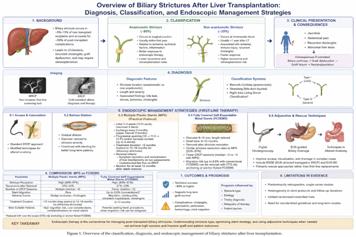
Figure 1: Overview of the classification, diagnosis, and endoscopic management of biliary strictures after liver transplantation. The illustration outlines the background and clinical importance of biliary strictures after liver transplantation, distinguishes anastomotic and non-anastomotic strictures, and highlights their differences in timing, pathogenesis, treatment response, recurrence, and prognosis. Please click here to view a larger version of this figure.
Conclusions
This review summarizes the etiological classification, imaging, and endoscopic diagnosis of biliary strictures after liver transplantation. It also examines the principal endoscopic interventions centered on ERCP, including balloon dilation, multiple plastic stents, and fully covered self-expandable metal stents, together with available evidence on long-term outcomes and complication prevention.
Anastomotic strictures generally respond well to stepwise endoscopic treatment and are associated with favorable stricture-resolution rates in published series. In contrast, ischemia-mediated non-anastomotic strictures are often diffuse, have a higher risk of long-term recurrence, and may respond only temporarily to endoscopic decompression. Modified stents, EUS-based approaches, and three-dimensional visualization may offer additional diagnostic or therapeutic options in selected patients, although supporting evidence remains limited. However, evidence-based standards remain lacking for stent indwelling duration, dilation parameters, and management of patients with altered anatomy. Most available evidence derives from single-center retrospective studies, while high-quality randomized controlled trials with stratified comparison groups and long-term follow-up remain limited.
Future research should focus on stratified management of refractory strictures, the long-term safety of biodegradable stents, and individualized peri-endoscopic infection prevention. Standardized endoscopic management workflows tailored to donor type, stricture classification, and underlying liver disease may improve the consistency of care and long-term outcomes of liver transplant recipients.
Disclosures
The authors declare no conflicts of interest. No commercial entities, stent manufacturers, or medical device companies provided financial support, consultancy fees, or other benefits related to the preparation or publication of this article. The authors report no proprietary or financial interests in any endoscopic equipment or biliary stent products discussed in this review.
Acknowledgements
This work was supported by clinical research funds from The First Affiliated Hospital of Guangzhou Medical University. The authors thank the clinical staff of the Department of Hepatobiliary Surgery and Liver Transplantation Center for their assistance with clinical data collection and literature collation. The authors also thank the patients with post-liver-transplantation biliary strictures who were included in the relevant clinical studies. Finally, the authors thank the editors and anonymous reviewers for their constructive comments and suggestions. ChatGPT (OpenAI) was used to assist in the preparation of Figure 1. The authors reviewed and approved the final figure and are responsible for its accuracy and content.
References
- Matar AJ, et al. Biliary complications following adult deceased donor liver transplantation: risk factors and implications at a high-volume US center. Transplant Direct. 2021;7(10):e754. doi:10.1097/TXD.0000000000001207.
- Yasen A, et al. Efficiency of percutaneous transhepatic cholangioscopy in the treatment of biliary complications after liver transplantation. HPB (Oxford). 2023;25(4):463–471. doi:10.1016/j.hpb.2023.01.010.
- Vingrovich O, et al. Biliary strictures post pediatric liver transplantation: incidence and risk factors in a single tertiary referral transplant center. Pediatr Transplant. 2024;28(3):e14727. doi:10.1111/petr.14727.
- López Álvarez M, et al. Endoscopic treatment of biliary complications after liver transplantation. Rev Esp Enferm Dig. 2020;112(8):605–608. doi:10.17235/reed.2020.6704/2019.
- Karakaya MF, et al. Management of biliary complications in liver transplant recipients with duct-to-duct anastomosis: a single-center experience. Turk J Gastroenterol. 2023;34(2):177–181. doi:10.5152/tjg.2023.22724.
- Khubutiya MSh, et al. Endoscopic intraluminal treatment of early biliary complications after orthotopic liver transplantation. Pirogov Russ J Surg. 2023;(8):13. doi:10.17116/hirurgia202308113.
- Macías-Gómez C, Dumonceau JM. Endoscopic management of biliary complications after liver transplantation: an evidence-based review. World J Gastrointest Endosc. 2015;7(6):606–616. doi:10.4253/wjge.v7.i6.606.
- Seehofer D, Eurich D, Veltzke-Schlieker W, Neuhaus P. Biliary complications after liver transplantation: old problems and new challenges. Am J Transplant. 2013;13(2):253–265. doi:10.1111/ajt.12034.
- Crismale JF, Ahmad J. Endoscopic management of biliary issues in the liver transplant patient. Gastrointest Endosc Clin N Am. 2019;29(2):237–256. doi:10.1016/j.giec.2018.11.003.
- Ito T, et al. Nonanastomotic biliary strictures after liver transplantation. Am Surg. 2020;86(10):1363–1367. doi:10.1177/0003134820964461.
- Taniai T, et al. Multimodal management for refractory biliary stricture after living donor liver transplantation. Transplant Proc. 2023;55(4):940–944. doi:10.1016/j.transproceed.2023.04.001.
- Xu YB, et al. Diagnostic value of magnetic resonance cholangiopancreatography for biliary complications in orthotopic liver transplantation: a meta-analysis. Transplant Proc. 2013;45(6):2341–2346. doi:10.1016/j.transproceed.2013.03.031.
- Sissingh NJ, et al. Intraductal fully covered self-expandable metal stent versus multiple plastic stents for treating biliary anastomotic strictures after liver transplantation. Gastrointest Endosc. 2023;97(4):704–712.e2. doi:10.1016/j.gie.2022.11.018.
- Jorgensen JE, et al. Is MRCP equivalent to ERCP for diagnosing biliary obstruction in orthotopic liver transplant recipients? A meta-analysis. Gastrointest Endosc. 2011;73(5):955–962. doi:10.1016/j.gie.2010.12.014.
- Zhang CC, et al. Scheduled endoscopic treatment of biliary anastomotic and nonanastomotic strictures after orthotopic liver transplantation. Gastrointest Endosc. 2023;97(1):42–49. doi:10.1016/j.gie.2022.08.034.
- Kimura K, et al. Intractable biliary strictures after living donor liver transplantation: a case series. Transplant Proc. 2021;53(5):1726–1730. doi:10.1016/j.transproceed.2021.04.015.
- Keane MG, et al. Diagnosis and management of benign biliary strictures post liver transplantation in adults. Transplant Rev. 2021;35(1):100593. doi:10.1016/j.trre.2020.100593.
- Patel VB, Musa RK, Patel N, Patel SD. Role of MRCP to determine the etiological spectrum, level and degree of biliary obstruction in obstructive jaundice. J Fam Med Prim Care. 2022;11(7):3436–3441. doi:10.4103/jfmpc.jfmpc_2362_21.
- Yu JF, Zhang DL, Wang YB, Hao JY. Digital single-operator cholangioscopy for biliary stricture after cadaveric liver transplantation. World J Gastrointest Oncol. 2022;14(5):1037–1049. doi:10.4251/wjgo.v14.i5.1037.
- Shin SP, et al. Endoscopic retrograde cholangiopancreatography training using a silicone simulator fabricated using a 3D printing technique (with videos). Sci Rep. 2025;15(1):2619. doi:10.1038/s41598-025-86755-9.
- Lopes L, Canena J. ERCP in Portugal: a wide survey on the prevention of post-ERCP pancreatitis and papillary cannulation techniques. GE Port J Gastroenterol. 2019;26(1):14–23. doi:10.1159/000487150.
- Hüsing-Kabar A, et al. Single-operator cholangioscopy for biliary complications in liver transplant recipients. World J Gastroenterol. 2017;23(22):4064–4071. doi:10.3748/wjg.v23.i22.4064.
- Khara HS, et al. Review of ERCP techniques in Roux-en-Y gastric bypass patients: highlight on the novel EUS-directed transgastric ERCP (EGDE) technique. Curr Gastroenterol Rep. 2021;23(7):10. doi:10.1007/s11894-021-00808-3.
- Bowen H, et al. The reasonable therapeutic modality for biliary duct-to-duct anastomotic stricture after liver transplantation: ERCP or PTC? Front Oncol. 2022;12:1035722. doi:10.3389/fonc.2022.1035722.
- Parlak E, et al. The classification of biliary strictures in patients with right-lobe liver transplant recipients and its relation to traversing the stricture with a guidewire. Transplantation. 2022;106(2):328–336. doi:10.1097/TP.0000000000003738.
- Han S, et al. Biliary and pancreatic stents. Gastrointest Endosc. 2023;97(6):1003–1004. doi:10.1016/j.gie.2023.03.016.
- Koksal AS, Eminler AT, Parlak E, Gurakar A. Management of biliary anastomotic strictures after liver transplantation. Transplant Rev. 2017;31(3):207–217. doi:10.1016/j.trre.2017.03.002.
- Brinkmann F, et al. Anchoring fins of fully covered self-expandable metal stents affect pull-out force and stent migration. Gastrointest Endosc. 2024;99(3):377–386.e3. doi:10.1016/j.gie.2023.10.036.
- Wang X, et al. Effects of clip anchoring on preventing migration of fully covered self-expandable metal stent in patients undergoing endoscopic retrograde cholangiopancreatography: a multicenter, randomized controlled study. Am J Gastroenterol. 2024;119(10):2028–2035. doi:10.14309/ajg.0000000000002813.
- Thiruvengadam NR, Saumoy M, Schneider Y, Kochman ML. Fully covered self-expanding stents are cost-effective at remediating biliary strictures in patients with chronic pancreatitis. Clin Gastroenterol Hepatol. 2023;21(2):552–554.e4. doi:10.1016/j.cgh.2022.02.019.
- Vanella G, et al. Fully versus partially-covered self-expandable metal stents for palliation of distal malignant biliary obstruction: a meta-analysis. Pancreatology. 2023;23:e79. doi:10.1016/j.pan.2023.06.605.
- Fan Z, et al. Endoscopic treatment of benign biliary stricture using different stents: a systematic review and meta-analysis. Videosurgery Miniinv. 2022;17(1):35–60. doi:10.5114/wiitm.2021.108418.
- Balderramo D, et al. Complications after ERCP in liver transplant recipients. Gastrointest Endosc. 2011;74(2):285–294. doi:10.1016/j.gie.2011.04.025.
- Jung Y. Medical dispute related to gastrointestinal endoscopy complications: prevention and management. Korean J Gastroenterol. 2019;73(6):315–321. doi:10.4166/kjg.2019.73.6.315.
- Zhang CMJ, Wang X. Suspected cerebrovascular air embolism during endoscopic esophageal varices ligation under sedation with fatal outcome: a case report. World J Clin Cases. 2022;10(1):371–380. doi:10.12998/wjcc.v10.i1.371.