This retrospective study of 101 PELD patients showed that preoperative MRI features, particularly Modic II (unfavorable) and DHI > 25% (protective), were closely associated with 1-year prognosis. These findings can guide surgical decisions and are not used as static predictive factors. Complete neural decompression remains the ultimate determining factor for success.
Research Article
Preoperative MRI Features and Outcomes for Lumbar Disc Herniation Treated with Percutaneous Endoscopic Lumbar Discectomy: A Retrospective Study
88 views
⸱
September 11th, 2026
In This Article
Summary
Abstract
Percutaneous endoscopic lumbar discectomy (PELD) is widely used for lumbar disc herniation (LDH), yet outcomes vary. Preoperative magnetic resonance imaging (MRI) features may predict prognosis, but systematic research on their correlation with surgical efficacy is lacking. The purpose of this study is to retrospectively analyze the relationship between preoperative MRI morphological characteristics and clinical outcomes in patients receiving PELD treatment for lumbar disc herniation, with the aim of providing a reference for preoperative evaluation, outcome prediction, and surgical strategy optimization. In this retrospective study, 101 single-level LDH patients undergoing PELD (2022.1–2024.9) with ≥1-year follow-up were included. Preoperative MRI parameters [Pfirrmann grade, Modic changes, herniation type/location/migration, canal occupation ratio, disc height index (DHI)] and clinical outcomes [Visual Analog Scale (VAS), Oswestry Disability Index (ODI), modified MacNab] were analyzed. Patients were grouped by 1-year MacNab scores. MRI features were compared between groups, with Pearson correlation and multivariate logistic regression used to identify predictors of a favorable prognosis. Of 101 patients, 76 (75.2%) had favorable outcomes and 25 (24.8%) unfavorable outcomes at 1-year follow-up. The unfavorable group showed significantly higher rates of disc extrusion/sequestration, larger herniation area, and Modic type II changes, but lower intervertebral height and foraminal area (P < 0.05). Canal occupation ratio and herniation area positively correlated with pain VAS and ODI, while DHI and foraminal area showed negative correlations. Modic type II changes independently predicted unfavorable prognosis, whereas DHI > 25% was a protective factor. Preoperative MRI features are closely associated with PELD outcomes, with Modic type II and reduced DHI identifying high-risk patients. This multidimensional MRI evaluation enables preoperative risk stratification and guides individualized surgical decision-making, including consideration of foraminoplasty or alternative approaches when adverse imaging features are present. Complete neural decompression, however, remains the ultimate determinant of success.
Introduction
Lumbar disc herniation (LDH) is primarily characterized by disc degeneration, annulus fibrosus rupture, and nucleus pulposus herniation compressing nerve roots or the cauda equina. Clinically, the condition is characterized by manifestations including sciatica, lower limb numbness and weakness, and intermittent claudication. These symptoms severely compromise patients’ daily life and working ability, thus placing a substantial economic burden on individuals, families, and society1,2. Although traditional open surgery for LDH allows direct removal of the herniated nucleus pulposus, it is associated with drawbacks, including substantial trauma, significant blood loss, delayed postoperative recovery, and disruption of spinal stability3. Percutaneous endoscopic lumbar discectomy (PELD) has evolved over more than two decades since Yeung and Tsou4 first reported the transforaminal endoscopic discectomy (YESS technique) in 1997, and has now become a mainstream minimally invasive approach for treating LDH. PELD utilizes a working channel approximately 6–7 mm in diameter to remove herniated disc tissue under direct endoscopic visualization, offering notable advantages such as minimal trauma, preserved spinal stability5,6. Numerous clinical studies have confirmed that PELD achieves excellent or good outcomes in 84%–94% of LDH cases, comparable to those of traditional open surgery7. However, clinical practice reveals that even with strict adherence to surgical indications and standardized procedures performed by the same surgical team, postoperative outcomes following PELD exhibit considerable inter-individual variability. While some patients experience significant pain relief and functional recovery, approximately 20%–30% report residual low back or leg pain, sensory abnormalities, suboptimal functional recovery, or even recurrence8,9. This phenomenon has emerged as a significant clinical challenge for spine surgeons. Recently, the concept of defining objective and subjective “end-points of decompression” has been proposed as a critical strategy to minimize surgical failures and optimize outcomes in transforaminal endoscopic lumbar surgery10.
Preoperative magnetic resonance imaging (MRI) is the preferred imaging modality for evaluating LDH, providing clear visualization of disc morphology, herniation extent, nerve compression, and the structure of the spinal canal and neural foramina. MRI morphological parameters not only aid in confirming diagnosis and surgical indications but may also offer predictive value for postoperative outcomes. The Pfirrmann grading system, based on T2-weighted MRI signal intensity, disc structure, disc height, and distinction between the nucleus pulposus and annulus fibrosus, classifies disc degeneration into grades 1–5 and is widely used in clinical assessment11. Modic changes refer to signal alterations in the vertebral endplates and adjacent bone marrow on MRI, categorized into type I (edematous), type II (fatty), and type III (sclerotic), closely associated with disc degeneration, low back pain, and postoperative recurrence12. An increasing number of studies have investigated the association between MRI morphological features and clinical outcomes following PELD. However, current research predominantly focuses on single or a few MRI parameters, lacking a systematic evaluation of multiple morphological indicators. Moreover, studies on the relationship between quantitative parameters—such as spinal canal occupation ratio, foraminal area, and disc height index (DHI)—and PELD outcomes remain insufficient13. Furthermore, recent advancements in minimally invasive spinal surgery have evolved from standalone endoscopic or channel techniques toward intelligent minimally invasive approaches integrating robotics, artificial intelligence, and imaging guidance. The integration of robotic-assisted systems has further addressed instrumentation challenges in confined surgical spaces, representing a critical advancement in minimally invasive spinal surgery technology14.
Based on the aforementioned research background and existing knowledge gaps, this single-center retrospective observational study aims to systematically collect clinical data and preoperative MRI morphological features from patients undergoing PELD for single-level LDH. A multidimensional MRI morphological evaluation system will be constructed, and in conjunction with one-year postoperative clinical outcomes, the associations between various MRI morphological parameters and PELD prognosis will be thoroughly analyzed to identify independent risk and protective factors influencing unfavorable postoperative outcomes. The primary objectives of this study include: determining the overall clinical efficacy of PELD for single-level LDH and the distribution of one-year prognostic outcomes; comparing differences in preoperative MRI morphological parameters between patients with favorable and unfavorable prognoses; identifying independent predictors of favorable postoperative outcomes through multivariate logistic regression analysis; and establishing a multidimensional preoperative MRI evaluation system that integrates both qualitative (Modic subtypes, herniation grade) and quantitative parameters (canal occupation ratio, DHI, foraminal area, herniation area) to enable more precise postoperative risk stratification and guide individualized surgical decision-making.
Protocol
This study was conducted in strict adherence to the principles of the Declaration of Helsinki15 and was approved by the Institutional Ethics Committee of North China Medical Health Group XingTai General Hospital (Approval No. ZCKT-2026-0018). All patients were informed of the study purpose, treatment protocols, and potential risks, and provided written informed consent. All study data were anonymized to ensure patient privacy.
Research subjects
This single-center retrospective observational study included patients with single-level LDH who underwent PELD in the orthopedics department of our hospital from January 2022 to September 2024. Among 123 initially screened patients, 101 were ultimately included after applying strict inclusion and exclusion criteria, all of whom completed at least one year of follow-up. Based on the modified MacNab criteria at one-year post-surgery, patients were classified into the favorable group (n = 76) and the unfavorable group (n = 25). The detailed process is shown in Figure 1.
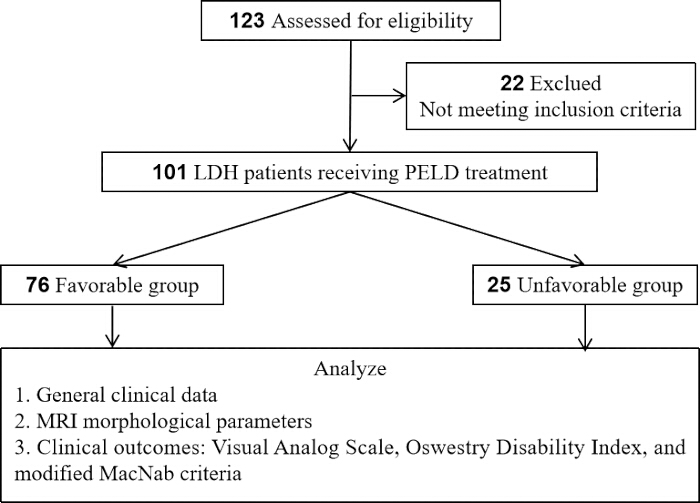
Figure 1: Research flowchart. Flowchart of patient selection and grouping. Of 123 assessed patients, 101 with single-level LDH undergoing PELD were included and divided into Favorable (n = 76) and Unfavorable (n = 25) groups based on 1-year modified MacNab criteria. The analytical framework covered general clinical data, MRI parameters, and clinical outcomes (VAS, ODI, MacNab). Please click here to view a larger version of this figure.
Inclusion criteria
Age between 18 and 75 years; diagnosed with single-level LDH at L3/4, L4/5, or L5/S1 based on clinical symptoms, physical examination, and MRI/CT findings; presence of definite symptoms and signs of nerve root compression, with no response to at least 6 weeks of systematic conservative treatment (including bed rest, medication, physical therapy, etc.); underwent PELD surgery; postoperative follow-up duration of at least 1 year with complete clinical data.
Exclusion criteria
Multilevel LDH or a history of previous lumbar surgery; concomitant lumbar spondylolisthesis, lumbar spinal stenosis, spinal tumors, spinal infections, or spinal fractures; presence of cauda equina syndrome requiring emergency surgery; poor preoperative MRI image quality precluding accurate measurement; severe comorbid medical conditions (e.g., cardiac, pulmonary, hepatic, or renal insufficiency) rendering patients unfit for surgery; loss to follow-up or incomplete clinical data.
Surgical procedure
All surgeries were performed by the same experienced team of spine surgeons. The surgical approach (transforaminal, TF, or interlaminar, IL) was determined based on a comprehensive evaluation of the herniation morphology and anatomical constraints. Specifically, the TF approach was preferentially adopted for foraminal, extraforaminal, or central/paracentral herniations located below the pedicle level. Conversely, the IL approach was selected for highly migrated or sequestered fragments, particularly those migrating cephalad to the L5 pedicle or in patients with a high iliac crest, as previously described by Choi et al.16. This differentiated selection strategy ensured that the surgical trajectory aligned optimally with the target lesion.
Procedures were conducted under local anesthesia with or without intravenous sedation, with patients positioned prone. The target disc space was localized using C-arm fluoroscopy. A puncture needle was advanced into the spinal canal through Kambin's triangle or the interlaminar space, followed by guidewire insertion and sequential dilation to establish a working channel. A working endoscope (diameter 6.9 mm, 30° viewing angle) was introduced, and herniated nucleus pulposus tissue was removed under direct visualization to achieve nerve root decompression. Annuloplasty and hemostasis were performed using a radiofrequency probe. The working channel was then withdrawn, and the skin incision was sutured.
In cases presenting with preoperative DHI ≤25% or severe foraminal narrowing on MRI, additional foraminoplasty using a reamer or high-speed drill was performed at the discretion of the attending surgeon to enlarge Kambin's working zone and facilitate channel placement. However, given the retrospective design, the execution of this foraminoplasty was neither strictly standardized nor systematically documented in the operative notes, which precludes a quantitative analysis of its specific impact on clinical outcomes in this cohort.
Observation indicators
In this study, T0, T1, T2, and T3 were defined as preoperative, 1 week postoperative, 3 months postoperative, and 1 year postoperative, respectively.
General clinical data
Patient data, including age, sex, body mass index (BMI), disease duration, smoking history, history of diabetes mellitus, history of hypertension, surgical level, and surgical approach, were collected.
Measurement of MRI morphological parameters
All patients underwent preoperative lumbar MRI, including sagittal T1WI, T2WI, and axial T2WI sequences. Two experienced radiologists independently measured the parameters in a double-blind manner, with the average values used for analysis. Pfirrmann grade: Assessed on sagittal T2WI based on disc signal intensity, structure, height, and distinction between nucleus pulposus and annulus fibrosus, graded 1–511. Modic changes: Evaluated as signal changes in vertebral endplates and adjacent bone marrow, classified as none, type I (hypointense on T1WI, hyperintense on T2WI), type II (hyperintense on T1WI, iso- or slightly hyperintense on T2WI), or type III (hypointense on both T1WI and T2WI)12. Herniation type: Classified on axial T2WI as bulging, protrusion, extrusion, or sequestration17. Herniation location: Categorized as central, paracentral, foraminal, or far lateral4. Extent of migration: Measured on sagittal T2WI as the vertical distance from the upper or lower margin of the herniated fragment to the disc level, classified according to Lee et al.18 as no migration (grade 0), low-grade migration (<50% of disc height, grades 1–2), or high-grade migration (≥50% of disc height, grades 3–4). Spinal canal occupation ratio: Calculated on axial T2WI at the level of maximal herniation as (anteroposterior diameter of the herniation / anteroposterior diameter of the spinal canal) × 100%19,20. DHI: Measured on sagittal T2WI as [(anterior disc height + posterior disc height) / 2] / [(anterior-posterior diameter of upper vertebral body + anterior-posterior diameter of lower vertebral body) / 2] × 100%21. Foraminal area: Manually delineated and measured on axial T2WI at the maximal foraminal level using ImageJ software22,23. Maximum herniation area: Measured as the cross-sectional area of the herniated fragment on axial T2WI at the level of maximal herniation24.
Clinical outcome evaluation
Pain Assessment: Pain intensity was evaluated using the Visual Analog Scale (VAS) for low back pain (VAS-Back) and leg pain (VAS-Leg), with scores ranging from 0 (no pain) to 10 (worst imaginable pain)25,26. Functional Disability: Lumbar function was assessed using the Oswestry Disability Index (ODI)27,28. Modified MacNab Criteria: Evaluated at the 1-year follow-up, outcomes were classified as excellent (complete symptom resolution, return to normal activities), good (occasional pain, no interference with work or daily life), fair (improved symptoms but activity limitations), or poor (no improvement or worsened symptoms)29,30. “Excellent” and “good” outcomes were defined as a favorable prognosis, while “fair” and “poor” outcomes were defined as an unfavorable prognosis. The modified MacNab criteria were selected as the primary outcome measure because they provide a global, patient-centered assessment of surgical success that integrates pain relief, functional recovery, and return to daily activities—domains that are collectively more relevant to clinical decision-making than any single continuous scale. Additionally, the MacNab classification has been widely utilized and validated in PELD literature29,30, facilitating direct comparison with prior studies.
Sample size estimation
Sample size estimation was performed using G*Power software31. As a retrospective observational study, the sample size was determined based on the total number of patients meeting the inclusion and exclusion criteria during the study period, and its adequacy was verified through post-hoc power analysis. Based on previous relevant studies8, assuming an incidence of unfavorable prognosis following PELD of approximately 25% and using disc extrusion/sequestration as the primary exposure factor, a minimum of 89 participants was required with a two-sided α = 0.05 and a power (1-β) of 80%. A total of 101 patients were ultimately included in this study, meeting the statistical requirements.
Statistical analysis
Statistical analyses were performed using SPSS. Normality of continuous variables was assessed by the Shapiro-Wilk test. Normally distributed data were presented as mean ± SD and compared using independent-samples and paired t-tests. Non-normally distributed data were shown as median (Q1, Q3) and analyzed via the Mann-Whitney U test. Categorical variables were compared using the chi-square test. Pearson correlation and multivariate logistic regression were applied. All tests were two-sided, and P < 0.05 was deemed statistically significant.
For multivariate logistic regression analysis, the dependent variable was encoded as favorable prognosis = 1 and unfavorable prognosis = 0, resulting in 76 positive events. The number of covariates included in the final model must comply with the EPV principle32. Used variance inflation factor (VIF) to evaluate multicollinearity among MRI parameters; a VIF value <5 is considered acceptable.
Results
General information about patients
A total of 101 patients were included in this study. As shown in Table 1, patients in the unfavorable group were significantly older (P < 0.05) and had a longer disease duration (P < 0.05). No statistically significant differences were observed between the two groups regarding sex, BMI, smoking history, comorbidities, surgical level, or surgical approach (P > 0.05).
| Variables | Favorable Group (n = 76) | Unfavorable Group (n = 25) | t/χ²/Z | P |
| Age (years) | 45.2 ± 11.8 | 51.6 ± 12.9 | 2.322 | 0.022 |
| Male | 42 (55.3) | 16 (64.0) | 0.587 | 0.444 |
| BMI (kg/m2) | 24.4 ± 3.0 | 25.4 ± 3.6 | 1.531 | 0.129 |
| Disease duration (months) | 9 (8, 13) | 15 (12, 17) | 4.174 | <0.001 |
| Smoking history | 18 (23.7) | 9 (36.0) | 1.457 | 0.227 |
| History of diabetes | 8 (10.5) | 5 (20.0) | 0.779 | 0.337 |
| History of hypertension | 12 (15.8) | 6 (24.0) | 0.396 | 0.529 |
| Operative segment | ||||
| L3/4 | 11 (14.5) | 1 (4.0) | ||
| L4/5 | 40 (52.6) | 16 (64.0) | ||
| L5/S1 | 25 (32.9) | 8 (32.0) | 2.18 | 0.336 |
| Operative approach | ||||
| Transforaminal approach | 59 (77.6) | 19 (76.0) | ||
| Interlaminar approach | 17 (22.4) | 6 (24.0) | 0.029 | 0.866 |
Table 1: Baseline characteristics[mean±SD, n (%), M(Q1, Q3)]. Baseline characteristics stratified by prognostic group. Data are mean ± SD, n (%), or median (Q1, Q3). The Unfavorable group was significantly older (P = 0.022) and had longer disease duration (P < 0.001). Other variables showed no significant intergroup differences. BMI, Body Mass Index.
Comparison of preoperative MRI morphological parameters
The preoperative MRI morphological parameters of 101 patients are shown in Table 2. There are significant differences (P < 0.05) between the two groups in terms of the proportions of Modic changes, herniation types; The DHI and foraminal area in the Favorable group were significantly higher than those in the Unfavorable group (P < 0.05), while the spinal canal occupation ratio and maximum herniation area in the Favorable group were significantly lower than those in the Unfavorable group (P < 0.05); There was no statistically significant difference (P > 0.05) in Pfirrmann grading, herniation location, and migration grades. The results suggest that the above parameters may be potential predictive indicators for the postoperative prognosis of PELD.
| Favorable Group (n = 76) | Unfavorable Group (n = 25) | t/χ² | P | |
| Pfirrmann Grade | ||||
| I-II | 28(36.8) | 5(20.0) | ||
| III | 32(42.1) | 10(40.0) | ||
| IV-V | 16(21.1) | 10(40.0) | 4.277 | 0.118 |
| Modic Changes | ||||
| None | 48(63.2) | 8(32.0) | ||
| Type I | 12(15.8) | 3(12.0) | ||
| Type II | 14(18.4) | 13(52.0) | ||
| Type III | 2(2.6) | 1(4.0) | 11.529 | 0.009 |
| Herniation Type | ||||
| Bulging | 8(10.5) | 1(4.0) | ||
| Protrusion | 44(57.9) | 7(28.0) | ||
| Extrusion | 20(26.3) | 12(48.0) | ||
| Sequestration | 4(5.3) | 5(20.0) | 11.605 | 0.009 |
| Herniation Location | ||||
| Central | 12(15.8) | 2(8.0) | ||
| Paracentral | 48(63.2) | 18(72.0) | ||
| Foraminal | 12(15.8) | 4(16.0) | ||
| Extraforaminal | 4(5.3) | 1(4.0) | 1.11 | 0.775 |
| Degree of Migration | ||||
| No Migration (Grade 0) | 25(32.9) | 6(24.0) | ||
| Low-Grade Migration (Grade 1–2) | 32(42.1) | 10(40.0) | ||
| High-Grade Migration (Grade 3–4) | 19(25.0) | 9(36.0) | 1.326 | 0.515 |
| Spinal Canal Occupation Rate (%) | 35.6 ± 6.9 | 47.2 ± 8.4 | 6.892 | <0.001 |
| Spinal Canal Occupation Rate > 40% | 30(39.5) | 14(56.0) | ||
| DHI (%) | 28.5 ± 5.2 | 24.5 ± 4.8 | 3.392 | 0.001 |
| DHI > 25% | 58(76.3) | 11(44.0) | ||
| Foraminal Area (cm²) | 1.1 ± 0.3 | 0.8 ± 0.3 | 4.029 | <0.001 |
| Foraminal Area > 1 cm² | 49(64.5) | 13(52.0) | ||
| Maximum Herniation Area (mm²) | 59.8 ± 11.9 | 86.0 ± 15.7 | 8.764 | <0.001 |
| Maximum Herniation Area > 75 mm² | 21(27.6) | 13(52.0) |
Table 2: Comparison of preoperative MRI morphological parameters [mean±SD, n (%)]. Preoperative MRI parameters compared between groups. Data are mean ± SD or n (%). Significant differences were observed in Modic changes, herniation type, migration grade, canal occupation ratio, DHI, foraminal area, and herniation area (all P < 0.05), but not in Pfirrmann grade or herniation location. DHI, Disc Height Index.
Changes in clinical outcome indicators
Table 3 presents the clinical outcomes of the 101 patients assessed using the modified MacNab criteria at the 1-year follow-up. The results showed excellent outcomes in 51 patients (50.5%) and good outcomes in 25 patients (24.8%), totaling 76 patients (75.2%) in the favorable group. Fair outcomes were observed in 16 patients (15.8%) and unfavorable prognosis in 9 patients (8.9%), comprising 25 patients (24.8%) in the unfavorable group, indicating overall favorable clinical efficacy.
| Outcome Grade | Number of Cases (n) | Percentage (%) | Definition of Clinical Outcome |
| Excellent | 51 | 50.50% | Favorable Group |
| Good | 25 | 24.80% | Favorable Group |
| Fair | 16 | 15.80% | Unfavorable Group |
| Poor | 9 | 8.90% | Unfavorable Group |
Table 3: One year postoperative clinical efficacy assessment based on modified MacNab criteria. One-year postoperative outcomes per modified MacNab criteria. Excellent/Good outcomes comprised the Favorable group (75.2%), while Fair/Poor comprised the Unfavorable group (24.8%).
Table 4 illustrates the dynamic changes in postoperative clinical outcomes. Preoperatively, there were no statistically significant differences between the two groups in VAS for low back pain, VAS for leg pain, or ODI scores (P > 0.05). At all postoperative time points, scores in both groups showed significant improvement compared to preoperative values (within-group P < 0.001), indicating the overall efficacy of PELD. However, between-group comparisons revealed that the favorable group exhibited significantly better VAS (back and leg) and ODI scores at all postoperative time points compared to the unfavorable group (P < 0.001). Additionally, the improvement in ODI at one year post-surgery was significantly greater in the favorable group than in the unfavorable group. These findings demonstrate that while surgery itself leads to significant improvement, patients stratified by preoperative MRI morphological features exhibit persistent differences in their clinical recovery trajectories, further validating the predictive value of preoperative MRI characteristics for long-term outcomes.
| Time | Favorable Group (n = 76) | Unfavorable Group (n = 25) | P (within-group) | P (between-group) | |
| VAS-Back | T0 | 6(6, 7) | 7(6, 7) | - | 0.212 |
| T1 | 3(3, 4) | 4(4, 5) | <0.001 | <0.001 | |
| T2 | 2(1, 2) | 3(2, 4) | <0.001 | <0.001 | |
| T3 | 1(1, 1) | 2(2, 3) | <0.001 | <0.001 | |
| VAS-Leg | T0 | 8(7, 9) | 8(7, 8) | - | 0.486 |
| T1 | 3(2, 3) | 5(4, 5) | <0.001 | <0.001 | |
| T2 | 2(1, 2) | 3(3, 4) | <0.001 | <0.001 | |
| T3 | 1(1, 1) | 3(2, 3) | <0.001 | <0.001 | |
| ODI(%) | T0 | 68.4 ± 9.6 | 70.2 ± 10.4 | - | 0.408 |
| T1 | 30.1 ± 7.4 | 42.4 ± 8.9 | <0.001 | <0.001 | |
| T2 | 18.7 ± 5.5 | 29.5 ± 6.4 | <0.001 | <0.001 | |
| T3 | 10.1 ± 4.0 | 21.8 ± 5.5 | <0.001 | <0.001 | |
| ODI Change Score | T3-T0 | 58.2 ± 10.9 | 48.5 ± 11.5 | - | <0.001 |
Table 4: Changes in clinical outcome indicators before and after surgery [mean ± SD, M(Q1, Q3)]. Longitudinal changes in VAS and ODI scores. Data are median (Q1, Q3) or mean ± SD. Both groups improved significantly postoperatively (within-group P < 0.001), but the Favorable group showed consistently better scores at all time points (between-group P < 0.001). Comparisons within groups are made through Wilcoxon signed-rank test, while comparisons between groups are made through Mann-Whitney U tests.
Correlation analysis between MRI morphological parameters and clinical outcomes Table 5 presents the Pearson correlation analysis, which revealed that spinal canal occupation ratio and maximum herniation area were significantly positively correlated with postoperative VAS for low back pain, VAS for leg pain, and ODI scores (r: 0.375–0.538, P < 0.001). This indicates that greater severity of spinal canal compromise and larger herniation volume are associated with more pronounced residual symptoms and poorer functional recovery. In contrast, DHI and foraminal area showed significant negative correlations with all clinical outcome measures (r: -0.637 to -0.240, P < 0.05). Among them, the correlation between intervertebral foramen area and ODI score was close to significant (P=0.053). These results suggesting that preserved intervertebral disc height and adequate foraminal space facilitate postoperative neural decompression and symptom relief. These correlation findings further quantify the strength of associations between MRI parameters and prognosis. DHI exhibited the strongest correlation with VAS-Back (r = -0.637), highlighting its particular predictive value for postoperative functional recovery and providing a quantitative basis for preoperative imaging assessment.
| VAS-Back | VAS-Leg | ODI | ||||
| MRI parameters | r | P | r | P | r | P |
| Spinal Canal Occupation Ratio | 0.501 | <0.001 | 0.52 | <0.001 | 0.404 | <0.001 |
| DHI | -0.637 | <0.001 | -0.608 | <0.001 | -0.323 | 0.001 |
| Foraminal Area | -0.24 | 0.016 | -0.298 | 0.002 | -0.193 | 0.053 |
| Maximum Herniation Area | 0.475 | <0.001 | 0.538 | <0.001 | 0.375 | <0.001 |
Table 5: Pearson correlation analysis between MRI morphological parameters and clinical outcome indicators at 1 year after surgery. Pearson correlations between MRI parameters and 1-year outcomes. Canal occupation ratio and herniation area correlated positively with VAS/ODI scores (r = 0.457–0.653), while DHI and foraminal area correlated negatively (r = –0.627 to –0.311), all P < 0.01.
Multivariate logistic regression analysis
Using the modified MacNab criteria at 1-year post-surgery as the dependent variable (favorable prognosis = 1, unfavorable prognosis = 0), variables with P < 0.1 in univariate analysis of MRI morphological parameters (Modic changes, herniation type, extent of migration, spinal canal occupation ratio, DHI, foraminal area, and maximum herniation area) were included in multivariate logistic regression analysis. Due to collinearity between herniation type and maximum herniation area, and because maximum herniation area as a continuous variable provides more information, maximum herniation area was included in the model instead of herniation type. As shown in Table 6, after multivariate adjustment, Modic type II changes (OR = 0.213, 95% CI: 0.062–0.728, P = 0.014) were identified as an independent risk factor for unfavorable prognosis, whereas DHI > 25% (OR = 5.154, 95% CI: 1.622–16.376, P = 0.005) was an independent protective factor for favorable prognosis. Table 6 shows that the VIFs of all covariates are less than 2, indicating no significant collinearity issue.
| 95% CI for OR | ||||||||
| B | SE | Wald | P | OR | Lower | Upper | VIF | |
| Modic changes type I | -0.542 | 0.853 | 0.404 | 0.525 | 0.581 | 0.109 | 3.095 | 1.082 |
| Modic changes type II | -1.547 | 0.627 | 6.082 | 0.014 | 0.213 | 0.062 | 0.728 | |
| Modic changes type III | -0.802 | 1.452 | 0.305 | 0.581 | 0.449 | 0.026 | 7.72 | |
| Low-grade migration | -0.539 | 0.707 | 0.583 | 0.445 | 0.583 | 0.146 | 2.329 | 1.073 |
| High-grade migration | -0.953 | 0.738 | 1.669 | 0.196 | 0.386 | 0.091 | 1.637 | |
| Spinal canal occupation ratio > 40% | -0.813 | 0.563 | 2.084 | 0.149 | 0.444 | 0.147 | 1.337 | 1.016 |
| DHI > 25% | 1.64 | 0.59 | 7.729 | 0.005 | 5.154 | 1.622 | 16.376 | 1.026 |
| Foraminal area > 1 cm² | 0.84 | 0.582 | 2.084 | 0.149 | 2.316 | 0.741 | 7.245 | 1.047 |
| Maximum herniation area > 75 mm² | -1.153 | 0.614 | 3.526 | 0.06 | 0.316 | 0.095 | 1.052 | 1.154 |
| Constant | 1.596 | 0.782 | 4.173 | 0.041 | 4.936 | - | - | |
Table 6: Multivariate logistic regression analysis of postoperative clinical outcomes. Multivariate logistic regression for predictors of favorable prognosis. Modic type II was an independent risk factor (OR = 0.213, P = 0.014), while DHI > 25% was a protective factor (OR = 5.154, P = 0.005). VIF < 2 indicated no collinearity (Hosmer-Lemeshow P = 0.933). Hosmer-Lemeshow test χ2=3.017, P=0.933, Nagelkerke R2 = 0.353.
Prognostic analysis of patients with different types of Modic changes
Table 7 presents the prognostic outcomes of patients stratified by different types of Modic changes. VAS scores of 4–6 indicate moderate pain, and ODI scores of 21%–40% indicate moderate disability. Patients with Modic type II changes exhibited the lowest rate of favorable prognosis (51.9%) and a relatively high rate of functional disability (33.3%).
| Modic Changes | n | Favorable prognosis | Moderate Back Pain | Moderate Leg Pain | Moderate Disability |
| None | 56 | 48(85.7) | 2(3.6) | 1(1.8) | 5(8.9) |
| I | 15 | 12(80.0) | 0 | 0 | 2(13.3) |
| II | 27 | 14(51.9) | 2(7.4) | 2(7.4) | 8(33.3) |
| III | 3 | 2(66.7) | 0 | 0 | 1(33.3) |
| χ²/F | - | 11.529 | 1.608 | 2.668 | 6.617 |
| P | - | 0.009 | 0.658 | 0.446 | 0.085 |
Table 7: Comparison of clinical outcomes with different types of Modic changes [n (%)]. Clinical outcomes by Modic change type. Data are n (%). The Modic II group had the lowest favorable prognosis rate (51.9%) and highest moderate disability rate (32.0%, P = 0.048).
DATA AVAILABILITY:
All data supporting the findings of this study are included within the article and its Supplementary File 1.
Supplementary File 1: Raw data. Please click here to download this file.
Discussion
This retrospective study analyzed 101 patients with single-level LDH who underwent PELD, aiming to investigate the correlation between preoperative MRI morphological features, clinical factors, and 1-year postoperative clinical outcomes. The findings demonstrated that PELD achieved favorable overall clinical efficacy for LDH, with an excellent-to-good rate (favorable group) of 75.2% at the 1-year follow-up. However, 24.8% of patients experienced unfavorable outcomes (unfavorable group). This result is highly consistent with recent multicenter large-sample studies reporting residual symptoms or recurrence rates ranging from 10% to 25% following PELD33,34. By comparing baseline data and morphological features between the two groups, this study confirmed that MRI morphological features, including Modic type II changes, are closely associated with unfavorable clinical outcomes.
To contextualize our findings within the existing body of literature, we summarized recent large-scale clinical series that investigated MRI predictors for outcomes after transforaminal PELD (Table 8)33,34,35. Compared to previous studies that predominantly focused on single parameters such as herniation type or Modic changes in isolation, our study provides a multidimensional morphological evaluation integrating qualitative (Modic subtypes, herniation grade) and quantitative metrics (DHI, foraminal area, and herniation cross-sectional area). This comprehensive approach allows for a more nuanced risk stratification, particularly highlighting the independent protective role of DHI > 25%, which has been underreported in earlier series. Therefore, the main contribution of this study is not only to identify individual MRI predictive factors, but also to establish a systematic and multidimensional preoperative evaluation framework that can accurately predict results and provide actionable guidance for tailoring surgical strategies based on the patient's specific imaging phenotype, including deciding to undergo intervertebral foramen shaping surgery or considering alternative methods.
| Author (Year) | N | MRI Parameters Evaluated | Key Prognostic Findings | Limitations |
| Shen et al. (2019) | 241 | Herniation type, Modic | Extrusion & Modic I as risk factors | No quantitative foraminal/DHI analysis |
| Li et al. (2021) | 582 | Canal occupation ratio | Occupation > 50% predicted poor outcome | Did not analyze Modic subtypes separately |
| Zhu et al. (2022) | 120 | Modic type II | Modic II linked to higher residual back pain | Focused only on Modic changes |
| Present study | 101 | Modic type, DHI, Foraminal area, Herniation area | Modic II (OR=0.213) risk; DHI>25% (OR=5.154) protective | Single-center, no post-op MRI, limited 1-year F/U |
Table 8: Summary of recent clinical series reporting MRI predictors for outcomes after transforaminal PELD. Comparison with recent PELD outcome studies. Unlike prior single-parameter studies, the present study integrates qualitative and quantitative MRI metrics, highlighting DHI > 25% as an independent protective factor. Shen et al. (2019)33; Li et al. (2021)34; Zhu et al. (2022)35.
Before further exploring the predictive value of individual MRI parameters, it is important to validate the clinical relevance of the outcome grouping itself. In our cohort, the MacNab-based classification demonstrated strong consistency with VAS and ODI changes. The favorable group (excellent/good MacNab) showed a mean ODI improvement from 68.4% preoperatively to 10.1% at 1 year (a reduction of 58.3 percentage points), whereas the unfavorable group improved from 70.2% to 21.8% (a reduction of 48.4 percentage points). These improvements substantially exceeded the established minimal clinically important difference (MCID) for ODI in lumbar disc surgery, generally accepted as a 10–15 percentage point reduction36. The marked between-group difference in ODI improvement (58.3% vs. 48.4%) further confirms that the modified MacNab criteria effectively distinguish patients with clinically meaningful functional recovery from those with suboptimal outcomes, reinforcing the validity of this grouping for subsequent prognostic analysis.
Univariate analysis revealed a significant association between disc extrusion/sequestration, maximum herniation area, and unfavorable postoperative outcomes, along with a strong positive correlation between maximum herniation area and postoperative pain and disability scores. These findings indicate that morphological features of disc herniation are closely related to functional recovery following surgery for LDH. From a pathophysiological standpoint, while a sequestered fragment may occasionally undergo spontaneous resorption37, the persistence of suboptimal outcomes following PELD is often rooted in structural pathologies extending beyond the herniated nucleus itself. Annular tears, cartilaginous endplate injuries, posterior ring apophyseal fractures (PRAF), and calcified disc components, all of which may be overlooked or inadequately addressed during standard endoscopic procedures, can impair disc healing, create non-resorbable fragments, and lead to incomplete decompression38,39. Furthermore, it is critical to recognize that the severity of clinical symptoms does not strictly correlate with herniation size40; rather, symptom severity and postoperative recovery are modulated by the local inflammatory response triggered by exposure of the nucleus pulposus to the epidural space. While some studies have identified extrusion/sequestration and fragment weight as predictors of residual symptoms and slower recovery41,42, others have found no direct correlation between herniation size and symptom severity39, reflecting the complex interplay of mechanical compression, anatomical disruption, and chemical radiculopathy. Nevertheless, as an objective baseline parameter, maximum herniation area retains predictive value for postoperative prognosis.
In this study, Modic type II changes were identified as an independent risk factor for unfavorable outcomes (OR = 0.213, P = 0.014), consistent with previous research35,43. While our findings establish a clear statistical association, the underlying mechanisms remain incompletely understood. Based on prior literature, several hypotheses have been proposed. First, Rajasekaran et al.38 demonstrated that endplate damage is the most common anatomical failure mode in lumbar disc herniation, impairing disc nutrition and predisposing to irreversible degeneration. Second, Kjaer et al.43 reported that Modic changes are closely associated with chronic low back pain, suggesting that endplate-derived pain may persist even after adequate nerve root decompression. Third, a meta-analysis by Luo et al.44 (n = 5,446) reported that Modic changes increased the risk of recurrence after PELD by 2.48-fold (OR = 2.48, 95% CI: 1.54–3.99). Additionally, Zhu et al.35 specifically found that patients with Modic type II changes had significantly higher rates of persistent low back pain and recurrence following PELD compared to those without. In summary, our study confirms the prognostic relevance of Modic type II changes as an imaging biomarker, but the mechanistic pathways linking these changes to unfavorable prognosis are multifactorial and not fully elucidated. Future prospective studies incorporating histologic or biochemical analyses are needed to clarify the causal mechanisms.
DHI and foraminal area were significantly reduced in the unfavorable group and showed negative correlations with postoperative outcomes, with DHI demonstrating the strongest correlation with low back pain VAS (r = -0.637). Disc height loss triggers a cascade of anatomical changes, such as facet subluxation, ligamentum flavum buckling, and secondary foraminal stenosis, thereby reducing Kambin's triangle and increasing surgical difficulty45,46. Moreover, the resulting stenosis is osseous or ligamentous in nature; PELD primarily targets soft disc tissue, and without adequate foraminoplasty, removing the herniated fragment alone cannot fully address the stenotic volume47,48. These findings support the routine use of power instruments for foraminoplasty or consideration of alternative approaches when DHI is severely reduced or foraminal stenosis is present.
This study inevitably has certain limitations. First, as a single-center retrospective observational study, inherent selection bias may exist, and the findings may not be representative of broader population characteristics. Second, the follow-up duration was limited to one year. While this captures short- to medium-term clinical outcomes, it is insufficient for evaluating long-term recurrence rates and adjacent segment degeneration, necessitating future studies with 5- to 10-year follow-up periods. Third, all MRI morphological parameters were manually measured on two-dimensional images by two radiologists, with averaged values used for analysis. Despite the double-blind design, subjective measurement errors and variations in image slice selection may have influenced the results. Fourth, we did not routinely perform postoperative MRI to verify the adequacy of decompression. Thus, we cannot definitively exclude the possibility that incomplete resection of occult calcified fragments, missed PRAF, or untreated secondary stenosis contributed to the unfavorable outcomes. Fifth, although two radiologists independently measured all MRI parameters, we did not formally calculate interobserver reliability coefficients (ICC or kappa). This represents a methodological limitation, as reproducibility of imaging measurements cannot be quantified. Sixth, this study did not incorporate potentially confounding factors such as individual history of heavy occupational labor and postoperative rehabilitation adherence into the multivariate model, as these variables are difficult to quantify, which may have had some impact on the findings. Seventh, this study has several additional methodological constraints that should be considered. The single-center retrospective design inherently carries selection bias, and the relatively modest sample size (101 patients) limits the generalizability of our findings. Moreover, the absence of an external validation cohort precludes assessment of the reproducibility and transportability of our proposed multidimensional MRI evaluation model. Finally, there is the issue of selection bias related to surgical indications. Because the same surgical team believes that all patients are suitable candidates for PELD, this may introduce confusion that cannot be fully adjusted in our analysis.
In summary, this study confirms that preoperative MRI features are significantly associated with 1-year clinical outcomes, yet these findings should not be interpreted deterministically. Instead, they serve as a roadmap for surgical decision-making. For patients presenting with Modic type II changes, DHI ≤ 25%, or large calcified/sequestered fragments, the surgeon must recognize the inherent technical limitations of standard transforaminal PELD. In this high-risk phenotype, achieving the endpoint of decompression, which is the ultimate determinant of surgical success, may require assisted foraminal reconstruction, switching to interlayer or UBE approaches, or even open microdissection. Therefore, preoperative MRI empowers the surgeon not merely to predict outcomes, but to critically assess whether the chosen approach can adequately address both the disc fragment and the concomitant secondary stenotic pathology.
Preoperative MRI morphological features, particularly Modic type II changes (independent risk factor) and DHI > 25% (protective factor), are closely linked to 1-year outcomes after PELD. However, the clinical value of these imaging biomarkers lies in their ability to guide proactive and adaptable surgical planning rather than serving as static prognosticators. The pivotal lesson for spine surgeons is to respect the limitations of their chosen approach and to prioritize the achievement of objective decompression endpoints. If standard PELD is unlikely to achieve these endpoints based on preoperative MRI traits (e.g., severe foraminal stenosis, calcified discs), alternative strategies such as UBE or fusion should be considered. Future prospective studies with mandatory post-operative imaging are warranted to validate whether adapting surgical aggressiveness based on these MRI features can effectively convert a predicted “unfavorable” prognosis into a favorable one.
Disclosures
The authors affirm that they do not have any financial conflicts of interest.
Materials
| Name | Company | Catalog Number | Comments |
|---|---|---|---|
| C-arm Fluoroscope | Philips | 718133 | Intraoperative localization of target disc space |
| G*Power | Heinrich-Heine-Universität Düsseldorf | Version 3.1.9.7 | Performed post-hoc power analysis for sample size. |
| ImageJ Software | National Institutes of Health (NIH) | Version 1.54r | Measure foraminal area & herniation area |
| MRI Scanner (3.0T Siemens Skyra) | Siemens Healthineers, Erlangen, Germany | syngo MR E11 | Preoperative lumbar MRI: sagittal T1WI, T2WI, and axial T2WI sequences using a dedicated spine coil. |
| Radiofrequency System | Elliquence | IEC6-SU170 | Intraoperative annuloplasty and hemostasis |
| Statistical Package for the Social Sciences (SPSS) | IBM | Version 25.0 | Used for all statistical analyses. |
References
- Hoy D, et al. The global burden of low back pain: estimates from the Global Burden of Disease 2010 study. Ann Rheum Dis. 2014;73(6):968-974.
- Konstantinou K, Dunn KM. Sciatica: review of epidemiological studies and prevalence estimates. Spine (Phila Pa 1976). 2008;33(22):2464-2472.
- Lan W, Cui H, Zhou D, Xiao X, Xiong H. Advances in minimally invasive surgical techniques for lumbar disc herniation: a comprehensive review. Front Surg. 2025;12.
- Yeung AT, Tsou PM. Posterolateral endoscopic excision for lumbar disc herniation: surgical technique, outcome, and complications in 307 consecutive cases. Spine (Phila Pa 1976). 2002;27(7):722-731.
- Ruetten S, Komp M, Merk H, Godolias G. Full-endoscopic interlaminar and transforaminal lumbar discectomy versus conventional microsurgical technique: a prospective, randomized, controlled study. Spine (Phila Pa 1976). 2008;33(9):931-939.
- Pan M, et al. Percutaneous endoscopic lumbar discectomy: indications and complications. Pain Physician. 2020;23(1):49-56.
- Choi KC, Kim JS, Park CK. Percutaneous endoscopic lumbar discectomy as an alternative to open lumbar microdiscectomy for large lumbar disc herniation. Pain Physician. 2016;19(2)-E300.
- Yao Y, et al. Risk factors for recurrent herniation after percutaneous endoscopic lumbar discectomy. World Neurosurg. 2017;100:1-6.
- Yin S, et al. Prevalence of recurrent herniation following percutaneous endoscopic lumbar discectomy: a meta-analysis. Pain Physician. 2018;21(4):337-350.
- Krishnan A, et al. End-points of decompression of in lumbar transforaminal endoscopic spine surgery: a narrative review of objective and subjective criteria to prevent failures. J Minim Invasive Spine Surg Tech. 2022;7(1):68-83.
- Pfirrmann CW, Metzdorf A, Zanetti M, Hodler J, Boos N. Magnetic resonance classification of lumbar intervertebral disc degeneration. Spine (Phila Pa 1976). 2001;26(17):1873-1878.
- Modic MT, Steinberg PM, Ross JS, Masaryk TJ, Carter JR. Degenerative disk disease: assessment of changes in vertebral body marrow with MR imaging. Radiology. 1988;166(1 Pt 1):193-199.
- Choi G, et al. Percutaneous endoscopic lumbar herniectomy for high-grade down-migrated L4-L5 disc through an L5-S1 interlaminar approach: a technical note. Minim Invasive Neurosurg. 2010;53(3):147-152.
- Guo Y, D X, Liu M, Liu D, Wang J. Robotic-assisted systems in minimally invasive neurosurgical techniques: a review. Neurosci Res Clin Pract. 2025;1(1):27-40.
- Wen B, Zhang G, Zhan C, Chen C, Yi H. The 2024 revision of the Declaration of Helsinki: a modern ethical framework for medical research. Postgrad Med J. 2025;101(1194):371-382.
- Choi KC, Kim JS, Lee DC, Park CK. Percutaneous endoscopic lumbar discectomy: minimally invasive technique for multiple episodes of lumbar disc herniation. BMC Musculoskelet Disord. 2017;18.
- Fardon DF, et al. Lumbar disc nomenclature: version 2.0: recommendations of the combined task forces of the North American Spine Society, the American Society of Spine Radiology and the American Society of Neuroradiology. Spine J. 2014;14(11):2525-2545.
- Lee S, et al. Percutaneous endoscopic lumbar discectomy for migrated disc herniation: classification of disc migration and surgical approaches. Eur Spine J. 2007;16(3):431-437.
- Kalidindi KKV, Sath S, Vishwakarma G, Chhabra HS. Magnetic resonance imaging findings in intervertebral disc herniation: comparison of canal compromise and canal size in patients with and without cauda equina syndrome. Surg Neurol Int. 2020;11:171.
- Korse NS, Kruit MC, Peul WC, Vleggeert-Lankamp CLA. Lumbar spinal canal MRI diameter is smaller in herniated disc cauda equina syndrome patients. PLoS One. 2017;12(10).
- Shiwen L, et al. MRI changes and clinical results of low energy semiconductor percutaneous laser disc decompression (LS-PLDD) for lumbar disc herniation in adolescents. Lasers Med Sci. 2025;40(1):481.
- Lee CK, Rauschning W, Glenn W. Lateral lumbar spinal canal stenosis: classification, pathologic anatomy and surgical decompression. Spine (Phila Pa 1976). 1988;13(3):313-320.
- Inufusa A, et al. Anatomic changes of the spinal canal and intervertebral foramen associated with flexion-extension movement. Spine (Phila Pa 1976). 1996;21(21):2412-2420.
- Yuan S, Tang Q, Wang X, Kong K. Significance of spinal canal and dural sac dimensions in predicting treatment of lumbar disc herniation. Acta Orthop Belg. 2014;80(4):575-581.
- Huskisson EC. Measurement of pain. Lancet. 1974;2(7889):1127-1131.
- Aun C, Lam YM, Collett B. Evaluation of the use of visual analogue scale in Chinese patients. Pain. 1986;25(2):215-221.
- Fairbank JC, Pynsent PB. The Oswestry Disability Index. Spine (Phila Pa 1976). 2000;25(22):2940-2952.
- Liu H, Tao H, Luo Z. Validation of the simplified Chinese version of the Oswestry Disability Index. Spine (Phila Pa 1976). 2009;34(11):1211-1216.
- Macnab I. Negative disc exploration: an analysis of the causes of nerve-root involvement in sixty-eight patients. J Bone Joint Surg Am. 1971;53(5):891-903.
- Gugliotta M, et al. Surgical versus conservative treatment for lumbar disc herniation: a prospective cohort study. BMJ Open. 2016;6(12).
- Kim N, Fischer AH, Dyring-Andersen B, Rosner B, Okoye GA. Research techniques made simple: choosing appropriate statistical methods for clinical research. J Invest Dermatol. 2017;137(10)-e178.
- Peduzzi P, Concato J, Kemper E, Holford TR, Feinstein AR. A simulation study of the number of events per variable in logistic regression analysis. J Clin Epidemiol. 1996;49(12):1373-1379.
- Shen Z, et al. Predictors for poor outcomes after percutaneous endoscopic lumbar discectomy: a retrospective study of 241 patients. World Neurosurg. 2019;126-e431.
- Li Y, et al. Adjuvant surgical decision-making system for lumbar intervertebral disc herniation after percutaneous endoscopic lumber discectomy: a retrospective nonlinear multiple logistic regression prediction model based on a large sample. Spine J. 2021;21(12):2035-2048.
- Zhu H, et al. Percutaneous endoscopic lumbar discectomy for lumbar disc herniation with Type II Modic changes. World Neurosurg. 2022;164-e149.
- Copay AG, et al. Minimum clinically important difference: current trends in the orthopaedic literature, Part II lower extremity—a systematic review. JBJS Rev. 2018;6.
- Krishnan P. Spontaneous complete absorption of a large prolapsed lumbar intervertebral disc with extrusion and cranial migration. J Neurosci Rural Pract. 2017;8(2):307-309.
- Rajasekaran S, Bajaj N, Tubaki V, Kanna RM, Shetty AP. ISSLS Prize winner: the anatomy of failure in lumbar disc herniation: an in vivo, multimodal, prospective study of 181 subjects. Spine (Phila Pa 1976). 2013;38(17):1491-1500.
- Dunsmuir RA, Nisar S, Cruickshank JA, Loughenbury PR. No correlation identified between the proportional size of a prolapsed intravertebral disc with disability or leg pain. Bone Joint J. 2022;104-B(6):715-720.
- Sriri H, et al. Postoperative chemical radiculitis mimicking recurrent disc herniation: a case report. Cureus. 2026;18(4).
- Yu Y, et al. Prognostic factors for residual symptoms following percutaneous endoscopic lumbar discectomy. Neurosurg Rev. 2024;47(1).
- Mariajoseph FP, et al. Relationship between herniated intervertebral disc fragment weight and pain in lumbar microdiscectomy patients. J Clin Neurosci. 2022;102:75-79.
- Kjaer P, Korsholm L, Bendix T, Sorensen JS, Leboeuf-Yde C. Modic changes and their associations with clinical findings. Eur Spine J. 2006;15(9):1312-1319.
- Luo M, et al. Risk factors for lumbar disc herniation recurrence after percutaneous endoscopic lumbar discectomy: a meta-analysis of 58 cohort studies. Neurosurg Rev. 2023;46(1):159.
- Yusof MI, Salim AA, Johari J, Rajagopal AR. Determination of the entry point for lower lumbar intradiscal procedure using transforaminal technique: cross-sectional study using magnetic resonance imaging. Spine Surg Relat Res. 2022;6(6):689-695.
- Sinha MB, Rachel John M, Abraham Poonuraparampil J, Prasad Sinha H. Dimensions of bony Kambin’s triangle pertaining to endoscopic procedure. Med J Armed Forces India. 2024;80-S134.
- Choi KC, et al. Advanced Techniques of Endoscopic Lumbar Spine Surgery. Springer Singapore; 2020:103-113.
- Nurmukhametov R, et al. Exploring pathways for pain relief in treatment and management of lumbar foraminal stenosis: a review of the literature. Brain Sci. 2024;14(8):740.