This study presents a pediatric case of Takayasu arteritis with intestinal inflammation and suspected Behcet’s disease. It highlights a multidisciplinary diagnostic workflow and a stepwise biologics treatment strategy for complex autoimmune overlap conditions.
Case Report
Takayasu Arteritis in a Child with Chronic Active Proctitis of Unspecified Etiology: A Case Report with Diagnostic Challenges and Literature Review
* These authors contributed equally
In This Article
Summary
Abstract
Takayasu arteritis (TAK) is a rare large-vessel vasculitis in children. A 14-year-old girl presented with intermittent fever, elevated inflammatory markers (C-reactive protein up to 117.63 mg/L and erythrocyte sedimentation rate of 93 mm/h), and computed tomography angiography demonstrating diffuse wall thickening of the aortic arch and its major branches. She had a previous diagnosis of ulcerative colitis; however, colonoscopy and biopsy revealed severe chronic active proctitis with glandular architectural irregularity and occasional cryptitis, but without crypt abscesses or granulomas, and the overall findings were insufficient to confirm ulcerative colitis or intestinal Behçet's disease. Behçet's disease was excluded because of the absence of oral or genital ulcers, ocular lesions, and skin manifestations. The patient met both the 2010 EULAR/PReS/PRINTO and 2022 ACR/EULAR classification criteria for TAK. Initial treatment with prednisone and adalimumab induced remission. A disease flare in June 2025 (C-reactive protein, 53.43 mg/L; erythrocyte sedimentation rate, 50 mm/h; and worsening carotid artery wall thickening) was controlled by increasing the prednisone dose and adding tocilizumab. At the 10-month follow-up, she remained asymptomatic, with stable vascular findings. This case highlights the importance of applying rigorous diagnostic criteria to avoid overdiagnosis of Behçet's disease in children with TAK and nonspecific intestinal inflammation.
Introduction
Takayasu arteritis (TAK) is a chronic granulomatous large-vessel vasculitis that primarily affects the aorta and its major branches. Although TAK is the most common large-vessel vasculitis in the pediatric population, its incidence in children is extremely low, estimated at approximately 0.4 per million person-years1. Compared with adult-onset disease, childhood-onset TAK is associated with more severe organ involvement, a higher relapse rate, and worse long-term outcomes2,3,4. The clinical presentation in children is often insidious and nonspecific, with unexplained fever, malaise, weight loss, and elevated acute-phase reactants being common early manifestations, frequently leading to diagnostic delays5,6.
The coexistence of TAK with inflammatory bowel disease (IBD), particularly ulcerative colitis (UC), has been increasingly recognized in the literature7,8,9. Epidemiological data suggest that UC occurs in approximately 6.4% of patients with TAK, and both conditions share genetic susceptibility loci, including HLA-B*52:01 and IL12B10. The two diseases may also share common immunopathological pathways, including B-cell dysregulation and the presence of anti-endothelial protein C receptor autoantibodies11. However, the co-occurrence of TAK with a history suggestive of UC in children is exceedingly rare, and diagnostic challenges arise when the intestinal pathology does not meet the classic histopathological criteria for UC.
Behçet's disease (BD) is a systemic vasculitis that can involve both large and small vessels and may present with gastrointestinal ulcers and vascular inflammation, creating diagnostic overlap with TAK and UC12. However, BD is defined by recurrent oral ulcers as a mandatory criterion, together with at least two of the following: genital ulcers, eye lesions, skin lesions, or a positive pathergy test13. When a child presents with both intestinal inflammation and large-vessel vasculitis, BD often enters the differential diagnosis. Strict adherence to validated classification criteria is therefore essential to avoid overdiagnosis and unnecessary treatment.
Herein, we report a 14-year-old female patient with a previous diagnosis of ulcerative colitis who presented with intermittent fever and was ultimately diagnosed with definite Takayasu arteritis and chronic active proctitis of unspecified etiology. BD was considered in the differential diagnosis but was rigorously excluded because of the absence of its core clinical features. This case highlights the diagnostic challenges in differentiating Takayasu arteritis from BD and ulcerative colitis in children and emphasizes the importance of applying validated classification criteria.
Case Presentation:
The patient was a 14-year-old girl who was admitted to the hospital on 15 January 2025 with a 20-day history of intermittent fever. The maximum recorded temperature was 38.1 °C, with an irregular fever pattern. During febrile episodes, she reported dizziness, nausea, and neck pain. Approximately one year before admission, she had been diagnosed with ulcerative colitis at the Seventh Medical Center of the Chinese PLA General Hospital because of hematochezia. She received a 10-day course of inpatient treatment and had been taking oral mesalazine granules (0.5 g twice daily) regularly for one year after discharge. During follow-up, stool examinations and liver and kidney function tests remained within the normal ranges.
On admission, her vital signs were as follows: temperature, 37.1 °C; heart rate, 112 beats/min; and respiratory rate, 24 breaths/min. Blood pressure was measured in the supine position using a standard mercury sphygmomanometer with an appropriately sized cuff, sequentially in all four limbs. On 18 January 2025, the measurements were 105/64 mmHg in the left upper limb, 106/64 mmHg in the right upper limb, 104/73 mmHg in the left lower limb, and 102/67 mmHg in the right lower limb. No persistent interlimb blood pressure difference exceeding 20 mmHg was observed. Peripheral pulses were palpable without deficits, and no vascular bruits were detected. Physical examination revealed no oral ulcers, genital ulcers, skin lesions (including erythema nodosum or folliculitis), or ocular abnormalities. A tender right cervical lymph node measuring approximately 1.5 × 1.0 cm with good mobility was palpated. Cardiopulmonary, abdominal, musculoskeletal, and neurological examinations were otherwise unremarkable. Serum amyloid A (SAA) was measured once at an outside hospital before admission, with a result of 307.71 mg/L (reference <10 mg/L); this test was not repeated during follow-up.
Diagnosis, Assessment, and Plan:
Laboratory evaluation was performed on samples collected on different dates. On 15 January 2025, a complete blood count demonstrated mild anemia (hemoglobin, 108 g/L; reference, 114–154 g/L) and thrombocytosis (platelet count, 463 × 109/L; reference, 150–407 × 109/L). The C-reactive protein (CRP) level was 93.19 mg/L (reference, <10 mg/L), and the erythrocyte sedimentation rate (ESR) was 90 mm/h (reference, 0–20 mm/h). On the same day, autoantibody testing was positive for perinuclear antineutrophil cytoplasmic antibody (pANCA), whereas antinuclear antibody (ANA), rheumatoid factor, and anti-cyclic citrullinated peptide antibody were negative. Complement C3 (2.060 g/L; reference, 0.850–1.930 g/L) and C4 (0.494 g/L; reference, 0.120–0.360 g/L) levels were elevated. Serum immunoglobulin levels (IgG, IgA, IgM, and IgE) were within the normal ranges. Infection screening was negative, and liver and kidney function tests were normal.
Computed tomography angiography (CTA) demonstrated diffuse wall thickening of the aortic arch and brachiocephalic trunk, and of the bilateral common carotid, subclavian, and axillary arteries, with luminal stenosis most severe in the subclavian arteries. No aneurysms were identified (Figure 1). Initial color Doppler ultrasonography performed on 18 January 2025 confirmed thickening of the bilateral common carotid arteries (left, 2.6 mm; right, 2.0 mm) and the left subclavian artery (1.3 mm). Colonoscopy performed on 6 February 2025 revealed rectal mucosal elevations with erosions. Histopathological examination of the rectal biopsy demonstrated severe chronic active proctitis, characterized by focal mucosal erosion/ulceration, glandular architectural irregularity, prominent lymphoplasmacytic and neutrophilic infiltration, and occasional cryptitis; no crypt abscesses or granulomas were identified. Biopsies from the terminal ileum, cecum, and sigmoid colon showed only mild chronic inflammatory changes without cryptitis or crypt abscesses. Whole-exome sequencing identified no pathogenic variants associated with monogenic autoinflammatory or autoimmune diseases.
The patient fulfilled the 2010 EULAR/PReS/PRINTO classification criteria for childhood-onset Takayasu arteritis, based on angiographic abnormalities of the aorta and its major branches, together with elevated acute-phase reactants2,14. Therefore, the diagnosis of Takayasu arteritis was established.
Behçet's disease (BD) was considered in the differential diagnosis because of the coexistence of intestinal inflammation (a previous diagnosis of ulcerative colitis and biopsy-confirmed proctitis) and large-vessel vasculitis. However, the patient did not fulfill the International Study Group (ISG) criteria for Behçet's disease15.
The previous diagnosis of ulcerative colitis was subsequently re-evaluated. Histopathological examination showed glandular architectural irregularity and active inflammation in the rectum, including occasional cryptitis, but no crypt abscesses or granulomas. Although mild chronic inflammatory changes were also present in the terminal ileum, cecum, and sigmoid colon, the overall distribution and histopathological pattern were insufficiently specific to confirm classic ulcerative colitis.
Histopathologically, the rectal lesion was diagnosed as severe chronic active proctitis. Because the overall clinical, endoscopic, and histopathological findings were insufficient to establish a specific etiology, the condition was ultimately classified as chronic active proctitis of unspecified etiology. This finding may represent a limited form of inflammatory bowel disease, nonspecific inflammation, or an intestinal manifestation of systemic inflammation.
Based on the integration of the clinical presentation, imaging findings, laboratory results, and histopathological examination, the final diagnoses were (1) definite Takayasu arteritis and (2) chronic active proctitis of unspecified etiology.
Protocol
This study strictly complied with the principles of the Declaration of Helsinki and was formally approved by the Ethics Committee of the Baoding Hospital of Beijing Children’s Hospital Capital Medical University (ethical batch number: 2026-23). Informed consent was obtained from all legal guardians.
1. Patient admission and clinical evaluation
- The patient was admitted to the pediatric ward with a 20-day history of intermittent fever.
- A comprehensive physical examination was performed, including assessment of vital signs (temperature, heart rate, and respiratory rate) and four-limb blood pressure measurements using a standard mercury sphygmomanometer with an appropriately sized cuff while the patient remained in the supine position.
- Peripheral pulses were palpated, and vascular bruits were assessed by auscultation over the carotid, subclavian, abdominal, and femoral arteries.
- A detailed medical history was obtained, focusing on the previous diagnosis of ulcerative colitis, duration of symptoms, medication history (mesalazine, 0.5 g twice daily for one year), and a review of systems with specific attention to oral and genital ulcers, skin lesions, ocular symptoms, joint complaints, and gastrointestinal symptoms.
2. Laboratory investigations
- Blood samples were collected by venipuncture into vacuum tubes (EDTA tubes for complete blood count, sodium citrate tubes for erythrocyte sedimentation rate, and serum separator tubes for chemistry and immunology).
- C-reactive protein was measured by immunoturbidimetry, and the erythrocyte sedimentation rate was determined using the Westergren method.
- A complete blood count was performed using an automated hematology analyzer.
- Autoantibody testing was performed. Perinuclear antineutrophil cytoplasmic antibody was detected by indirect immunofluorescence, antinuclear antibody by indirect immunofluorescence, rheumatoid factor by immunoturbidimetry, and anti-cyclic citrullinated peptide antibody by immunoblot assay.
- Complement C3 and C4 concentrations were measured by nephelometry.
- Infection screening was performed using a tuberculosis T-cell spot test (enzyme-linked immunospot assay), a respiratory virus nucleic acid panel (polymerase chain reaction), and anti-streptolysin O testing (immunoturbidimetry).
- Liver and kidney function tests were performed using an automated biochemical analyzer.
3. Imaging evaluation
- Computed tomography angiography was performed using a 64-slice multidetector CT scanner. The scan included the cervical, subclavian, thoracic, abdominal, renal, mesenteric, and iliac arteries. A nonionic iodinated contrast agent (iohexol, 350 mg I/mL) was administered intravenously at 1.8 mL/kg body weight.
- Vascular wall thickening, luminal stenosis, and aneurysm formation were evaluated by an experienced radiologist.
- Serial color Doppler ultrasonography was performed using an ultrasound system equipped with a 7–15 MHz linear transducer to evaluate the cervical vessels.
4. Gastrointestinal assessment
- Colonoscopy was performed using a video colonoscope after standard bowel preparation with polyethylene glycol and under conscious sedation with midazolam and fentanyl. The entire colon, from the rectum to the terminal ileum, was examined.
- Biopsy specimens were obtained from the terminal ileum, cecum, sigmoid colon, and rectum using standard biopsy forceps. At least one to two biopsy specimens were collected from each site and immediately fixed in 10% neutral-buffered formalin.
- Tissue specimens were embedded in paraffin, sectioned, and stained with hematoxylin and eosin.
NOTE: Histopathological examination was performed to assess epithelial changes, crypt architecture, inflammatory infiltrates (lymphocytes, plasma cells, and neutrophils), and the presence or absence of crypt abscesses or granulomas.
5. Genetic testing
- Whole-exome sequencing was performed to evaluate for monogenic autoinflammatory and autoimmune diseases.
6. Diagnostic integration
- Clinical, laboratory, imaging, and histopathological findings were integrated.
- Infectious diseases, monogenic disorders, and other systemic vasculitides were excluded based on the clinical, laboratory, imaging, and genetic findings.
- The patient was evaluated according to the 2010 EULAR/PReS/PRINTO classification criteria for Takayasu arteritis and the International Study Group criteria for Behçet's disease.
7. Treatment initiation
- Oral prednisone was initiated at 2 mg/kg/day (maximum 60 mg once daily) for induction therapy. The dose was maintained for 4 weeks, then tapered by 5 mg every 2 weeks to 25 mg once daily, followed by 5 mg every 4 weeks.
- Adalimumab was administered subcutaneously at 40 mg every 2 weeks.
- Mesalazine was continued at 0.5 g twice daily. Calcium carbonate D3 (3 g/day), alfacalcidol (10 µg/day), and omeprazole (10 mg twice daily) were administered to reduce glucocorticoid-related adverse effects.
8. Monitoring and follow-up
- Inflammatory markers (C-reactive protein and erythrocyte sedimentation rate) were measured at each follow-up visit (2 weeks, 1 month, monthly for 6 months, and every 2–3 months thereafter) using the methods described in steps 2.1 and 2.2.
- Serial vascular ultrasonography was performed at 1 month, 2 months, and every 2–3 months thereafter to evaluate treatment response by measuring common carotid and subclavian artery wall thickness as described in step 3.3.
- Clinical symptoms, including fever, neck pain, headache, dizziness, nausea, blood pressure changes, and gastrointestinal symptoms, were assessed at each follow-up visit.
9. Management of disease flare
- Disease recurrence was identified based on recurrent neck pain, elevated inflammatory markers (C-reactive protein and erythrocyte sedimentation rate), and increased vascular wall thickness on ultrasonography.
- The prednisone dose was increased to 1.5 mg/kg/day.
- Tocilizumab was administered at 8 mg/kg every 4 weeks.
10. Outcome evaluation
- Normalization of inflammatory markers was monitored by serial measurements of C-reactive protein and erythrocyte sedimentation rate, as described in step 8.1.
- Stability of vascular lesions was assessed by serial vascular ultrasonography, as described in step 8.2.
Results
Treatment was initiated with oral prednisone (2 mg/kg/day; maximum, 60 mg once daily) and subcutaneous adalimumab (40 mg every 2 weeks) after baseline computed tomography angiography demonstrated diffuse wall thickening of the aortic arch and its major branches (Figure 1). The fever resolved within several days after treatment initiation.
On 4 February 2025, the C-reactive protein (CRP) level had decreased to 1.38 mg/L (reference, <10 mg/L), and the erythrocyte sedimentation rate (ESR) was 15 mm/h (reference, 0–20 mm/h). By 26 February 2025, the CRP level had decreased further to <0.50 mg/L, and the ESR had decreased to 9 mm/h. On 3 April 2025, the CRP level remained <0.50 mg/L, and the ESR was 5 mm/h, with both values within the normal ranges. Serial vascular ultrasonography demonstrated progressive improvement. On 19 March 2025, the left common carotid artery wall thickness measured 2.2 mm, and the right common carotid artery wall thickness measured 1.1 mm. On 27 April 2025, the left common carotid artery wall thickness had decreased to 1.0 mm, the right common carotid artery wall thickness had decreased to 0.9–1.0 mm, and the left subclavian artery wall thickness had decreased to 0.9 mm.
On 10 May 2025, the CRP level increased to 6.22 mg/L, and the ESR increased to 15 mm/h, suggesting mild disease reactivation. On 8 June 2025, the CRP level increased further to 15.91 mg/L, whereas the ESR remained within the normal range at 17 mm/h. On 13 June 2025, vascular ultrasonography demonstrated a left common carotid artery wall thickness of 1.0 mm, with no obvious thickening of the right common carotid artery, and a left subclavian artery wall thickness of 0.7–0.9 mm. However, recurrent neck pain developed, and vascular bruits became audible on auscultation during June 2025. On 6 July 2025, the CRP level increased to 53.43 mg/L, and on 15 July 2025, the ESR increased to 50 mm/h. Follow-up ultrasonography performed on 16 July 2025 demonstrated worsening vascular involvement, with the left common carotid artery wall thickness increasing to 2.6 mm and the right common carotid artery wall thickness increasing to 1.5 mm. The left subclavian artery wall thickness measured 1.4 mm, with a luminal diameter of 2.4 mm, whereas the right subclavian artery wall thickness measured 1.3 mm, with a luminal diameter of 2.8 mm. Following evidence of disease recurrence, the prednisone dose was increased to 1.5 mg/kg/day, and tocilizumab was initiated at 8 mg/kg intravenously every 4 weeks. Neck pain resolved rapidly after treatment adjustment.
On 31 July 2025, the CRP level had decreased to 1.16 mg/L, and the ESR had decreased to 5 mm/h. By 14 August 2025, the CRP level had decreased further to <0.20 mg/L. Follow-up ultrasonography performed on 28 August 2025 demonstrated improvement, with the left common carotid artery wall thickness measuring 1.6–1.9 mm, the right common carotid artery wall thickness measuring 1.0–1.4 mm, the left subclavian artery wall thickness measuring 1.4 mm, and the right subclavian artery wall thickness measuring 1.1 mm.
On 25 September 2025, the CRP level remained <0.20 mg/L. At the follow-up visit on 3 November 2025, the patient was asymptomatic with a CRP level of <0.20 mg/L and an ESR of 15 mm/h. Vascular ultrasonography performed on the same day demonstrated bilateral common carotid artery wall thickening of approximately 1.7 mm, involving the subclavian arteries and portions of the axillary arteries, although the degree of thickening was markedly reduced compared with that observed during the active disease phase. A subsequent ultrasonographic examination performed on 29 March 2026 demonstrated persistent mild wall thickening, with the left common carotid artery measuring 1.1–1.4 mm, the right common carotid artery measuring 1.0–1.4 mm, and the brachiocephalic trunk measuring 0.8–1.0 mm, without significant thickening of the subclavian arteries. At the final follow-up, the patient remained clinically stable without fever or pain, and inflammatory markers remained within the normal ranges.
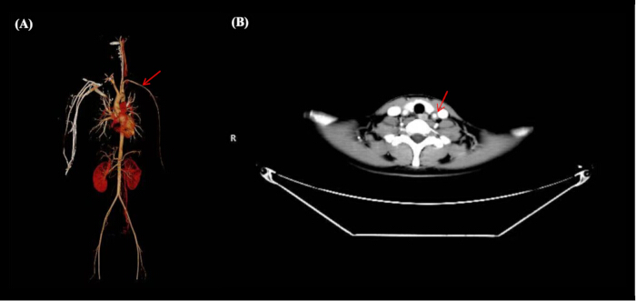
Figure 1: Computed tomography angiography findings demonstrating large-vessel involvement. (A) Three-dimensional volume-rendered image of whole-body computed tomography angiography. Diffuse concentric wall thickening and varying degrees of luminal stenosis were observed in the aortic arch, brachiocephalic trunk, bilateral common carotid arteries, subclavian arteries, and axillary arteries, with the most severe stenosis within the bilateral subclavian arteries. No aneurysm or arterial dilatation was detected, consistent with Takayasu arteritis. (B) Axial contrast-enhanced computed tomography angiography image at the neck level demonstrating wall thickening and mild stenosis of the bilateral common carotid arteries. The axial source image permitted detailed evaluation of cervical vascular involvement. Please click here to view a larger version of this figure.
Discussion
A pediatric case of definite Takayasu arteritis (TAK) in a 14-year-old girl with a previous diagnosis of ulcerative colitis (UC) and biopsy-proven chronic active proctitis was presented. The coexistence of intestinal inflammation and large-vessel vasculitis initially raised the possibility of Behçet's disease (BD), but strict application of validated classification criteria enabled confident exclusion of BD. Several important lessons emerge from this case.
The diagnosis of TAK was established according to the 2010 EULAR/PReS/PRINTO classification criteria, which require mandatory angiographic abnormalities of the aorta or its major branches together with at least one of five supplementary criteria14. The patient fulfilled the mandatory criterion based on computed tomography angiography findings of diffuse wall thickening of the aortic arch and its major branches and also met the supplementary criterion of elevated acute-phase reactants (CRP, 117.63 mg/L; ESR, 93 mm/h). The EULAR/PReS/PRINTO criteria were specifically developed for childhood-onset TAK and have remained the standard classification framework for pediatric TAK over the past decade15. Notably, despite extensive vascular involvement, the patient had no pulse deficits or significant interlimb blood pressure differences. In early or active inflammatory disease, vessel wall thickening may not yet produce hemodynamically significant stenosis, and bilateral symmetric involvement may preserve normal blood pressure. The dynamic changes in arterial wall thickness documented by serial ultrasonography, with worsening during disease flares and improvement following treatment, further supported the diagnosis and illustrated the value of ultrasonography as a noninvasive tool for disease monitoring.
The exclusion of Behçet's disease represents a key aspect of this case. Although BD can involve both the gastrointestinal tract and large vessels, the diagnosis requires recurrent oral ulcers as a mandatory criterion according to the International Study Group (ISG) criteria and the International Criteria for Behçet's Disease (ICBD)13,16,17. The patient had no oral ulcers during repeated physical examinations throughout hospitalization and follow-up and did not develop genital ulcers, ocular lesions, or characteristic skin manifestations such as erythema nodosum or folliculitis. Pathergy testing was not performed. Therefore, BD was excluded with high confidence. This case underscores that isolated large-vessel vasculitis, even when accompanied by nonspecific intestinal inflammation, does not justify a diagnosis of BD in the absence of its defining mucocutaneous manifestations12.
The previous diagnosis of ulcerative colitis also required re-evaluation based on the histopathological findings obtained during the current admission. Ulcerative colitis is histologically characterized by continuous mucosal inflammation extending proximally from the rectum and is associated with crypt abscesses, crypt architectural distortion, and diffuse inflammatory cell infiltration18,19. In contrast, severe chronic active inflammation was most prominent in the rectum, where glandular architectural irregularity and occasional cryptitis were present, but no crypt abscesses or granulomas were identified. Biopsies from the terminal ileum, cecum, and sigmoid colon showed only mild chronic inflammatory changes without cryptitis or crypt abscesses. Although some histopathological abnormalities overlapped with features that may occur in inflammatory bowel disease, the overall distribution and histological pattern were insufficiently specific to confirm classic ulcerative colitis. Intestinal BD typically presents with deep, punched-out ulcers, most commonly in the ileo-caecal region, and may demonstrate submucosal vasculitis, features that were likewise absent20. Accordingly, the rectal lesion was histopathologically diagnosed as severe chronic active proctitis. After integration of the clinical, endoscopic, and histopathological findings, the intestinal condition was classified as chronic active proctitis of unspecified etiology. Mesenteric ischemia secondary to TAK was considered; however, computed tomography angiography demonstrated no stenosis of the mesenteric arteries. Alternative explanations include a limited form of inflammatory bowel disease or an unrelated inflammatory process.
The treatment response observed in this patient is consistent with the current understanding of TAK pathophysiology. Tumor necrosis factor-α (TNF-α) and interleukin-6 (IL-6) are central mediators of vascular inflammation in TAK, and biologic therapies targeting these cytokines have emerged as effective treatment options, particularly for patients with refractory disease or those requiring glucocorticoid-sparing strategies21,22,23. The patient achieved initial remission following treatment with prednisone and adalimumab (anti-TNF-α therapy). Although a disease flare subsequently occurred, increasing the glucocorticoid dose and adding tocilizumab (an anti-IL-6 receptor monoclonal antibody) resulted in rapid clinical and laboratory improvement, with normalization of CRP and ESR levels and stabilization of vascular wall thickening on follow-up ultrasonography. This stepwise biologic strategy, beginning with anti-TNF-α therapy and escalating to anti-IL-6 therapy for refractory disease, is supported by recent multicenter studies and systematic reviews24,25. The favorable therapeutic response also supports the diagnosis of TAK, as activation of both the TNF-α and IL-6 signaling pathways has been demonstrated in active disease26.
The value of serial color Doppler ultrasonography for monitoring disease activity also deserves emphasis. In this patient, carotid artery wall thickness closely paralleled both clinical symptoms and inflammatory marker levels, increasing during disease flares and decreasing following treatment. The 2025 Chinese Expert Consensus for the Diagnosis and Treatment of Pediatric Takayasu Arteritis recommends color Doppler ultrasonography as a preferred imaging modality for monitoring disease activity because of its noninvasive nature, absence of radiation exposure, good reproducibility, and ability to evaluate vascular wall thickness in real time2. The longitudinal ultrasonographic findings presented in this case clearly illustrate the clinical utility of this monitoring strategy.
The preparation of this case report and literature review followed the principles of structured medical writing described by Kasapçopur27. Several limitations should be acknowledged. First, HLA-B51 and HLA-B52 genotyping was not performed. Although the absence of recurrent oral ulcers excludes BD regardless of HLA status, genotyping could have provided additional immunogenetic information28. Second, imaging evaluation was limited to computed tomography angiography and color Doppler ultrasonography. Contrast-enhanced magnetic resonance imaging, which can evaluate vessel wall edema as a marker of active inflammation, was not performed, and the degree of luminal stenosis was estimated qualitatively rather than quantified angiographically. Third, a formal Numano angiographic classification was not assigned, and a standardized disease activity instrument such as the Indian Takayasu Arteritis Score (ITAS) was not used for longitudinal assessment. These limitations restricted detailed disease classification and quantitative assessment of disease activity.
This case highlights the importance of applying validated classification criteria and carefully re-evaluating previous diagnoses in children presenting with large-vessel vasculitis and intestinal inflammation. The patient fulfilled the 2010 EULAR/PReS/PRINTO classification criteria for definite Takayasu arteritis, whereas Behçet's disease was confidently excluded because the mandatory diagnostic features were absent. Histopathological re-evaluation did not provide sufficient evidence to confirm the previous diagnosis of ulcerative colitis. The rectal biopsy was consistent with severe chronic active proctitis, and integration of the clinical, endoscopic, and histopathological findings supported classification of the intestinal condition as chronic active proctitis of unspecified etiology. Together, these findings emphasize the importance of integrating clinical assessment, vascular imaging, and histopathological evaluation to avoid misdiagnosis and to guide appropriate management of pediatric patients with Takayasu arteritis and nonspecific intestinal inflammation.
Disclosures
The authors have nothing to disclose.
Acknowledgements
The authors have no acknowledgments.
Materials
| Name | Company | Catalog Number | Comments |
|---|---|---|---|
| Adalimumab injection | AbbVie | N/A | Anti-TNF-α monoclonal antibody, 40 mg subcutaneous every 2 weeks |
| Alfacalcidol | Teva (or equivalent) | N/A | Vitamin D analogue for bone metabolism support |
| Autoantibody detection system | EUROIMMUN (or equivalent) | N/A | Detection of pANCA and other autoantibodies |
| Calcium carbonate D3 granules | Bayer (or equivalent) | N/A | Supplement for bone protection during steroid therapy |
| Colonoscopy system | Olympus | N/A | Endoscopic evaluation of intestinal lesions |
| Color Doppler ultrasound system | Philips / GE Healthcare | N/A | Used for dynamic monitoring of vascular wall thickness |
| CT angiography (CTA) system | Siemens / GE Healthcare | N/A | Imaging system for evaluation of large-vessel vasculitis |
| Histopathology processing system | Leica Biosystems | N/A | Tissue processing and microscopic evaluation |
| Laboratory blood analyzer | Beckman Coulter / Sysmex | N/A | Used for CBC, CRP, ESR and biochemical tests |
| Mesalazine sustained-release granules | Dr. Falk Pharma (or equivalent) | N/A | Maintenance therapy for intestinal inflammation |
| Omeprazole capsules | AstraZeneca (or equivalent) | N/A | Proton pump inhibitor for gastric protection |
| PCR detection kits (viral panel) | Various suppliers | N/A | Detection of EBV, influenza, RSV, Mycoplasma |
| Prednisone tablets | Pfizer (or equivalent) | N/A | Oral glucocorticoid used for induction and flare control |
| Tocilizumab injection | Roche | N/A | Anti-IL-6 receptor monoclonal antibody, 8 mg/kg IV every 4 weeks |
| T-SPOT.TB test kit | Oxford Immunotec | N/A | Tuberculosis screening |
| Whole-exome sequencing platform | Illumina | N/A | Genetic testing for monogenic disease exclusion |
References
- Aeschlimann FA, Yeung RSM, Laxer RM. An update on childhood-onset Takayasu arteritis. Front Pediatr. 2022;10:872313.
- Rheumatology Group, Pediatrics Branch, Chinese Medical Association; Immunology Group, Pediatrics Branch, Chinese Medical Association; Rheumatology, Immunology and Allergy Group, Pediatrics Branch, Chinese Medical Doctor Association; Childhood Rheumatology and Immunology Alliance. Expert consensus on diagnosis and treatment of childhood Takayasu arteritis (2025). Chin J Pediatr. 2025;63(12):1278-1286.
- Millan P, Gavcovich TB, Abitbol C. Childhood-onset Takayasu arteritis. Curr Opin Pediatr. 2022;34(2):223-228.
- Sahin S, et al. Childhood-onset Takayasu arteritis: A 15-year experience from a tertiary referral center. Int J Rheum Dis. 2019;22(1):132-139.
- Karabacak M, et al. Childhood-onset versus adult-onset Takayasu arteritis: A study of 141 patients from Turkey. Semin Arthritis Rheum. 2021;51(1):192-197.
- Somashekar A, Leung YT. Updates in the diagnosis and management of Takayasu's arteritis. Postgrad Med. 2023;135(Suppl 1):14-21.
- Pang X, Yang H, Wang C, Tian S. Exploring the causal relationship between Takayasu arteritis and inflammatory bowel disease using Mendelian randomization. Immunol Res. 2024;72(4):707-713.
- İlgen U, Emmungil H, Tezel HA. On the coexistence of Takayasu arteritis and inflammatory bowel disease. J Clin Rheumatol. 2021;27(8 Suppl):S864.
- Terao C, et al. Two susceptibility loci to Takayasu arteritis reveal a synergistic role of the IL12B and HLA-B regions in a Japanese population. Am J Hum Genet. 2013;93(2):289-297.
- Khalaji A, Mahdian T, Eftekharsadat AT, Jafarpour M. Sudden onset of symptoms in concurrent Takayasu arteritis and ulcerative colitis: A case report with neurological complications. Clin Case Rep. 2024;12(3):e8673.
- Shirai T. Common autoantibody among Takayasu arteritis and ulcerative colitis: A possible pathophysiology that includes the gut-vessel connection in vascular inflammation. JMA J. 2023;6(3):265-273.
- Lavalle S, et al. Behçet's disease: Pathogenesis, clinical features, and treatment approaches: A comprehensive review. Medicina (Kaunas). 2024;60(4):562.
- International Study Group for Behçet's Disease. Criteria for diagnosis of Behçet's disease. Lancet. 1990;335(8697):1078-1080.
- Ozen S, et al. EULAR/PRINTO/PRES criteria for Henoch-Schönlein purpura, childhood polyarteritis nodosa, childhood Wegener granulomatosis and childhood Takayasu arteritis: Ankara 2008. Part II: Final classification criteria. Ann Rheum Dis. 2010;69(5):798-806.
- de Graeff N, et al. European consensus-based recommendations for the diagnosis and treatment of rare pediatric vasculitides: The SHARE initiative. Rheumatology (Oxford). 2019;58(4):656-671.
- Davatchi F, et al. Behcet's disease: Epidemiology, clinical manifestations, and diagnosis. Expert Rev Clin Immunol. 2017;13(1):57-65.
- International Team for the Revision of the International Criteria for Behçet's Disease. The International Criteria for Behçet's Disease (ICBD): A collaborative study of 27 countries on the sensitivity and specificity of the new criteria. J Eur Acad Dermatol Venereol. 2014;28(3):338-347.
- Voelker R. What is ulcerative colitis? JAMA. 2024;331(8):716.
- Bhalla A, et al. Inflammatory bowel disease in children: Current diagnosis and treatment strategies. Cureus. 2025;17(2):e78462.
- Skef W, Hamilton MJ, Arayssi T. Gastrointestinal Behçet's disease: A review. World J Gastroenterol. 2015;21(13):3801-3812.
- Regola F, et al. Novel therapies in Takayasu arteritis. Front Med (Lausanne). 2021;8:814075.
- Mekinian A, et al. Efficacy and safety of TNF-α antagonists and tocilizumab in Takayasu arteritis: Multicentre retrospective study of 209 patients. Rheumatology (Oxford). 2022;61(4):1376-1384.
- Salvarani C, et al. Rescue treatment with tocilizumab for Takayasu arteritis resistant to TNF-α blockers. Clin Exp Rheumatol. 2012;30(1 Suppl 70):S90-S93.
- Sener S, et al. Treatment of childhood-onset Takayasu arteritis: Switching between anti-TNF and anti-IL-6 agents. Rheumatology (Oxford). 2022;61(12):4885-4891.
- Kang L, et al. Systematic review and meta-analysis of the current literature on tocilizumab in patients with refractory Takayasu arteritis. Front Immunol. 2023;14:1084558.
- Wen D, et al. Biomarkers in Takayasu arteritis. Int J Cardiol. 2023;380:413-417.
- Kasapçopur Ö. Tips for writing a medical review. Turk Arch Pediatr. 2026;61(2):92-94.
- de Menthon M, et al. HLA-B51/B5 and the risk of Behçet's disease: A systematic review and meta-analysis of case-control genetic association studies. Arthritis Rheum. 2009;61(10):1287-1296.