This review synthesizes evidence on mitochondrial quality control (biogenesis, dynamics, and mitophagy) across spermatogenesis stages, linking metabolic shifts and structural remodeling to male infertility and identifying methodological gaps to guide future research.
Review Article
Mitochondrial Quality Control: A Gatekeeper of Spermatogenesis
In This Article
Summary
Abstract
Male infertility is rising worldwide. Spermatogenesis is energetically demanding and tightly coupled to mitochondrial quality control (MQC)—including biogenesis, fusion–fission dynamics, and mitophagy. Here we synthesize current evidence across developmental stages of germ cells, highlighting stage-specific shifts from glycolysis-supported ATP production to oxidative phosphorylation (OXPHOS)-dominated metabolism and remodeling of mitochondrial architecture into the midpiece sheath. We summarize genetic and functional data implicating OPA1/MFN1-2 in inner/outer membrane fusion, DRP1/FIS1/MFF in fission and sheath organization, and the roles of PINK1–Parkin-dependent and -independent mitophagy in mitochondrial quality control, while acknowledging that cytoplasm removal and acrosome maintenance during spermiogenesis may involve general autophagy rather than mitophagy specifically. Mitochondrial dysfunction—excess reactive oxygen species (ROS), mitochondrial membrane potential (MMP) collapse, dysregulated dynamics, or defective mitophagy—impairs spermatogenesis and sperm function. We also identify methodological gaps that limit translation, particularly the lack of standardized phenotyping and human genetic evidence. Clarifying MQC causality and druggability may support future diagnostic or therapeutic research in male infertility.
Introduction
Sperm quality is declining worldwide, and the incidence of male infertility caused by abnormal sperm quality is increasing1. One recent meta-analysis showed that between 1973 and 2011, sperm counts among men from North America, Europe, Australia, and New Zealand declined by 50–60%2, and this decline has accelerated since 20003. The accelerated decline in sperm quality has been reported worldwide2,3,4,5,6. The reasons for this decline require further study and targeted measures to protect male fertility.
Spermatogenesis occurs in the seminiferous tubules of the testis. It is a wave-like process involving specific combinations of spermatogenic cells along the long axis of the seminiferous tubules, ultimately leading spermatogonial stem cells (SSCs) to proliferate and differentiate into spermatozoa7. Spermatogenesis in mammals is one of the most complex and lengthy differentiation processes in biology. In humans, a full spermatogenic cycle spans 64–74 days, with each seminiferous epithelial cycle lasting 16 days. This intricate process involves significant changes in the shape, number, and distribution of mitochondria8.
Mitochondrial quality control (MQC)—the coordinated processes of mitochondrial biogenesis, fusion, fission, and selective mitophagy—maintains organelle integrity under varying bioenergetic demands8,9. In the testis, MQC is stage-specific and supports the transition from proliferating spermatogonia to meiosis and spermiogenesis, culminating in the assembly of a tightly packed mitochondrial sheath in the sperm midpiece9. Figure 1 presents the mechanism of MQC during spermatogenesis. Disruption of MQC components can lead to defective spermatogenesis and subfertility across model organisms and humans. This review integrates current mechanistic insights and outlines translational opportunities.
Review and Perspective
Spermatogenesis and mitochondrial energy metabolism
Spermatogenesis comprises three sequential stages: mitotic proliferation, meiosis, and spermiogenesis, involving the proliferation of spermatogonia, their differentiation into spermatocytes, meiotic division, maturation of spermatids, and spermiation into the seminiferous lumen. Spermatogonia are categorized into reserve and renewing types. Some spermatogonia are retained as stem cells, while others participate in spermatogenesis through differentiation. Additionally, some undergo programmed cell death, such as apoptosis. Diploid primary spermatocytes undergo two rounds of meiotic division to differentiate into haploid spermatids. During spermiogenesis, round spermatids no longer divide and transform into highly elongated spermatids, which eventually develop into mature spermatozoa.
Spermatogenesis requires coordinated interactions between germ cells (including spermatogonia, spermatocytes, spermatids, and spermatozoa) and supporting Sertoli cells, as well as the maintenance of blood–testis barrier integrity10,11. The blood–testis barrier, formed by tight junctions between adjacent Sertoli cells, divides the seminiferous epithelium into basal and adluminal compartments, thereby creating and maintaining a specialized microenvironment essential for germ cell development11,12. Consequently, spermatogenesis—from spermatogonial stem cells to mature spermatozoa—takes place within this unique testicular microenvironment. The process of sperm production heavily relies on mitochondrial function13. As the energy metabolism center in eukaryotic cells, mitochondria are considered essential for sperm production. The generation of sperm is highly dependent on the energy supply and signaling functions of mitochondria. In developing germ cells, mitochondria not only provide essential ATP for spermatogenesis but also influence the process by participating in intracellular signaling, cell cycle regulation, and programmed cell death13. In Leydig cells, mitochondria play a crucial role in steroid hormone production, and mitochondrial ATP synthesis is necessary for testosterone production in these testicular interstitial cells14. In addition, as the only organelle with independent DNA, mitochondrial DNA is closely related to sperm quality. Therefore, understanding mitochondrial status and its regulatory mechanisms is important for revealing the process of spermatogenesis and maintaining male fertility.
Energy metabolism is crucial for spermatogenesis, which requires a significant amount of ATP. The two primary pathways for ATP production are mitochondrial oxidative phosphorylation (OXPHOS) and glycolysis. ATP supply is stage-dependent; while OXPHOS becomes increasingly important during meiosis and midpiece formation, glycolysis substantially contributes to ATP production, particularly in mature spermatozoa. Glycolysis also supports sperm maturation, motility, and passage through the female reproductive tract by providing a flexible and rapid energy supply.
However, in spermatogonia and early spermatocytes, mitochondria are relatively small and exhibit limited OXPHOS activity. As spermatogenesis progresses to later stages, such as spermatocytes, spermatids, and spermatozoa, mitochondria undergo remodeling and their OXPHOS activity becomes more spatially concentrated and efficient. Additionally, because of the blood–testis barrier, available substrates vary across stages. Spermatogonia, residing in the basal compartment of the seminiferous epithelium, can access circulating glucose and produce energy through glycolysis. In contrast, spermatocytes and spermatids in the adluminal compartment are separated from the blood and interstitium by the blood–testis barrier and rely on Sertoli cells to absorb glucose through glucose transporters (GLUTs), converting it into pyruvate via glycolysis. Pyruvate is then converted into lactate by lactate dehydrogenase (LDH). Lactate is transported to spermatocytes via monocarboxylate transporters (MCTs), where it is converted back into pyruvate by LDH, which can then be converted into acetyl-CoA by pyruvate dehydrogenase (PDH) for entry into the tricarboxylic acid (TCA) cycle. Finally, pyruvate is transported to mitochondria via the mitochondrial pyruvate carrier to promote OXPHOS.
Impairment of spermatogenesis by mitochondrial dysfunction
During spermiation, mature spermatozoa are released from Sertoli cells into the lumen of the seminiferous tubules, followed by transport to the epididymis. This process requires a substantial energy supply from mitochondria. When mitochondrial function is impaired, spermatogenesis is hindered. Key signs of mitochondrial dysfunction include excessive production of reactive oxygen species (ROS), changes in mitochondrial membrane potential (MMP), imbalances in mitochondrial fission and fusion dynamics, impaired mitochondrial biogenesis, defective mitophagy, and calcium ion homeostasis disruption.
The process by which mitochondria produce ATP through OXPHOS is also accompanied by ROS production. Mitochondria are the main source of ROS, which are mainly generated at respiratory complexes I and III of the mitochondrial electron transport chain (ETC)15. Under normal physiological conditions, appropriate levels of ROS are involved in signaling pathways regulating spermatogenesis. However, excessive ROS can lead to redox imbalance, causing oxidative stress and impairing spermatogenesis. Therefore, the balance between mitochondrial energy metabolism and ROS production is a key factor in ensuring spermatogenesis.
Efficient mitochondrial OXPHOS requires the entry of respiratory carbon substrates (e.g., pyruvate, fatty acids) through the mitochondrial outer membrane into the intermembrane space. These substrates are then transported to the matrix via carriers located in the inner mitochondrial membrane, where they participate in the TCA cycle. In the TCA cycle, these carbon substrates undergo oxidative breakdown, producing nicotinamide adenine dinucleotide (NADH) and flavin adenine dinucleotide (FADH2). These products donate electrons to the ETC and drive OXPHOS for ATP synthesis from ADP and inorganic phosphate (Pi). Interruptions in the TCA cycle or ETC pathways can impair sperm production16. MMP, as an indicator of cellular energy status and mitochondrial function, has been shown to be associated with sperm motility17 and overall fertilizing capability18.
Under mitochondrial stress, fission can segregate damaged mitochondrial segments, whereas fusion can promote content mixing and functional complementation. Damaged or excess mitochondria may then be degraded and recycled through mitophagy. Together, mitochondrial biogenesis, fission, fusion, and mitophagy constitute mitochondrial quality control (MQC), which supports mitochondrial function and remodeling19.
Spermatogenesis involves the differentiation of spermatogonia into spermatozoa, which not only results in significant changes in cell morphology but also requires the support and regulation of energy metabolism. As the powerhouses of cells, mitochondria play a crucial role in key aspects of spermatogenesis, including energy supply, signal transduction, and programmed cell death. During spermatogenesis, the metabolic demands of germ cells vary at different stages of development. Mitochondrial number and morphology undergo significant changes during spermatogenesis. During sperm formation, many mitochondria are discarded, while retained mitochondria are organized in the sperm midpiece to form a mitochondrial sheath around the axoneme and associated flagellar structures. Therefore, during spermatogenesis, mitochondria in various testicular cells continuously undergo fusion and fission, requiring an efficient and precise MQC mechanism to ensure their normal function and support the high energy demands of spermatogenesis. As sperm are produced, both the morphology and number of mitochondria undergo significant changes, and MQC plays a critical role in this process. The key target proteins involved in MQC are listed in Table 1.
Spermiogenesis and mitochondrial dynamics
Mitochondria are highly dynamic organelles whose morphology, size, and subcellular distribution are continually remodeled through coordinated fusion and fission—a process collectively termed mitochondrial dynamics. Under normal physiological conditions, fusion and fission exist in a dynamic equilibrium that maintains mitochondrial homeostasis, facilitates functional complementation, and ensures proper mitochondrial inheritance during cell division. Disruption of this balance—whether by genetic ablation, post-translational modification defects, or pathological stress—impairs mitochondrial function, compromises energy production, and ultimately hinders spermatogenesis. The precise regulation of mitochondrial dynamics is particularly critical in germ cells, as spermatogenesis entails dramatic metabolic shifts and extensive morphological remodeling that demand stage-specific adaptation of mitochondrial architecture and activity.
Mitochondrial fusion is mediated by MFN1 and MFN2 on the outer mitochondrial membrane and OPA1 on the inner membrane, facilitating content mixing, functional complementation, and maintenance of mitochondrial DNA integrity. Fusion is not merely a defensive response but a context-dependent mechanism that supports mitochondrial inheritance and metabolic adaptation during germ cell differentiation. During spermatogonial differentiation, MFN expression increases, and loss of either MFN1 or MFN2 leads to DNA oxidation and apoptosis in spermatogonia and spermatocytes, resulting in male infertility20,21. MFN1 mutations cause testicular atrophy in mice, with marked reduction in spermatocytes and failed sperm production, whereas undifferentiated spermatogonia remain unaffected, suggesting that MFN1 is required for meiotic progression20. MFN2, in contrast, regulates both mitochondrial and endoplasmic reticulum functions, playing a distinct role in spermatogenesis21.
Mitochondrial fission is orchestrated by DRP1, which is recruited to the outer mitochondrial membrane via adaptor proteins including FIS1, MFF, MID49, and MID51. Upon recruitment, DRP1 assembles into ring-like oligomers that constrict and divide mitochondria in a GTP-dependent manner. The process involves three main steps: DRP1 activation by phosphorylation, recruitment to the outer membrane via interactions with FIS1, MFF, MID49, and MID51, and assembly into a ring structure that encircles and constricts mitochondria, consuming GTP and producing two separate organelles. Fission is not simply a mechanism for clearing depolarized mitochondria or meeting increased energy demands; rather, it is a coordinated process that regulates mitochondrial distribution, segregation of damaged components, inheritance during cell division, and remodeling of mitochondrial architecture. Mice lacking DRP1 are embryonic lethal22,23; MFF-deficient mice exhibit reduced fertility and sperm count, providing a valuable model for studying mitochondrial fission in male germ cell development24. Mitochondrial fission is essential for proper formation of the mitochondrial sheath during spermiogenesis.
Spermatogenesis is energetically demanding, relying primarily on glycolysis in spermatogonia and OXPHOS in spermatocytes and spermatids. OXPHOS is particularly critical during meiosis, and its efficiency depends on intact mitochondrial dynamics25,26. Following meiosis, MFF and other fission proteins regulate mitochondrial fission and participate in the formation of the sperm mitochondrial sheath24. During spermiogenesis, some mitochondria are selectively retained and remodeled into the sheath, while excess mitochondria and cytoplasmic components are eliminated via autophagy27. Proteins such as OPA1, DRP1, and MFF may represent candidate research targets for enhancing sperm function, but their therapeutic relevance requires further validation. Further investigation of MQC in human spermatogenesis may facilitate future diagnostic or therapeutic research in male infertility.
Spermatogenesis and mitochondrial autophagy
Mitophagy plays a critical role in spermatogenesis28. Mitophagy primarily involves the engulfment of damaged mitochondria by autophagosomes. The autophagosomes containing damaged mitochondria then fuse with lysosomes, and the mitochondria are degraded through lysosomal action, thereby clearing damaged mitochondria and achieving quality control. The classic pathway of mitochondrial autophagy is the PINK1/Parkin pathway, and PINK1/Parkin-independent pathways have also been described29.
Autophagy helps maintain acrosome integrity, spermatid polarization, and cytoplasmic component degradation during spermatogenesis, although these processes reflect general autophagy rather than selective mitophagy unless mitochondrial clearance is specifically demonstrated. Studies have found that mitophagy is closely related to mitochondrial dynamics, and mitochondrial fission may facilitate mitophagy to remove excess or damaged mitochondria30. FIS1, although a mitochondrial fission protein, is also involved in mitophagy during spermatogenesis31. RAB7A is involved in mitophagosome formation and autophagosome maturation32. FIS1 interacts with mitochondrial GTPase-activating protein TBC1D1533,34. TBC1D15 regulates RAB7A activity, which is involved in the fusion of vesicles and lysosomes and the maturation of autophagosomes34,35. When FIS1 is inactivated, RAB7A can remain active on the mitochondrial surface, leading to dysregulated autophagosome membrane growth and tubulation; LC3 aggregation can be observed by optical microscopy35. Mitophagy can also occur independently of DRP1-mediated mitochondrial fission, so fission should not be treated as obligatory for mitochondrial clearance35.
During spermiogenesis, spermatids are stripped of excess cellular components, and studies have found that autophagosomes are most abundant in spermatids36. The core autophagy gene Atg7 has been linked to spermatid polarization and cytoplasm removal during spermatogenesis; however, early germ cell types appear less affected in this context27,36. In addition, mitophagy is enhanced in damaged testicular models, including Leydig cells29, spermatogonial stem cells/progenitors37,38, Sertoli cells39, and immature rat testis exposed to toxicants40. In spermatogenesis, mitochondrial autophagy is linked to apoptosis and other modes of programmed cell death, thereby affecting sperm production in the testis41,42.
Bibliometric context
A focused bibliometric analysis of the Web of Science Core Collection from 1970 to 2023 was performed using the search terms “spermatogenesis” and “MQC”; non-mammalian studies and irrelevant records were excluded, and VOSviewer 1.6.20 was used for visualization43. After screening, 1,019 articles from 1,217 institutions in 66 countries and 5,705 authors were retained. Publications increased steadily (Figure 2A). The United States and China contributed the most articles (212 and 196, respectively), and the United States had the highest citation count (12,295; Figure 2B). Institutional and author networks are summarized in Figure 2C,D.
Keyword co-occurrence analysis (frequency > 7) indicated emphasis on sperm motility, oxidative stress, apoptosis/programmed cell death, DNA damage, lipid peroxidation, and sperm quality (Figure 3). These bibliometric patterns support, but do not replace, the mechanistic review: the field is concentrated around oxidative damage and motility outcomes, whereas direct human evidence linking stage-specific MQC pathways to infertility remains limited.
Conclusions
With the advancement of biotechnology, research on male fertility has shifted from descriptive morphology and motility toward the molecular and cellular mechanisms of spermatogenesis. Mitochondria are central to this process: they generate ATP, support remodeling during meiosis and spermiogenesis, and are highly sensitive to oxidative and environmental insults44. To maintain functionality under such stress, mitochondria rely on MQC, which integrates biogenesis, fusion–fission dynamics, and mitophagy. Disruption of these pathways impairs germ-cell development, sperm midpiece architecture, and ultimately fertility. In conclusion, the available evidence supports a stage-specific model in which mitochondrial dynamics and mitophagy help coordinate energy production, organelle remodeling, and removal of damaged components during spermatogenesis. However, direct human evidence and standardized phenotyping remain limited, so MQC-related proteins should be viewed as candidate research targets that require further validation rather than established therapeutic targets.
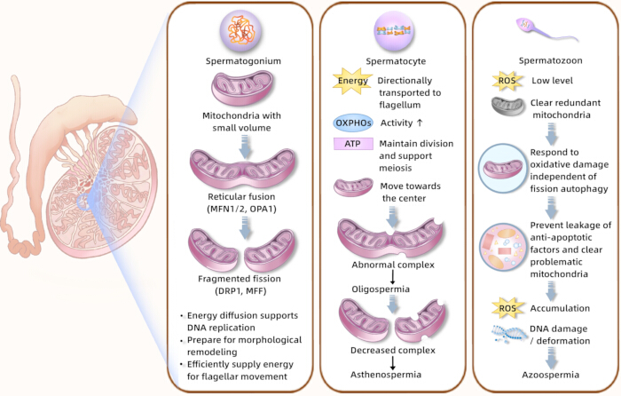
Figure 1. The mechanism of mitochondrial quality control during spermatogenesis. Stage-resolved energy metabolism and mitochondrial remodeling in spermatogenesis. The schematic shows Sertoli–germ cell metabolic coupling, glucose transporters, monocarboxylate transporters, ATP production, the transition from glycolysis-supported metabolism to oxidative phosphorylation during meiosis and spermiogenesis, and reactive oxygen species-related mitochondrial damage. Abbreviations: ATP = adenosine triphosphate; OXPHOS = oxidative phosphorylation; ROS = reactive oxygen species. Please click here to view a larger version of this figure.
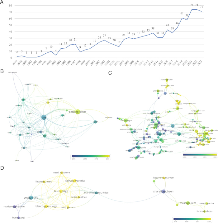
Figure 2. National, institutional, author publications and collaborative analysis. (A) Annual number of publications in the field of spermatogenesis and MQC. (B) Temporal distribution map of international collaborative publications in the field of spermatogenesis and MQC. (C) Temporal distribution map of institutional collaborations in the field of spermatogenesis and MQC. (D) Temporal distribution map of author publications in the field of spermatogenesis and MQC. The nodes indicate the number of publications; node color indicates publication time from earlier to more recent; and lines represent collaborative relationships, with thicker lines indicating closer collaboration. Abbreviation: MQC = mitochondrial quality control. Please click here to view a larger version of this figure.
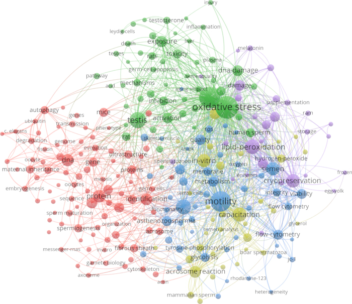
Figure 3. High-frequency keyword co-occurrence chart. The nodes represent high-frequency keywords; node size represents frequency; node color indicates clustering, with the same color indicating more closely related topics; and lines represent relationships between keywords, with thicker lines indicating closer relationships. Please click here to view a larger version of this figure.
| MQC process | Target protein |
| Mitochondrial fusion | Optic atrophy 1 (OPA1) |
| Mitofusin 1 (MFN1) | |
| Mitofusin 2 (MFN2) | |
| Mitochondrial fission | Dynamin-related protein 1 (DRP1) |
| Mitochondrial fission 1 protein (FIS1) | |
| Mitochondrial fission factor (MFF) | |
| Mitophagy | PTEN-induced putative kinase 1 (PINK1) |
| Ubiquitin-protein ligase Parkin (E3 ubiquitin-protein ligase Parkin) |
Table 1: Key target proteins in mitochondrial quality control. The table lists representative proteins involved in mitochondrial fusion, mitochondrial fission, and mitophagy during spermatogenesis.
Disclosures
The authors have no conflicts of interest to declare.
Acknowledgements
This work was supported by the Ningxia Natural Science Foundation Project (2025AAC030481, 2024AAC03715), National Natural Science Foundation of China (82560939, 82505601), Ningxia Key Research and Development Program (2025FRG05005, 2025BEG2008), Beijing Natural Science Foundation (7254520), China Postdoctoral Science Foundation (2025T181076), Young Elite Scientists Sponsorship Program by China Association of Science and Technology (YESS20240193), Young Talents Support Program of the China Association of Chinese Medicine (CACM-2024-QNRC2-B17), High-Level Key Discipline Construction Project of Traditional Chinese Medicine by the National Administration of Traditional Chinese Medicine (zyyzdxk-2023238).
References
- Esteves SC. Evolution of the World Health Organization semen analysis manual: where are we? Nat Rev Urol. 2022;19(7):439-46.
- Levine H, et al. Temporal trends in sperm count: a systematic review and meta-regression analysis of samples collected globally in the 20th and 21st centuries. Hum Reprod Update. 2023;29(2):157-76.
- Cipriani S, et al. Trend of change of sperm count and concentration over the last two decades: a systematic review and meta-regression analysis. Andrology. 2023;11(6):997-1008.
- Lv MQ, et al. Temporal trends in semen concentration and count among 327 373 Chinese healthy men from 1981 to 2019: a systematic review. Hum Reprod. 2021;36(7):1751-75.
- Li Y, et al. Trends in sperm quality by computer-assisted sperm analysis of 49 189 men during 2015-2021 in a fertility center from China. Front Endocrinol (Lausanne). 2023;14:1194455.
- Bonde JP, te Velde ER. Male factor infertility: declining sperm counts: the never-ending story. Nat Rev Urol. 2017;14(11):645-6.
- Hermo L, Pelletier RM, Cyr DG, Smith CE. Surfing the wave, cycle, life history, and genes/proteins expressed by testicular germ cells. Part 1: background to spermatogenesis, spermatogonia, and spermatocytes. Microsc Res Tech. 2010;73(4):241-78.
- Costa J, et al. Mitochondria quality control and male fertility. Biology (Basel). 2023;12(6):827.
- Varuzhanyan G, Chan DC. Mitochondrial dynamics during spermatogenesis. J Cell Sci. 2020;133(14):jcs235937.
- Wu S, Yan M, Ge R, Cheng CY. Crosstalk between Sertoli and germ cells in male fertility. Trends Mol Med. 2020;26(2):215-31.
- Mruk DD, Cheng CY. The mammalian blood-testis barrier: its biology and regulation. Endocr Rev. 2015;36(5):564-91.
- Ma Y, Ma QW, Sun Y, Chen XF. The emerging role of extracellular vesicles in the testis. Hum Reprod. 2023;38(3):334-51.
- Vertika S, Singh KK, Rajender S. Mitochondria, spermatogenesis, and male infertility: an update. Mitochondrion. 2020;54:26-40.
- Midzak A, et al. Mitochondrial protein import and the genesis of steroidogenic mitochondria. Mol Cell Endocrinol. 2011;336(1-2):70-9.
- Brand MD. Mitochondrial generation of superoxide and hydrogen peroxide as the source of mitochondrial redox signaling. Free Radic Biol Med. 2016;100:14-31.
- Boguenet M, et al. Mitochondria: their role in spermatozoa and in male infertility. Hum Reprod Update. 2021;27(4):697-719.
- Barbagallo F, et al. Evaluation of sperm mitochondrial function: a key organelle for sperm motility. J Clin Med. 2020;9(2):363.
- Gallon F, Marchetti C, Jouy N, Marchetti P. The functionality of mitochondria differentiates human spermatozoa with high and low fertilizing capability. Fertil Steril. 2006;86(5):1526-30.
- Gan Z, Fu T, Kelly DP, Vega RB. Skeletal muscle mitochondrial remodeling in exercise and diseases. Cell Res. 2018;28(10):969-80.
- Zhang J, et al. GASZ and mitofusin-mediated mitochondrial functions are crucial for spermatogenesis. EMBO Rep. 2016;17(2):220-34.
- Chen W, et al. MFN2 plays a distinct role from MFN1 in regulating spermatogonial differentiation. Stem Cell Reports. 2020;14(5):803-17.
- Ishihara N, et al. Mitochondrial fission factor Drp1 is essential for embryonic development and synapse formation in mice. Nat Cell Biol. 2009;11(8):958-66.
- Wakabayashi J, et al. The dynamin-related GTPase Drp1 is required for embryonic and brain development in mice. J Cell Biol. 2009;186(6):805-16.
- Varuzhanyan G, et al. Mitochondrial fission factor (Mff) is required for organization of the mitochondrial sheath in spermatids. Biochim Biophys Acta Gen Subj. 2021;1865(5):129845.
- Miao J, et al. MFN1 and MFN2 are dispensable for sperm development and functions in mice. Int J Mol Sci. 2021;22(24):13507.
- Arslan NP, Keles ON, Gonul-Baltaci N. Effect of titanium dioxide and silver nanoparticles on mitochondrial dynamics in mouse testis tissue. Biol Trace Elem Res. 2022;200(4):1650-8.
- Shang Y, et al. Autophagy regulates spermatid differentiation via degradation of PDLIM1. Autophagy. 2016;12(9):1575-92.
- Vargas JNS, et al. The mechanisms and roles of selective autophagy in mammals. Nat Rev Mol Cell Biol. 2023;24(3):167-85.
- Yi L, et al. Cadmium-induced apoptosis of Leydig cells is mediated by excessive mitochondrial fission and inhibition of mitophagy. Cell Death Dis. 2022;13(11):928.
- Losón OC, Song Z, Chen H, Chan DC. Fis1, Mff, MiD49, and MiD51 mediate Drp1 recruitment in mitochondrial fission. Mol Biol Cell. 2013;24(5):659-67.
- Varuzhanyan G, et al. Fis1 ablation in the male germline disrupts mitochondrial morphology and mitophagy, and arrests spermatid maturation. Development. 2021;148(16):dev199686.
- Tan EHN, Tang BL. Rab7a and mitophagosome formation. Cells. 2019;8(3):224.
- Onoue K, et al. Fis1 acts as a mitochondrial recruitment factor for TBC1D15 that is involved in regulation of mitochondrial morphology. J Cell Sci. 2013;126(1):176-85.
- Yamano K, et al. Mitochondrial Rab GAPs govern autophagosome biogenesis during mitophagy. Elife. 2014;3:e01612.
- Yamashita SI, et al. Mitochondrial division occurs concurrently with autophagosome formation but independently of Drp1 during mitophagy. J Cell Biol. 2016;215(5):649-65.
- Yang P, et al. In vivo autophagy and biogenesis of autophagosomes within male haploid cells during spermiogenesis. Oncotarget. 2017;8(34):56791-801.
- Li T, et al. YTHDF2 controls hexavalent chromium-induced mitophagy through modulating Hif1α and Bnip3 decay via the m6A/mRNA pathway in spermatogonial stem cells/progenitors. Toxicol Lett. 2023;377:38-50.
- Lv Y, et al. Melatonin attenuates chromium (VI)-induced spermatogonial stem cell/progenitor mitophagy by restoration of METTL3-mediated RNA N6-methyladenosine modification. Front Cell Dev Biol. 2021;9:684398.
- Eid N, Ito Y, Otsuki Y. Enhanced mitophagy in Sertoli cells of ethanol-treated rats: morphological evidence and clinical relevance. J Mol Histol. 2012;43(1):71-80.
- Hong Y, et al. X-box binding protein 1 caused an imbalance in pyroptosis and mitophagy in immature rats with di-(2-ethylhexyl) phthalate-induced testis toxicity. Genes Dis. 2024;11(2):935-51.
- Su Y, et al. Mutual promotion of apoptosis and autophagy in prepubertal rat testes induced by joint exposure of bisphenol A and nonylphenol. Environ Pollut. 2018;243:693-702.
- Quan C, et al. Prenatal bisphenol A exposure leads to reproductive hazards on male offspring via the Akt/mTOR and mitochondrial apoptosis pathways. Environ Toxicol. 2017;32(3):1007-23.
- Chen C, Song M. Visualizing a field of research: a methodology of systematic scientometric reviews. PLoS One. 2019;14(10):e0223994.
- Yang M, et al. Melatonin promotes male reproductive performance and increases testosterone synthesis in mammalian Leydig cells. Biol Reprod. 2021;104(6):1322-36.