This protocol is intended to demonstrate how clinicians can evaluate the torque-velocity relationship following anterior cruciate ligament reconstruction.
A subscription to JoVE is required to view this content. Sign in or start your free trial.
Method Article
Quantifying Recovery After Anterior Cruciate Ligament Reconstruction Using the Torque- Velocity Relationship
In This Article
Summary
Abstract
Following anterior cruciate ligament reconstruction (ACLR), patients often display persistent deficits in quadriceps muscle strength that reduce quality of life and lead to long-term functional impairment. Historically, post-operative assessments of muscle function have focused on maximal isometric torque or peak torque at a single isokinetic velocity, neglecting the variety of demands placed on a muscle group across activities and the precision needed to guide clinical decision-making. The goal of this demonstration is to illustrate how isokinetic testing at different angular velocities can be used to characterize the torque-velocity relationship following ACLR. The protocol includes participant preparation, dynamometer setup, range-of-motion configuration, and alignment of key testing components, including chair position and dynamometer arm length. Strength assessment consists of isometric knee extension testing and concentric isokinetic knee extension and flexion testing at 60°/s, 180°/s, and 240°/s. For the two-point method, torque-velocity is calculated as the difference in normalized torque production between a slow and a fast testing velocity (e.g., 60°/s and 180°/s). For full multi-velocity characterization, peak torque values obtained at each velocity are exported to a spreadsheet or statistical software and analyzed using regression techniques. Previous results have demonstrated that patients following ACLR have knee extensor torque-velocity relationships of the involved limb about half the magnitude of the contralateral limb, using the two-point method, and a lesser slope than the contralateral limb using multiple velocity characterization. The torque-velocity relationship may identify persistent deficits not captured by single-velocity testing and can be used to monitor recovery over time.
Introduction
Anterior cruciate ligament injuries are among the most common in sports, resulting in billions of dollars in healthcare costs in the US annually1. Despite advances in surgical technique and clinical care, following anterior cruciate ligament reconstruction (ACLR), patients regularly develop persistent quadriceps muscle weakness2,3. Deficits in quadriceps muscle function are associated with decreased quality of life, decreased ability to complete activities of daily living, and decreased overall function4,5,6,7. Given the importance of quadriceps function, test batteries including objective assessment of quadriceps are a key piece of multidimensional test batteries used during return-to-sport decision-making8.
Current clinical assessments of muscle function evaluate maximal strength as a measure of muscle capacity, but muscle performance is also influenced by additional physiological principles8,9,10. The force-velocity relationship is a basic physiological principle defined by the inverse relationship between contraction velocity and force production11,12. This model has been demonstrated in a number of different in vitro and in vivo studies, as well as in human whole muscle, via the torque-velocity relationship12,13. The torque-velocity relationship has previously been characterized via isokinetic dynamometry and has been shown to be altered in patients with a history of ACLR as well as due to immobilization, aging, and other pathologies14,15,16,17,18. Given the widespread use of testing at one or two velocities for clinical use, this method was developed to monitor changes in an underlying physiological principle of muscle.
The use of isometric testing and isokinetic testing following ACLR is widespread and widely accepted19,20. Focusing solely on torque production at a single velocity via isometric or isokinetic testing at a single velocity may not be representative of the range of muscular demands during activities of daily living and high-level sport14,21,22. The two-point method of characterizing the torque-velocity relationship has been found reliable and may be clinically useful as long as the chosen velocities have a difference in angular velocity of at least 90°/s15,23. This protocol demonstrates two velocity isokinetic tests for clinical use and the use of multiple velocities to characterize the torque-velocity relationship in greater detail. While this protocol focuses on the analysis of peak torque production, the dynamometer will also allow full export of time-series data for time-series analyses. Peak torque is a measure of maximal force or power generation, but variables such as rate of torque development, time to peak torque, and angle of peak torque demonstrate additional methods to determine participants’ ability to generate force quickly and respond to varying potential demands24,25,26,27.
Access restricted. Please log in or start a trial to view this content.
Protocol
All representative data were collected following institutional review board approval, and informed consent was obtained from all participants. The protocol below has been performed on participants aged at least 12 years, and 4 months post-ACLR, with no restrictions based on graft type. Exclusion criteria were pregnancy, serious infection near the lower limb, known muscular abnormalities, and a history of cardiopulmonary disorder.
1. Method 1: Participant preparation and dynamometer setup
NOTE: A consistent dynamometer setup is critical to ensure valid, reproducible torque evaluation across sessions and participants. As recommended by the manufacturer, the dynamometer should be calibrated monthly. The torque, velocity, and position data in this protocol were exported at 100 Hz, and no additional smoothing or filtering was applied beyond the manufacturer’s signal processing. This protocol should not be completed before a patient has completed open-chain knee extension during rehabilitation.
- Begin all procedures with the uninvolved limb.
- Have the participant complete a standardized warm-up consisting of approximately 5 min of treadmill walking at a standardized pace of 3.0 mph.
- Turn the dynamometer on and attach the dynamometer arm.
- Position the chair at a back angle of 85° and set the dynamometer rotation to 40° consistent with manufacturer standards.
- Seat the participant with hips flexed to approximately 85° and instruct them to maintain an upright posture and avoid grabbing the handles of the dynamometer during testing.
- Adjust the back translation so that the popliteal fossa is positioned approximately 3–4 cm anterior to the edge of the seat.
- Adjust chair position and dynamometer arm orientation until the mechanical axis aligns with the lateral femoral condyle.
- Position the dynamometer arm resistance pad approximately 5 cm proximal to the lateral malleolus on the anterior portion of the lower leg28,29.
- Secure the test limb to the dynamometer arm. Ensure the shank remains neutral to avoid rotation when securing it to the dynamometer arm with a strap. Stabilize the participant using straps across the torso and pelvis.
- Secure the testing thigh with a strap to prevent hip flexion and secure the contralateral limb using the available restraint. In addition to restraints, patients must be cued to only try to extend and flex the tested limb for the duration of the test.
- Passively move the limb through a full range of motion arc to ensure that no additional adjustments are needed. Record chair position, dynamometer position, and arm length for future assessment.
2. Method 2: Setting the range of motion
NOTE: Use software-based stops and manual stops.
- Set anatomical zero at 180°/full knee extension. If the participant lacks full knee extension, use the dynamometer offset function to account for the limitation. Enter the difference from the current limb position to the anatomical zero degrees into the dynamometer offset. Range-of-motion limitations may alter waveform analysis and peak torque angle.
- Set the dynamometer limit of extension at 175° (5° knee flexion from anatomical zero or maximum available knee extension).
- Set the dynamometer limit of flexion at 70° (110° knee flexion from anatomical zero or maximum available knee flexion if 110° cannot be reached).
- Adjust the manual stops accordingly.
- Limb weight should be measured as close to maximal knee extension as possible without compromising participant comfort and not exceeding. Instruct the patient to relax and weigh the limb 3–4 times in quick succession until the weight appears stable.
NOTE: If limb weight fluctuates less than 1.5 Nm, consider the weight stable. Once the weight is accepted, the software will automatically apply gravity correction. Corrected values should be used for all analyses.
3. Method 3: Assessing strength
NOTE: Consistency in cuing and instructions is critical. Instruct patients to kick as hard and fast as possible. Provide verbal encouragement.
- Begin testing with the uninvolved limb
- Provide standardized verbal instructions such as: "Kick as hard and fast as possible," "Pull your leg back down as hard and fast as possible," and "You are always moving the machine, and it is not moving you."
- Provide strong verbal encouragement during all trials.
4. Method 4: Isometric testing
- Perform isometric knee extension testing at a joint angle of 90 degrees of knee flexion.
- Begin with three familiarization repetitions. Instruct the participant to kick out and maintain a contraction at 25% of their perceived maximum, followed by 50% and 75%, with 30 seconds of rest between contractions. This rest interval is consistent with other investigations utilizing isokinetic and isometric dynamometry24,26.
- Instruct the participant to kick out as hard and fast as possible and to maintain their contraction for 3–5 seconds.
- Perform 2–3 maximal trials with 30 seconds of rest between trials.
5. Method 5: Isokinetic testing
- Perform concentric isokinetic knee extension and flexion testing at the following velocities: 60°/s, 180°/s, 240°/s.
NOTE: Dynamometer settings pair agonist and antagonist motions by default during concentric isokinetic testing, but these results will focus on the knee extensors, as that is where previous deficits have been identified. - At each velocity,
- Perform 4 consecutive submaximal practice repetitions, progressing from low to near maximal effort (25–75%).
- Perform 8 maximal effort repetitions.
- Provide real-time visual feedback displaying the torque vs time or torque vs position curves and consistent verbal encouragement.
- Allow 30–60 seconds of rest between tests at each new velocity. This rest interval is consistent with other investigations utilizing isokinetic and isometric dynamometry24,26. Single-velocity testing often uses rest intervals of 30 seconds, but investigations utilizing multiple velocity testing have demonstrated that rest intervals of 60 seconds appear sufficient to reduce the potential effects of fatigue24,30,31. If participants show signs of excessive fatigue, consider site-specific increases in rest time.
- Repeat the entire protocol on the ACLR limb.
- Instruct participants to discontinue testing if they experience sharp pain or discomfort.
6. Method 6: Data processing and outcome measures
NOTE: For clinical use, assess symmetry in peak torque production across all velocities and confirm decreases in torque as contraction velocity increases. If the coefficient of variation across trials for any isokinetic velocity exceeds 10%, retest if time allows, unless the patient reports pain.
- Identify peak torque (the highest instantaneous torque value) for each velocity and contraction type from the repetition that produced the highest peak torque.
- Normalize torque values to body mass (Nm/kg).
- Identify a primary set of two velocities for torque-velocity calculation using the two-point method (e.g., 60°/s and 180°/s). For two-point calculations, calculate the torque-velocity as the difference in normalized torque production at the slow and fast velocities.
7. Method 7: Multi-velocity torque-velocity characterization (optional)
- Export peak torque values for each tested condition, including isometric (0°/s) and all isokinetic velocities (60°/s, 180°/s, 240°/s).
- Enter the data into the Microsoft Excel spreadsheet software:
- Column A: Angular Velocity
- Column B: Peak Torque Involved (Nm)
- Column C: Peak Torque Uninvolved (Nm)
NOTE: Raw and normalized torque values produce proportional slopes within an individual participant. For comparison between participants, body mass and other individualized factors may be included as covariates in statistical models.
- Create an additional row using the SLOPE function in the spreadsheet software using =SLOPE(torque_values, velocity_values)
- Create an additional row to analyze symmetry using =(involved_limb_slope/uninvolved_limb_slope)*100 (Figure 1).
- Record the resulting slope value
- If desired, export the complete torque, position, velocity, and time-series data for subsequent analysis. Exported data may be used to calculate additional variables, such as the rate of torque development (change in torque over time) and other torque-time characteristics26,32.
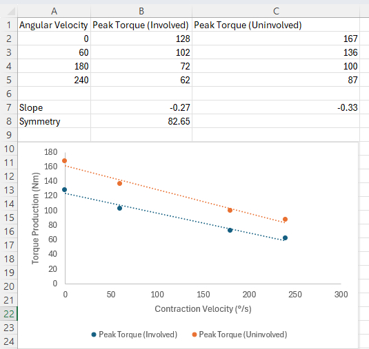
Figure 1. Simplified representative torque-velocity dataset and quick analysis workflow. Peak torque values were exported into spreadsheet software and plotted against angular velocity using a linear regression line. This represents simplified data entry for a single participant and should be expanded with additional columns for each participant and testing session for longitudinal assessment. Please click here to view a larger version of this figure.
Access restricted. Please log in or start a trial to view this content.
Results
Successful completion of this protocol should result in the highest torque output at zero velocity (isometric) and a progressive decline in torque with increasing isokinetic contraction velocities12. In a previous cohort, when examining patients with a history of ACLR, this progressive decline was greater in uninvolved limbs than in ACLR limbs14. Individual torque-time or torque-position graphs should demonstrate relatively consistent contractions. The ideal coefficient of ...
Access restricted. Please log in or start a trial to view this content.
Discussion
This protocol describes a clinically feasible method for evaluating the quadriceps torque-velocity relationship using multimodal dynamometry following ACLR. Persistent quadriceps weakness is common across age groups and ACLR graft types36,37. Traditional test batteries for patients returning to sport post-surgery focus on maximal strength but may not fully assess quadriceps function across a range of contractile demands 14. Single-velocity testing at slow isokine...
Access restricted. Please log in or start a trial to view this content.
Disclosures
The authors have no conflicts of interest or disclosures to report.
Materials
| Name | Company | Catalog Number | Comments |
|---|---|---|---|
| Computer workstation | Any commercial manufacturer | N/A | Used to operate dynamometer software and export data |
| Digital Inclinometer | Any commercial manufacturer | N/A | Used to verify anatomical zero position if desired |
| HUMAC NORM Testing and Rehabilitation System | CSMi Solutions | https://csmisolutions.com/ | Computerized multimode dynamometer used for isometric and isokinetic knee strength testing |
| Spreadsheet software | Microsoft | Excel (version dependent) | Used for data entry and data processing of torque data |
| Treadmill | Any commercial manufacturer | N/A | Used for standardized warm-up prior to testing |
References
- Mather RC, et al. Societal and economic impact of anterior cruciate ligament tears. J Bone Joint Surg Am. 2013;95(19):1751-1759. doi:10.2106/JBJS.L.01705.
- Bodkin SG, Norte GE, Hart JM. Corticospinal excitability can discriminate quadriceps strength indicative of knee function after ACL reconstruction. Scand J Med Sci Sports. 2019;29(5):716-724. doi:10.1111/sms.13394.
- Fukunaga T, Johnson CD, Nicholas SJ, McHugh MP. Muscle hypotrophy, not inhibition, is responsible for quadriceps weakness during rehabilitation after anterior cruciate ligament reconstruction. Knee Surg Sports Traumatol Arthrosc. 2019;27(2):573-579. doi:10.1007/s00167-018-5166-1.
- Garcia SA, Moffit TJ, Vakula MN, Holmes SC, Montgomery MM, Pamukoff DN. Quadriceps muscle size, quality, and strength and self-reported function in individuals with anterior cruciate ligament reconstruction. J Athl Train. 2020;55(3):246-254. doi:10.4085/1062-6050-38-19.
- Akazawa N, et al. Longitudinal relationship between intramuscular adipose tissue of the quadriceps and activities of daily living in older inpatients. J Cachexia Sarcopenia Muscle. 2021;12(6):2231-2237. doi:10.1002/jcsm.12842.
- Hunnicutt JL, McLeod MM, Slone HS, Gregory CM. Quadriceps neuromuscular and physical function after anterior cruciate ligament reconstruction. J Athl Train. 2020;55(3):238-245. doi:10.4085/1062-6050-516-18.
- Nilius A, Dewig DR, Johnston CD, Pietrosimone BG, Blackburn JT. Quadriceps composition and function influence downhill gait biomechanics >1 year following anterior cruciate ligament reconstruction. Clin Biomech. 2024;114:106229. doi:10.1016/j.clinbiomech.2024.106229.
- Barfod KW, Feller JA, Clark R, Hartwig T, Devitt BM, Webster KE. Strength testing after anterior cruciate ligament reconstruction: a prospective cohort study investigating overlap of tests. J Strength Cond Res. 2019;33(11):3145-3150. doi:10.1519/JSC.0000000000002491.
- Barker KL, Jenkins C, Pandit H, Murray D. Muscle power and function two years after unicompartmental knee replacement. Knee. 2012;19(4):360-364. doi:10.1016/j.knee.2011.05.006.
- Allison SJ, Brooke-Wavell K, Folland JP. Multiple joint muscle function with ageing: the force-velocity and power-velocity relationships in young and older men. Aging Clin Exp Res. 2013;25(2):159-166. doi:10.1007/s40520-013-0024-y.
- Hill AV. The heat of shortening and the dynamic constants of muscle. Proc R Soc Lond B Biol Sci. 1938;126(843):136-195. doi:10.1098/rspb.1938.0050.
- Alcazar J, Csapo R, Ara I, Alegre LM. On the shape of the force-velocity relationship in skeletal muscles: the linear, the hyperbolic, and the double-hyperbolic. Front Physiol. 2019;10. doi:10.3389/fphys.2019.00769.
- Froese EA, Houston ME. Torque-velocity characteristics and muscle fiber type in human vastus lateralis. J Appl Physiol. 1985;59(2):309-314. doi:10.1152/jappl.1985.59.2.309.
- Thompson XD, et al. Knee extensor torque-velocity relationships following anterior cruciate ligament reconstruction. Clin Biomech. 2023;108:106058. doi:10.1016/j.clinbiomech.2023.106058.
- Thompson XD, Resch JE, Kuenze C, Brockmeier SF, Hart J. Torque-velocity is associated with voluntary activation and corticospinal excitability in healthy knees, but not following anterior cruciate ligament reconstruction. J Sport Rehabil. 2026:1-6. doi:10.1123/jsr.2025-0237.
- Clark DJ, Condliffe EG, Patten C. Activation impairment alters muscle torque-velocity in the knee extensors of persons with post-stroke hemiparesis. Clin Neurophysiol. 2006;117(10):2328-2337. doi:10.1016/j.clinph.2006.07.131.
- Labarque VL, 't Eijnde BO, Van Leemputte M. Effect of immobilization and retraining on torque-velocity relationship of human knee flexor and extensor muscles. Eur J Appl Physiol. 2002;86(3):251-257. doi:10.1007/s00421-001-0530-z.
- Harries UJ, Bassey EJ. Torque-velocity relationships for the knee extensors in women in their 3rd and 7th decades. Eur J Appl Physiol Occup Physiol. 1990;60(3):187-190. doi:10.1007/BF00839157.
- Simonsson R, Piussi R, Högberg J, Sundberg A, Hamrin Senorski E. Rehabilitation and return to sport after anterior cruciate ligament reconstruction. Clin Sports Med. 2024;43(3):513-533. doi:10.1016/j.csm.2023.07.004.
- Almeida GPL, Albano TR, Melo AKP. Hand-held dynamometer identifies asymmetries in torque of the quadriceps muscle after anterior cruciate ligament reconstruction. Knee Surg Sports Traumatol Arthrosc. 2019;27(8):2494-2501. doi:10.1007/s00167-018-5245-3.
- Andersen LL, et al. Changes in the human muscle force-velocity relationship in response to resistance training and subsequent detraining. J Appl Physiol. 2005;99(1):87-94. doi:10.1152/japplphysiol.00091.2005.
- Gregor RJ, Edgerton VR, Perrine JJ, Campion DS, DeBus C. Torque-velocity relationships and muscle fiber composition in elite female athletes. J Appl Physiol. 1979;47(2):388-392. doi:10.1152/jappl.1979.47.2.388.
- Drigny J, Calmès A, Reboursière E, Hulet C, Gauthier A. Changes in the force-velocity relationship of knee muscles after anterior cruciate ligament reconstruction using the isokinetic 2-point model. Int J Sports Physiol Perform. 2023;1(aop):1-9. doi:10.1123/ijspp.2023-0108.
- Grbic V, Djuric S, Knezevic OM, Mirkov DM, Nedeljkovic A, Jaric S. A novel two-velocity method for elaborate isokinetic testing of knee extensors. Int J Sports Med. 2017;38(10):741-746. doi:10.1055/s-0043-113043.
- Davis H, et al. Quadriceps rate of torque development and disability in individuals with anterior cruciate ligament reconstruction. Clin Biomech. 2017;46:52-56. doi:10.1016/j.clinbiomech.2017.04.011.
- White MS, Graham MC, Janatova T, Hawk GS, Thompson KL, Noehren B. Effect of sampling rate, filtering, and torque onset detection on quadriceps rate of torque development and torque steadiness. Sensors (Basel). 2024;24(13):4250. doi:10.3390/s24134250.
- Thompson XD, Gabler CM, Mattacola CG. The relationship between rate of torque development and vertical jump performance: possible implications for ACL injury? Athl Train Sports Health Care. 2021;13(3):136-143. doi:10.3928/19425864-20200205-01.
- Hansen EM, McCartney CN, Sweeney RS, Palimenio MR, Grindstaff TL. Hand-held dynamometer positioning impacts discomfort during quadriceps strength testing: a validity and reliability study. Int J Sports Phys Ther. 2015;10(1):62-68.
- Lesnak J, Anderson D, Farmer B, Katsavelis D, Grindstaff TL. Validity of hand-held dynamometry in measuring quadriceps strength and rate of torque development. Int J Sports Phys Ther. 2019;14(2):180-187.
- Dauty M, Rochcongar P. Reproducibility of concentric and eccentric isokinetic strength of the knee flexors in elite volleyball players. Isokinetics Exerc Sci. 2001;9(2-3):129-132. doi:10.3233/IES-2001-0076.
- Bottaro M, Russo AF, de Oliveira RJ. The effects of rest interval on quadriceps torque during an isokinetic testing protocol in elderly. J Sports Sci Med. 2005;4(3):285-290.
- Giordano KA, Cich M, Oliver GD. Length-tension differences between concentric and eccentric shoulder rotation strength. J Strength Cond Res. 2024;38(2):253-258. doi:10.1519/JSC.0000000000004625.
- Thompson XD, et al. The torque-velocity relationship and second ACL injury risk. J Athl Train. 2026;1(aop). doi:10.4085/1062-6050-0217.25.
- Thompson XD, et al. The clinical utility of strength measures in predicting patient progression following ACLR. Orthop J Sports Med. 2026;14(1):23259671251400776. doi:10.1177/23259671251400776.
- Webster KE, Feller JA. Who passes return-to-sport tests, and which tests are most strongly associated with return to play after anterior cruciate ligament reconstruction? Orthop J Sports Med. 2020;8(12). doi:10.1177/2325967120969425.
- Weaver AP, Walaszek MC, Roman DP, Harkey MS, Kuenze C. The impact of meniscal tear type and surgical treatment on quadriceps strength: a study of adolescent patients post anterior cruciate ligament reconstruction. Am J Sports Med. 2023;51(9):2357-2365. doi:10.1177/03635465231177626.
- Huber R, et al. Knee extensor and flexor strength before and after anterior cruciate ligament reconstruction in a large sample of patients: influence of graft type. Physician Sportsmed. 2019;47(1):85-90. doi:10.1080/00913847.2018.1526627.
- Janicijevic D, Knezevic OM, Garcia-Ramos A, Cvetic D, Mirkov DM. Isokinetic testing: sensitivity of the force-velocity relationship assessed through the two-point method to discriminate between muscle groups and participants' physical activity levels. Int J Environ Res Public Health. 2020;17(22). doi:10.3390/ijerph17228570.
- Roberts D, Kuenze C, Saliba S, Hart JM. Accessory muscle activation during the superimposed burst technique. J Electromyogr Kinesiol. 2012;22(4):540-545. doi:10.1016/j.jelekin.2012.01.008.
- Rendos NK, Harriell K, Qazi S, Regis RC, Alipio TC, Signorile JF. Variations in verbal encouragement modify isokinetic performance. J Strength Cond Res. 2019;33(3):708-716. doi:10.1519/JSC.0000000000002998.
- Jevsevar DS, Riley PO, Hodge WA, Krebs DE. Knee kinematics and kinetics during locomotor activities of daily living in subjects with knee arthroplasty and in healthy control subjects. Phys Ther. 1993;73(4):229-239; discussion 240-242. doi:10.1093/ptj/73.4.229.
- Mentiplay BF, Banky M, Clark RA, Kahn MB, Williams G. Lower limb angular velocity during walking at various speeds. Gait Posture. 2018;65:190-196. doi:10.1016/j.gaitpost.2018.06.162.
- Thompson XD, et al. Disagreement in pass rates between strength and performance tests in patients recovering from anterior cruciate ligament reconstruction. Am J Sports Med. 2022;50(8):2111-2118. doi:10.1177/03635465221097712.
Access restricted. Please log in or start a trial to view this content.
Reprints and Permissions
