A 14-year-old boy developed a lower limb necrotizing soft tissue infection from which Corynebacterium minutissimum was isolated. Staged debridement, local antibiotic-loaded cement placement, and pedicled fascial flap reconstruction achieved wound healing. The case highlights diagnostic uncertainty when a skin commensal is recovered from a necrotic wound.
Case Report
Lower Limb Necrotizing Soft Tissue Infection Associated with Corynebacterium minutissimum in an Adolescent: A Case Report and Surgical Management
* These authors contributed equally
In This Article
Summary
Abstract
Corynebacterium minutissimum (C. minutissimum) is usually considered a skin commensal and is best known as the causative organism of erythrasma. Although invasive infections caused by this organism are rare, its clinical significance can be difficult to determine when it is isolated from wound specimens. Here, we present the case of a 14-year-old boy with progressive ulceration, swelling, malodor, and soft tissue destruction of the left lower leg. Intraoperative exploration revealed areas of nonviable fascia and adjacent soft tissue with purulent exudation, and a diagnosis of necrotizing soft tissue infection (NSTI) was made based on intraoperative findings. Surgical debridement was performed to remove visibly nonviable tissue, followed by temporary wound management using antibiotic-loaded bone cement. A deep purulent specimen obtained during surgery grew C. minutissimum, whereas anaerobic and fungal cultures showed no growth. After local infection control was achieved, the patient underwent repeat debridement and pedicled fascial flap reconstruction, with satisfactory wound healing and no evidence of recurrent infection during follow-up. Although the isolation of C. minutissimum was considered clinically relevant in this case, its role as the sole causative organism could not be confirmed because only one positive culture was available and additional microbiological evidence was lacking. This case illustrates the challenges in interpreting uncommon skin-associated organisms isolated from complex wounds and emphasizes that microbiological results should be assessed together with specimen characteristics, operative findings, clinical course, and treatment response. Early surgical debridement and staged reconstruction remain essential for achieving infection control and functional recovery in patients with NSTI.
Introduction
Corynebacterium minutissimum is a non-diphtheriae Corynebacterium species that is best known as the causative organism of erythrasma. It typically causes superficial infections involving moist intertriginous areas and is therefore often regarded as a low-virulence skin commensal or colonizing organism rather than an invasive pathogen1. However, improvements in microbiological identification techniques and the accumulation of clinical reports have shown that C. minutissimum can occasionally be recovered from deep-seated infections, including bacteremia, postoperative intra-abdominal infection, osteomyelitis, and septic arthritis2,3,4. These observations suggest that, under selected clinical circumstances, this organism may contribute to invasive disease, although its clinical significance should always be interpreted in the context of microbiological and clinical findings.
Necrotizing soft tissue infection is a rapidly progressive surgical emergency associated with substantial morbidity and mortality. Early diagnosis remains challenging because clinical manifestations may be nonspecific during the initial stage, while delayed surgical intervention is consistently associated with worse outcomes5,6. The condition is most commonly caused by β-hemolytic streptococci, Staphylococcus aureus, Gram-negative bacteria, or polymicrobial infection7,8. In contrast, reports involving C. minutissimum are exceedingly rare. When this organism is isolated from wound specimens, distinguishing true infection from colonization or contamination may be difficult, particularly in the absence of microbiological evidence from normally sterile sites or repeated cultures.
We report the case of an adolescent with a lower limb necrotizing soft tissue infection in whom C. minutissimum was isolated from a wound specimen obtained during the initial surgical procedure2,9. Rather than attributing the infection solely to this organism, this report describes how the microbiological findings were interpreted alongside the operative findings, clinical course, and treatment response. In addition, the case demonstrates a staged surgical strategy consisting of radical debridement, temporary antibiotic-loaded bone cement coverage, and delayed pedicled fascial flap reconstruction for the management of a complex lower limb wound.
Case presentation
A 14-year-old boy was admitted to the First Affiliated Hospital, Zhejiang University School of Medicine in December 2025 because of a 5-day history of progressive ulceration of the left lower leg. The lesion had enlarged rapidly and was accompanied by erythema, swelling, malodor, and purulent discharge. He denied fever, chills, abdominal pain, nausea, vomiting, or other systemic symptoms. His medical history was unremarkable, with no history of diabetes mellitus, immunodeficiency, chronic skin disease, or previous surgery involving the affected limb. After the initial operation and satisfactory control of the local infection, he was readmitted 34 days later for planned wound reconstruction.
On admission, the white blood cell count was 6.58 × 10⁹/L, with 53.2% neutrophils and 40.7% lymphocytes. Routine coagulation tests, liver and renal function tests, and preoperative infection screening were all within normal limits. The rapidly progressive soft tissue destruction raised concern for a necrotizing soft tissue infection, and immediate surgical exploration was performed without delaying treatment for additional imaging.
At surgery, areas of nonviable superficial fascia and adjacent soft tissue were identified, together with purulent exudation. Radical debridement was carried out until healthy, bleeding tissue was encountered. The wound was temporarily covered with antibiotic-loaded bone cement. No antimicrobial agent had been administered before intraoperative specimen collection. Before wound irrigation and antimicrobial administration, a deep purulent specimen was collected under sterile conditions for Gram staining, aerobic bacterial culture, anaerobic culture, and fungal culture. Gram staining revealed no visible organisms or leukocytes. Aerobic culture grew C. minutissimum, whereas anaerobic and fungal cultures were negative. The microbiological findings were interpreted together with the intraoperative findings and the subsequent clinical course.
The postoperative course was uncomplicated. The patient received regular wound care, nutritional support, and analgesia. The wound gradually became clean, with no evidence of progressive soft tissue necrosis. Thirty-four days after the initial operation, he returned for the planned second-stage procedure. At readmission, the white blood cell count had decreased to 5.29 × 10⁹/L, with 50.2% neutrophils and 44.5% lymphocytes. The wound bed showed healthy granulation tissue, and local infection was considered adequately controlled for definitive reconstruction.
During the second operation, the antibiotic-loaded bone cement was removed and the wound was reassessed. Small areas of residual nonviable tendon were identified and excised, followed by additional debridement until viable tissue was exposed. A pedicled fascial flap was then harvested and transferred to provide durable coverage of the defect. Recovery after reconstruction was uneventful. The flap survived completely, the wound healed satisfactorily, and no clinical evidence of recurrent infection was observed during follow-up.
Diagnosis, assessment, and plan
The diagnosis of necrotizing soft tissue infection (NSTI) was based on the clinical progression and intraoperative findings. The patient presented with rapidly progressive ulceration of the left lower leg accompanied by erythema, swelling, malodor, and purulent discharge. Although systemic inflammatory responses were limited and the white blood cell count remained within the normal range, the rapid progression of local tissue destruction raised concern for a deep soft tissue infection requiring urgent surgical assessment.
Routine preoperative examinations, including complete blood count, coagulation tests, liver and renal function tests, and infection screening, were performed. No significant laboratory abnormalities were identified. The diagnosis was not based on the Laboratory Risk Indicator for Necrotizing Fasciitis (LRINEC) score or another formal scoring system, as no such scoring system was used to guide clinical decision-making in this case. Given the potential for rapid progression and irreversible tissue damage associated with NSTI, surgical exploration was performed promptly based on the clinical findings without delaying treatment for additional diagnostic procedures or imaging.
During surgical exploration, purulent exudation and areas of nonviable superficial fascia and adjacent soft tissue were identified, supporting the diagnosis of NSTI. Before surgery, differential diagnoses included severe cellulitis, deep soft tissue abscess, and other complicated wound infections. However, the intraoperative finding of fascial necrosis and extensive tissue destruction supported NSTI as the most appropriate diagnosis.
The initial treatment strategy consisted of urgent radical debridement of all nonviable tissue, collection of deep intraoperative specimens for microbiological analysis, and temporary wound management using antibiotic-loaded bone cement. This staged approach was selected to achieve adequate infection control while preserving viable tissues and allowing reconstruction after stabilization of the wound condition. After local infection control was achieved and a healthy wound bed had developed, repeat debridement and pedicled fascial flap reconstruction were performed to provide definitive soft tissue coverage.
Protocol
This study was conducted in accordance with the Declaration of Helsinki and approved by the Ethics Committee of the First Affiliated Hospital, Zhejiang University School of Medicine (No. 2024-0844). Written informed consent was obtained from the patient’s legal guardian for participation and publication. The details of all the materials and equipment used are listed in the Table of Materials.
1. Initial evaluation and surgical decision-making
- Clinical assessment
- The patient was evaluated for rapidly progressive ulceration of the left lower leg.
- The wound was examined for size, local erythema, swelling, malodor, purulent discharge, and tissue necrosis. Distal neurovascular status was assessed.
- The patient’s medical history, previous treatment, and relevant risk factors were reviewed.
- Laboratory evaluation
- Preoperative laboratory examinations were performed, including a complete blood count, coagulation tests, liver and renal function tests, and infection-related investigations.
- The laboratory results were reviewed for evidence of a systemic inflammatory response. Although the white blood cell count remained within the normal range, the rapid progression of local tissue destruction raised concern for necrotizing soft tissue infection (NSTI).
- Surgical planning
- The diagnosis of NSTI was based on clinical progression and wound characteristics rather than the Laboratory Risk Indicator for Necrotizing Fasciitis (LRINEC) score or another formal scoring system.
- Emergency surgical exploration was undertaken without waiting for additional diagnostic procedures because of the risk of further tissue destruction.
2. Initial surgical exploration and debridement
- Surgical preparation and anesthesia
- The initial surgical procedure was performed under intravenous general anesthesia with endotracheal intubation.
- Standard skin preparation and sterile draping were completed before surgical exploration.
- Intraoperative assessment
- The wound was explored, and the involved skin, subcutaneous tissue, fascia, and surrounding soft tissues were examined.
- Purulent exudation and areas of nonviable fascia and adjacent soft tissue were identified, supporting the diagnosis of NSTI.
- Specimen collection and microbiological examination
- No antimicrobial agent had been administered before intraoperative specimen collection.
- A deep purulent specimen was collected under sterile conditions before wound irrigation and antimicrobial administration. The specimen was submitted for microbiological examination.
- Aerobic bacterial culture and identification, anaerobic culture, and fungal culture were performed.
- Aerobic culture identified C. minutissimum., whereas anaerobic and fungal cultures showed no growth.
NOTE: Details of the identification platform, culture media, incubation conditions, and incubation duration were unavailable in the clinical records.
3. First-stage wound management
- Surgical debridement
- Surgical debridement was performed to remove visibly necrotic and nonviable tissue.
- Viable functional structures, including vessels, nerves, and tendons, were preserved whenever possible.
- Temporary wound management
- After debridement, the wound was repeatedly irrigated and reassessed.
- Antibiotic-loaded bone cement was applied to provide local antimicrobial delivery and temporary coverage of the wound defect.
4. Second-stage reconstruction
- Timing and assessment before reconstruction
- Planned second-stage reconstruction was performed 34 days after the initial debridement.
- The decision to proceed with definitive reconstruction was based on improvement of the local wound condition, control of infection progression, and the presence of viable tissue suitable for soft tissue coverage.
- Repeat debridement
- The second-stage procedure was performed under intravenous general anesthesia with endotracheal intubation.
- The previously placed antibiotic-loaded bone cement was removed, and the wound bed was reassessed.
- Residual nonviable tissue, including localized necrotic tendon tissue, was removed until a viable wound bed was obtained.
- Pedicled fascial flap reconstruction
- A pedicled fascial flap was designed and rotated to cover the exposed bone defect.
- The flap provided vascularized soft tissue coverage after infection control and allowed reconstruction of the complex wound defect.
- After flap rotation and inset, the wound was closed, and a drainage strip was placed.
NOTE: The operative records documented the use of a pedicled fascial flap but did not specify its detailed anatomical origin.
5. Postoperative management and follow-up
- Wound and flap monitoring
- The postoperative wound was monitored by clinical examination and photographic documentation.
- Wound healing was evaluated according to wound closure, epithelialization, drainage, and soft tissue stability.
- Flap viability was assessed according to flap color, texture, perfusion, and the presence of necrosis or congestion.
- Infection surveillance and functional assessment
- Recurrence of infection was assessed by evaluating local redness, swelling, purulent discharge, wound breakdown, and other clinical signs of infection.
- Functional recovery was evaluated according to lower limb function and the patient’s ability to perform daily activities.
- Follow-up outcome
- The patient was followed for 3 months after reconstruction.
- At the final follow-up, the reconstructed wound remained stable without evidence of recurrent infection or wound breakdown.
Results
The patient underwent emergency surgical exploration because of progressive soft tissue destruction of the left lower leg. The overall clinical course, including initial presentation, staged surgical treatment, microbiological evaluation, reconstruction, and follow-up, is summarized in Figure 1. Intraoperative exploration identified purulent exudation and nonviable soft tissue requiring surgical debridement. Debridement was performed to remove visibly nonviable tissue. Available photographic documentation from the initial surgical procedure is shown in Figure 2.
Deep wound specimens collected during the initial surgical procedure were submitted for microbiological examination. Aerobic bacterial culture and identification revealed C. minutissimum. Anaerobic culture showed no growth, and fungal culture showed no fungal growth. The microbiological findings were interpreted together with the clinical presentation, surgical findings, and treatment response.
After the initial debridement, temporary wound management was performed using antibiotic-loaded bone cement. The wound condition was monitored during the interval before definitive reconstruction. Thirty-four days after the initial procedure, second-stage surgery was performed after local infection control and improvement of the wound bed. During this procedure, the previously placed bone cement was removed, residual nonviable tissue was excised, and a pedicled fascial flap was transferred to cover the exposed bone defect. Available photographic documentation of the second-stage procedure is shown in Figure 3.
Postoperatively, the flap remained viable without evidence of vascular compromise. The patient was followed for 3 months after reconstruction. During follow-up, the wound achieved stable soft tissue coverage without recurrent infection, wound breakdown, or additional surgical intervention. Flap viability was maintained, and the patient was able to perform daily activities with satisfactory functional recovery. The clinical appearance of the reconstructed wound at the 3-month follow-up is presented in Figure 4.
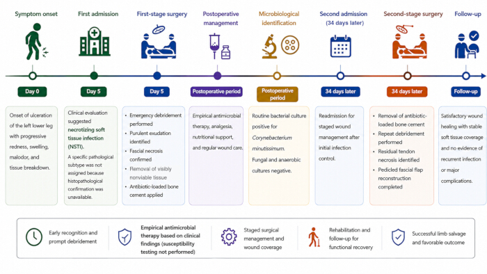
Figure 1: Timeline of clinical presentation, staged surgical management, and postoperative recovery. The timeline summarizes the clinical course of the patient with lower limb necrotizing soft tissue infection associated with C. minutissimum. The patient initially presented with progressive ulceration and local soft tissue infection of the left lower leg. Emergency surgical exploration and radical debridement were performed, followed by temporary wound management with antibiotic-loaded bone cement. Microbiological examination of the intraoperative specimen identified C. minutissimum. After local infection control and improvement of the wound condition, the patient underwent repeat debridement and pedicled fascial flap reconstruction 34 days after the initial debridement. Follow-up demonstrated stable wound healing without clinical evidence of recurrent infection. Please click here to view a larger version of this figure.
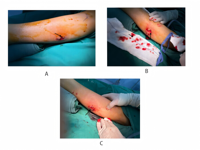
Figure 2: Clinical and intraoperative findings during the initial surgical procedure. Gross clinical photographs; scale bars are not applicable. (A) Initial focal wound lesion of the left lower leg (arrow). (B) Purulent material encountered during surgical exploration (arrows). (C) Appearance of the affected area during the initial surgical procedure (arrow). Please click here to view a larger version of this figure.
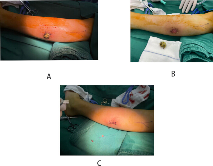
Figure 3: Second-stage debridement and pedicled fascial flap reconstruction. Gross clinical intraoperative photographs; scale bars are not applicable. (A) Appearance of the wound 34 days after the initial debridement and temporary antibiotic-loaded bone cement placement, before definitive reconstruction. (B) Intraoperative removal of the antibiotic-loaded bone cement 34 days after its placement and reassessment of the wound bed. Residual nonviable tissue was identified and excised before reconstruction. (C) Appearance of the wound after completion of the second-stage reconstructive procedure. Please click here to view a larger version of this figure.
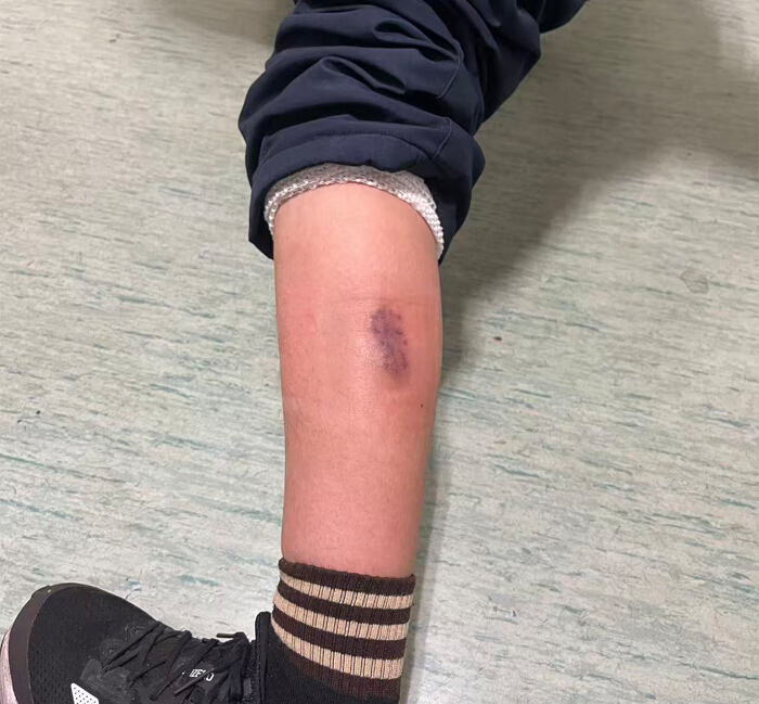
Figure 4: Postoperative wound healing after staged surgical treatment. Gross clinical photograph obtained at the 3-month postoperative follow-up after staged debridement and soft tissue reconstruction. The reconstructed wound demonstrated stable soft tissue coverage without evidence of recurrent infection or wound breakdown. The affected limb was preserved, with satisfactory functional recovery. Please click here to view a larger version of this figure.
Discussion
C. minutissimum is commonly regarded as a skin commensal and is best known as the causative organism of erythrasma10. Reports of invasive infection remain uncommon and have mainly involved bacteremia, postoperative wound infection, osteomyelitis, and septic arthritis3,11. In our patient, C. minutissimum was isolated from a deep intraoperative specimen obtained from an area of extensive fascial and soft tissue necrosis. No organisms were recovered from anaerobic or fungal cultures. Although these findings suggest that C. minutissimum may have contributed to the infectious process, they are insufficient to establish it as the sole pathogen2.
The microbiological findings should therefore be interpreted with caution. Only a single intraoperative specimen yielded C. minutissimum, while Gram staining showed neither leukocytes nor visible bacteria12. Several factors may account for the discrepancy between the negative Gram stain and positive culture, including a low bacterial burden and limitations in specimen sampling. Prior antimicrobial exposure was not considered a possible explanation in this case because no antimicrobial agent had been administered before the intraoperative specimen was collected11. In addition, repeat cultures, histopathological examination, molecular identification, and antimicrobial susceptibility testing were not available. Under these circumstances, the culture result should be considered alongside the operative findings, clinical progression, and response to treatment rather than being interpreted in isolation.
Diagnosis in this patient relied primarily on the clinical presentation and intraoperative findings13. Rapid progression of the wound and intraoperative evidence of fascial involvement and purulent soft tissue infection supported the diagnosis of necrotizing soft tissue infection14. By contrast, systemic inflammatory findings were relatively mild, and the white blood cell count remained within the normal range at presentation. This observation highlights that severe soft tissue infection may occur even in the absence of marked laboratory abnormalities. Because histopathological confirmation was unavailable, we considered the diagnosis of necrotizing soft tissue infection more appropriate than assigning a more specific pathological subtype.
Successful management depended primarily on timely surgical treatment. The initial operation focused on debridement of all nonviable tissue, followed by temporary wound management using antibiotic-loaded bone cement to control the local wound environment before definitive reconstruction15. The planned staged approach was also documented in the operative records. After local infection was controlled and a healthy wound bed had developed, repeat debridement and pedicled fascial flap reconstruction were performed. Delaying definitive reconstruction until tissue viability became clear reduced the risk of persistent infection and allowed stable soft tissue coverage.
The role of antimicrobial therapy in this case remains uncertain. No antimicrobial agent had been administered before intraoperative specimen collection. The available inpatient records did not provide sufficient information to reliably reconstruct the specific systemic antimicrobial regimen administered between specimen collection and discharge. Following surgical debridement and local infection control, the patient was discharged on January 2, 2026, with faropenem sodium granules (0.1 g orally once daily; 12 doses prescribed, corresponding to a 12-day course if taken as directed). The final culture report identifying C. minutissimum became available on January 6, 2026, after discharge; therefore, faropenem was prescribed empirically rather than selected on the basis of the final culture result. No subsequent modification of antimicrobial therapy was documented in the available medical records. Antimicrobial susceptibility testing was not performed, and it was therefore not possible to determine whether the isolated C. minutissimum was susceptible to faropenem. The favorable clinical outcome was more likely attributable to surgical debridement, repeated assessment of tissue viability, and staged reconstruction than to any specific antimicrobial agent. Future cases would benefit from obtaining multiple deep tissue specimens, repeat cultures during subsequent procedures, and antimicrobial susceptibility testing to better define the microbiological significance of unusual isolates and guide antimicrobial selection.
Several limitations should be acknowledged. This is a single case, and a causal relationship between C. minutissimum and the infection cannot be established with certainty. Only one deep intraoperative specimen yielded C. minutissimum, whereas Gram staining showed neither leukocytes nor visible bacteria. In addition, repeat microbiological sampling, histopathological examination, molecular confirmation, and antimicrobial susceptibility testing were unavailable. Previously reported invasive C. minutissimum infections have mainly included bacteremia, postoperative abdominal infection, osteomyelitis, septic arthritis, and other deep-seated infections, whereas involvement in necrotizing soft tissue infection has rarely been described2. Although the pathogenic role of C. minutissimum remains uncertain in the present case, this report highlights the need to interpret unusual microbiological findings together with specimen quality, operative findings, and the overall clinical course. It also supports the principle that early radical debridement, followed by staged reconstruction once local infection has been controlled, remains the foundation of successful management for complex necrotizing soft tissue infections.
Disclosures
The authors declare that they have no competing interests.
Acknowledgements
This study received no specific grant from any funding agency in the public, commercial, or not-for-profit sectors.
Materials
| Name | Company | Catalog Number | Comments |
|---|---|---|---|
| Aerobic bacterial culture and identification system | Institutional clinical microbiology laboratory | N/A | Aerobic culture and identification of C. minutissimum. |
| Anaerobic culture system | Institutional clinical microbiology laboratory | N/A | Anaerobic culture of the deep purulent specimen. |
| Clinical photography system | Institutional clinical photography system | N/A | Perioperative and follow-up wound documentation. |
| Drainage strip | Institutional operating room supplier | N/A | Postoperative drainage after flap inset and wound closure. |
| Endotracheal tube | Institutional operating room supplier | N/A | Airway management during general anesthesia. |
| Fungal culture system | Institutional clinical microbiology laboratory | N/A | Fungal culture of the deep purulent specimen. |
| Gram staining reagents | Institutional clinical laboratory | N/A | Direct Gram staining of the deep purulent specimen. |
| Pedicled fascial flap reconstruction instruments | Hospital central sterile supply department | Hand-surgery instrument pack No. 012 | Flap harvest, rotation, inset, and wound closure. |
| Skin preparation solution | Shanghai Likang Disinfectant Hi-Tech Co., Ltd. (hospital-common product) | 0.5% povidone-iodine solution, 500 mL/bottle; batch number varies | Preoperative skin antisepsis. |
| Sterile drapes | Hospital central sterile supply department | Institutional surgical pack | Maintenance of the sterile surgical field. |
| Sterile specimen container | Institutional clinical laboratory supplier | N/A | Collection and transport of the deep purulent specimen. |
| Surgical debridement instruments | Hospital central sterile supply department | Hand-surgery instrument packs No. 010 and No. 012 | Removal of visibly necrotic and nonviable tissue. |
| Wound irrigation solution | Cisen Pharmaceutical Co., Ltd. (hospital-common product) | 0.9% sodium chloride solution, 500 mL/bag; batch number varies | Repeated irrigation of the wound after debridement. |
References
- Forouzan P, Cohen PR. Erythrasma revisited: diagnosis, differential diagnoses, and comprehensive review of treatment. Cureus. 2020;12(9):e10733.
- Shin JY, Lee WK, Seo YH, Park YS. Postoperative abdominal infection caused by Corynebacterium minutissimum. Infect Chemother. 2014;46(4):261-263.
- Ansari A et al. Corynebacterium minutissimum as a rare cause of tibial osteomyelitis: a case report and literature review. Cureus. 2024;16(12):e75885.
- Akkaya S et al. First case report of Corynebacterium minutissimum septic arthritis associated with an indwelling device. Future Microbiol. 2025;20(3):183-188.
- Wei XK, Huo JY, Yang Q, Li J. Early diagnosis of necrotizing fasciitis: imaging techniques and their combined application. Int Wound J. 2024;21(1):e14379.
- Goh T, Goh LG, Ang CH, Wong CH. Early diagnosis of necrotizing fasciitis. Br J Surg. 2014;101(1):e119-e125.
- Guliyeva G, Huayllani MT, Sharma NT, Janis JE. Practical review of necrotizing fasciitis: principles and evidence-based management. Plast Reconstr Surg Glob Open. 2024;12(1):e5533.
- Misiakos EP, et al. Current concepts in the management of necrotizing fasciitis. Front Surg. 2014;1:36.
- Aperis G, Moyssakis I. Corynebacterium minutissimum endocarditis: a case report and review. J Infect. 2007;54(2):e79-e81.
- Sebaratnam DF, Lee S. Corynebacterium minutissimum infection: erythrasma. Med J Aust. 2017;207(5):189.
- Dalal A, Likhi R. Corynebacterium minutissimum bacteremia and meningitis: a case report and review of literature. J Infect. 2008;56(1):77-79.
- Miller JM, et al. Guide to utilization of the microbiology laboratory for diagnosis of infectious diseases: 2024 update by the Infectious Diseases Society of America (IDSA) and the American Society for Microbiology (ASM). Clin Infect Dis. 2024:ciae104.
- Stevens DL, et al. Practice guidelines for the diagnosis and management of skin and soft tissue infections: 2014 update by the Infectious Diseases Society of America. Clin Infect Dis. 2014;59(2):e10-e52.
- Zhou L, Li H, Luo G. Consensus on the diagnosis and treatment of adult necrotizing fasciitis (2025 edition). Burns Trauma. 2025;13:tkaf031.
- McDermott J, et al. Necrotizing soft tissue infections: a review. JAMA Surg. 2024;159(11):1308-1315.