Endoscopic ear surgery is a safe, effective, and versatile approach to address multiple middle ear diseases, offering comparable audiological outcomes to traditional microscopic techniques while reducing morbidity and improving visualization of difficult middle ear regions.
Review Article
Endoscopic Surgery for Chronic Middle Ear Disease: A Narrative Review of Surgical Techniques and Results
In This Article
Summary
Abstract
Endoscopic ear surgery (EES) is transforming contemporary otologic practice by enabling minimally invasive transcanal access and enhanced visualization of middle ear anatomy. This narrative review summarizes current technical considerations, clinical applications, and recent evidence regarding EES in tympanoplasty, ossiculoplasty, and cholesteatoma surgery. Technical factors influencing safety and efficacy include the use of angled endoscopes, the management of thermal risk from light sources, hemostasis, underwater endoscopic techniques, and emerging digital image enhancement systems. For type I tympanoplasty, endoscopic and microscopic approaches achieve comparable graft uptake and hearing outcomes, while EES may reduce operative time, postoperative pain, wound-related complications, and approach-related morbidity, particularly compared with postauricular microscopic surgery. Endoscopic ossiculoplasty also demonstrates audiological outcomes comparable to microscopic reconstruction, with potential benefits in operative efficiency and postoperative recovery in both adult and pediatric populations. In cholesteatoma surgery, EES provides wide-angle visualization and access to hidden recesses, and meta-analyses suggest lower residual or recurrent disease rates compared with traditional microscopic approaches. However, disease extension, particularly mastoid involvement, remains a key determinant of surgical strategy. Overall, EES is a safe, effective, and versatile technique for chronic middle ear disease, offering excellent clinical and audiological outcomes with minimal surgical morbidity.
Introduction
Over the last two decades, endoscopic ear surgery (EES) has reshaped otologic practice by improving visualization of the middle ear. Technical advancements enabled a totally endoscopic transcanal approach to middle ear diseases. Although EES was initially adopted by only a few pioneering groups, it has progressively gained popularity and is now widely used in otologic reference centers worldwide. Beyond its established middle ear applications, the feasibility of EES for accessing selected lateral skull base corridors, including infracochlear, suprageniculate, and transpromontorial approaches, has been investigated1. Potential new indications and technical modifications of endoscopic and endoscope-assisted lateral skull base approaches continue to be explored, including expanded transpromontorial and hearing-preserving multiportal strategies2,3.
Tarabichi and Arsiwala describe three successive waves of the endoscopic evolution: an initial stage in which the endoscope was used purely for diagnosis and documentation, a second phase characterized by adjunct use to assist disease removal, while the microscope remained the principal surgical tool, and a third wave in which the endoscope became central to the surgical procedure4. In this review, EES is defined as a fully endoscopic transcanal procedure, distinct from endoscope-assisted surgery in which the endoscope complements a primarily microscopic or transmastoid approach. The main advantages of EES include enhanced visualization of middle ear structures5, reduced need for canaloplasty6,7, improved cosmetic outcomes6,7, reduced postoperative pain8, and shorter operative time6,9. When compared with the traditional microscopic technique, EES has shown similar results regarding postoperative Air-Bone Gap (ABG) and graft uptake in tympanoplasty10. EES has also been associated with lower rates of residual or recurrent cholesteatoma11. Additionally, the shared visualization provided by endoscopy offers educational advantages, allowing real-time instruction and demonstrating superior outcomes for teaching middle ear anatomy and surgical skills compared to microscopic techniques12. However, EES also has limitations, particularly the need for a one-handed surgical technique, risk of thermal injury, loss of depth perception, and more challenging hemostasis within a narrow surgical field, which will be discussed in this article.
This narrative review provides an integrated overview of EES for chronic middle ear disease. It is organized into two complementary sections: the first addresses general technical and safety considerations relevant across endoscopic procedures, while the second summarizes procedure-specific evidence for tympanoplasty, ossiculoplasty, and cholesteatoma surgery.
A literature search was conducted in PubMed/MEDLINE using the following search string: (endoscopic ear surgery OR transcanal endoscopic ear surgery OR TEES) AND (tympanoplasty OR myringoplasty OR ossiculoplasty OR cholesteatoma). Titles and abstracts were screened for relevance, followed by full-text assessment of potentially eligible articles. The search was expanded by manual screening of the reference lists of relevant publications and by the inclusion of clinically relevant studies identified through the senior author’s expertise. Given the narrative design of the review, the search aimed to identify the most relevant literature rather than capture every available study systematically.
Review and Perspective
Part I. General technical and safety considerations
Overall safety
Although safety concerns were raised during the early adoption of the technique, subsequent clinical experience has demonstrated the safety and reliability of exclusive EES. In a large retrospective series of 825 exclusive EES procedures, Marchioni et al. found no major intraoperative complications, including dural, vascular, or persistent facial nerve injury. Minor intraoperative complications occurred in 4.1% of cases, while early postoperative complications were reported in only 1.3%. Delayed complications affected less than 1% of patients, supporting the overall safety and reliability of EES when performed by experienced surgeons13.
Angled endoscopes
The choice of endoscope angle has an important impact on the visualization of middle ear subregions. While most procedures are performed with 0° and 30° endoscopes, some surgeons take advantage of more angulated scopes, such as 45°, 70°, and even 90°, to further enhance their field of view. In Figure 1, we show a typical setup with a 0-degree endoscope, the surgeon’s position, and the screen. Bennett et al. demonstrated, using a three-dimensional model derived from temporal bone scans, that a 0-degree endoscope provides significantly better visualization than the microscope in nearly all middle ear subregions, with the exception of the antrum. Angled endoscopes further increase the visible surface area compared with both straight endoscopes and microscopy, regardless of whether the ossicles are present or absent. Among angled scopes, the 45° endoscope offers superior visualization of several subregions, supporting its value for inspection of anatomically concealed areas such as the epitympanum, retrotympanum, and other recesses that may be difficult to assess with conventional microscopic line-of-sight visualization5.
The use of a 70° angled endoscope during transcanal EES is less frequent than other angled scopes, but has also been described. Some authors have reported its utility for the removal of pars flaccida cholesteatoma14. However, it is important to underline that in some anatomical configurations of the retrotympanic space, a full view cannot be achieved even with the use of 70° angled scopes, and this should be considered a limitation of transcanal EES15. Extreme caution should be exercised when introducing angled endoscopes to avoid injury to delicate middle ear structures. This is particularly important near the stapes, where inadvertent contact may transmit trauma to the inner ear.
Thermal safety
Thermal safety is an important technical consideration in EES, as illumination is delivered in close proximity to middle ear structures. Excessive light intensity may increase the risk of thermal injury16. Commonly cited recommendations suggest setting xenon light intensity at 50% of maximum power during EES17. When using an LED light source, some authors have advocated reducing light intensity to approximately 30% of maximum output18. Importantly, even the combined use of laser and endoscopic illumination during procedures such as endoscopic stapes surgery has been reported to be safe when appropriate laser settings and surgical technique are applied, with complication rates comparable to those observed with the microscopic approach19,20.
Management of bleeding
Bleeding remains a key challenge in endoscopic ear surgery, as even small amounts of blood can impair visualization in the narrow operative field, soil the endoscope, and threaten continuation of the procedure. Therefore, effective hemostatic strategies are crucial to maintain visualization, avoid conversion to a microscopic approach, and support procedural safety. The posterosuperior external auditory canal has been reported as the most frequent bleeding site, and diluted epinephrine injection in this area before skin incision has therefore been recommended21. In the external auditory canal, persistent bleeding sources can be addressed using monopolar or bipolar cautery. During elevation of the tympanomeatal flap, when suction is required, direct suction on the skin flap should be avoided. Instead, a diluted epinephrine-soaked cottonoid may be interposed to promote vasoconstriction and reduce the risk of iatrogenic flap tearing. As the middle ear is approached, hemostatic techniques should be used more conservatively because the risk of iatrogenic injury increases21. Careful application of cottonoid, patience, and repeated cleaning of the surgical field are therefore essential for the progression of the surgery. Alicandri-Ciufelli et al. retrospectively evaluated 90 cases of transcanal EES and found that epinephrine administered by diluted local injection into the external auditory canal skin and by topical 1:1000-soaked cottonoids was not associated with major cardiovascular events, facial palsy, or surgery-limiting bleeding22.
Underwater technique
Underwater endoscopic ear surgery (UWEES) is a technique in which endoscopic ear procedures are performed under continuous fluid irrigation, creating a clear operative field and allowing improved visualization of middle ear structures. It was first proposed in 2014 by Yamauchi and colleagues23 to overcome some limitations of conventional endoscopic ear surgery, particularly impaired visualization due to bleeding and bone dust, as well as the need for repeated endoscope cleaning. This approach has subsequently been used in various surgical approaches, involving the external, middle, and inner ear. In a systematic review of 9 studies including 173 ears, Acharya et al. showed that one of the key indications for UWEES is labyrinthine fistula repair, but it has also been applied to canaloplasty, attic and mastoid cholesteatoma, and superior semicircular canal dehiscence plugging. Regarding operative data, they reported that bone conduction thresholds remained largely stable (+0.5 dB change in canaloplasty, shifts generally <10 dB in labyrinthine fistula patients), that ABG improved in cholesteatoma cases, including from 24.8 to 18.2 dB in attic disease and by 20.1 dB and 11.7 dB in antrum/mastoid involvement. While analyzing semicircular canal dehiscence repair, UWEES showed better long-term high-frequency bone conduction outcomes than microscopy (−5 vs. +16 dB) with comparable ABG closure (10–12 dB)24. These results underline the potential advantages of this technique in selected indications.
Digital image enhancement
To improve endoscopic visualization and disease detection, digital technologies have been developed to enhance the image signal displayed on the surgical screen25. Digital image enhancement (DIE) has also been applied intraoperatively in endoscopic cholesteatoma surgery26,27. In a questionnaire-based study involving 51 ear surgeons who assessed 12 intraoperative images, Spectra B, part of the Storz Professional Image Enhancement System (SPIES), demonstrated the highest sensitivity for detecting residual cholesteatoma compared with Clara and Spectra A25. These findings suggest that Spectra B may be a useful adjunct during EES to reduce the risk of overlooked cholesteatoma remnants, although further clinical validation is needed before routine use can be recommended.
Part II. Procedure-specific applications and outcomes
Endoscopic Type I tympanoplasty
Current evidence suggests that endoscopic type I tympanoplasty achieves graft uptake rate and audiological outcomes comparable to microscopic type I tympanoplasty. Advantages include reduced surgical morbidity and improved patient-centered outcomes. Crotty et al. performed a meta-analysis of nine randomized trials including 540 patients comparing endoscopic and microscopic type I tympanoplasty. Both approaches showed comparable graft success rates (89.6% endoscopic vs 91.1% microscopic) and hearing improvement (mean post-operative ABG difference between the two groups 0.57 dB) with matched grafting material. However, endoscopic tympanoplasty was associated with a significantly shorter operative time, by approximately 25 min, suggesting greater procedural efficiency10. Consistent with these findings, a 2024 systematic review and meta-analysis of 43 articles, including randomized controlled trials, retrospective studies, and prospective studies, suggested that the endoscopic approach should be the procedure of choice in type I tympanoplasty. This was based on a comparable graft success intake (Odds Ratio (OR) 0.831) and improvement of ABG (difference in means -0.666 dB), shorter operative time (20 min), lower rates of canaloplasty (OR 0.065), higher self-rated cosmetic results (OR 87.323), and lower post-operative pain (difference in means in visual analog pain scale -2.513 points)28.
However, the magnitude of benefit appears to depend on the type of microscopic approach. Although surgical time often favors the endoscopic approach, a subgroup analysis comparing transcanal microscopic tympanoplasty with endoscopic tympanoplasty in a meta-analysis by Gkrinia found no significant difference in operative time9. This finding is consistent with a 2025 single-blinded prospective randomized study, which showed no significant difference between transcanal microscopic and transcanal endoscopic type I tympanoplasty in ABG closure, speech recognition threshold, operative time, or patient-reported outcome measures, including pain29. These investigations suggest that the avoidance of the postauricular approach is key to lower perioperative morbidity. Given the improved visualization capabilities of the endoscope, more cases can be treated using a transcanal technique than with a microscopic approach, in which the anterior part of the tympanic membrane is often difficult to reach. When compared specifically with postauricular microscopic tympanoplasty, endoscopic techniques show fewer post-operative complications with lower rates of postoperative wound infection (OR -1.72), dysgeusia (OR -1.47), otitis externa (OR -1.96), and auricular numbness (OR -2.56)9.
Similar evidence is emerging in the pediatric population, although available data remain more limited. In a 2025 comparative study of 118 patients aged 3–15 years, closure rates (91.76% vs 69.7%) and audiometric outcomes (median -9 dB post-operative PTA vs -5 dB) were higher after type I tympanoplasty using EES compared with traditional techniques30. A similar trend was shown by Fink et al. in a retrospective case-control study on 78 ears of 50 children undergoing type I-III tympanoplasty, although without reaching statistical significance31. Specifically, for type I tympanoplasty, the graft intake rate was 91.66% for EES, compared to 58.33% in the microscopic technique.
In type I tympanoplasty, the underlay technique is historically the mainstay and has shown reliable anatomical and functional outcomes with the endoscopic approach. For example, a 2018 prospective randomized study of 60 patients comparing the underlay technique between the endoscopic and microscopic approaches found graft intake rates of 90% and 96.6%, respectively32. Excellent results have also been described with over-under techniques, in which the graft is positioned over the malleus handle and under the annulus. In a comparative study with 95 patients, the over-under technique showed a significantly higher tympanic closure rate (94.4%) compared to the underlay technique (80.6%)33. For myringoplasty, several endoscopic push-through techniques have been reported, allowing tympanic membrane closure without elevation of the tympanomeatal flap34.
Overall, these findings support endoscopic type I tympanoplasty as an excellent technique regarding surgical outcome, with a favorable safety profile and low perioperative morbidity.
Endoscopic ossiculoplasty
Endoscopic ossiculoplasty achieves audiological outcomes comparable to microscopic ossiculoplasty35,36, while potentially reducing approach-related morbidity. In a 2022 comparative study, Coleman et al. found no significant difference in ABG reduction between EES and microscopic ossiculoplasty, with both techniques achieving a mean postoperative ABG of less than 20 dB37. A large multicentric study published in 2025 supported the use of endoscopic ossiculoplasty, reporting a mean postoperative ABG of 19.94 dB and a graft success rate of 94.2%17. A retrospective study of 79 cases found significantly shorter surgery duration for the endoscopic technique compared to the microscopic technique, on average by 30 min38.
In a series of 60 patients, Fink and colleagues report an overall graft uptake rate of 98.3% and significant pre- to postoperative ABG reduction after incus interposition, partial ossicular replacement prosthesis (PORP), dense-cortical-bone PORP, total ossicular replacement prosthesis (TORP), and stapes-shoe TORP procedures39. Soloperto and colleagues described a series of 74 adult and pediatric patients who underwent endoscopic ossiculoplasty and showed a significant improvement in postoperative ABG, with an average ABG closure of 7.85 dB without a significant difference between TORP and PORP techniques40. Fink et al. also provided insight into reconstruction materials, suggesting more durable outcomes with bony or cement PORP than with titanium prostheses, with limited significance due to the limited and asymmetric sample size17.
Pediatric data are also encouraging, although available studies remain more limited. Kwinter et al. reported similar hearing outcomes in children undergoing TORP ossiculoplasty with either endoscopic (median ABG reduction of 18 dB) or microscopic techniques (median ABG reduction of 15 dB). They showed, however, that the endoscopic group had shorter operative times by an average of 33 min, less postoperative pain (mean difference of 3 on a visual analog pain scale), and reduced opioid prescription rates41. Similarly, Molinari and colleagues, in a pediatric series of 77 children, supported endoscopic ossiculoplasty as a safe technique for hearing improvement in pediatric patients. In their study, they showed significant improvement in average ABG, achieving a mean post-operative ABG of 18.48 dB from a baseline of 29.52 dB in PORP. However, in a TORP setting, their results showed no significant improvement in the ABG after the operation. The authors reported a preference for autologous PORP, whereas TORP hearing outcomes were better with titanium prostheses42.
Overall, the available evidence supports endoscopic ossiculoplasty as a versatile, safe, and effective alternative to microscopic ossiculoplasty in adult and pediatric patients.
Endoscopic cholesteatoma surgery
Endoscopic ear surgery has gained increasing relevance in cholesteatoma management because it provides wide-angle visualization of the middle ear and enables the use of angled endoscopes to access the hidden recesses.
Several meta-analyses have demonstrated favorable disease control with EES compared with microscopic surgery. A meta-analysis comparing microscopic surgery and EES reported significantly lower recurrence and residual disease rates with the endoscopic approach (relative risk of 0.51 favoring EES), whereas no significant differences were observed in auditory outcomes, graft uptake rates, or operative time11. Similarly, a more recent meta-analysis of 1134 cases from 13 studies also reported lower residual cholesteatoma rates with EES compared with microscopic surgery in both adult and pediatric populations (relative risk of 0.65 favoring EES)43. Pediatric-specific evidence also supports these findings, with a meta-analysis of 14 studies demonstrating reduced residual cholesteatoma rates in children treated endoscopically compared with traditional microscopic techniques (relative risk of 0.48, favoring EES)44. However, surgical technique alone does not fully explain recurrence risk in pediatric cholesteatoma. James et al. demonstrated that, after controlling for patient age and extension of the cholesteatoma, surgical approach was not independently associated with outcome, whereas cholesteatoma extension and mastoid involvement emerged as the major prognostic factors45.
Endoscopic approaches have also shown promising results in congenital cholesteatoma. A multicenter series of 65 congenital cholesteatomas showed a residual rate of cholesteatoma of 12%, supporting EES as a feasible and effective approach for the management of congenital cholesteatoma confined to the middle ear without mastoid extension46. Disease extension remains a key determinant of surgical strategy. Deep mastoid extension is one of the main limiting factors for successful EES, whereas attic-limited cholesteatoma is particularly suitable for this approach47.
Presutti et al. suggested that preserving the mastoid air cell system during cholesteatoma surgery might reduce recurrence rates in patients with acquired attic cholesteatoma. This was supported in their univariate analysis comparing transcanal EES with canal wall up mastoidectomy with an odds ratio of 0.405 in favor of EES. However, this association was not confirmed in multivariate analysis48.
A key element of successful and complete cholesteatoma removal is access to the epitympanum. Atticotomy and antrotomy may be performed using either conventional drilling or ultrasonic bone removal techniques47. When drilling is used, larger portions of bone can be removed using an underwater technique followed by low-speed cutting burrs, or with cutting or coarse diamond burrs at low speed and minimal irrigation. When ultrasonic devices are employed, larger bony areas can be removed with a curved tip under continuous underwater conditions, potentially reducing thermal injury to adjacent bone and soft-tissue structures47. Scutum removal facilitates exposure of the epitympanum, and visualization of the posterior epitympanum can be further enhanced using 45° endoscopes. Nevertheless, challenges associated with scutum reconstruction after an enlarged transcanal endoscopic atticotomy may limit the applicability of this approach for cholesteatoma removal and may favor a transmastoid microscopic approach in selected cases.
To address these limitations, Ayache explored the possibility of endoscopic epitympanic obliteration by 45S5 Bioactive Glass, arguing that this procedure offers immediate benefits by facilitating the scutum reconstruction procedure49. A recent meta-analysis of 12 studies in mastoid obliteration, including 583 patients, by Canali et al. suggests that S53P4 Bioactive Glass is a safe material associated with low postoperative complication rates. The most commonly reported complication was transient or persistent otorrhea, which varied in duration and severity and occurred in 16% of cases, whereas surgical wound infection was reported in 2%. Extrusion of granules into the external auditory canal was reported in only eight patients, corresponding to a pooled proportion of 1%50. Interestingly, robotic applications have been described to improve the technical setup of EES, thereby facilitating bimanual dissection51.
Overall, current evidence supports EES as an effective approach for cholesteatoma management, particularly for disease confined to the middle ear or attic. However, disease extension remains the principal determinant of surgical strategy. In many cases, optimal management may require combining endoscopic and microscopic techniques to achieve complete disease eradication while minimizing surgical morbidity.
Given the cumulative evidence, we can summarize that all cases of chronic otitis media are suitable candidates for EES. Where indicated, an additional mastoidectomy can complement the endoscopic management of middle ear disease.
Conclusions
Endoscopic ear surgery has become an integral component of modern otologic practice, offering a safe, effective, and minimally invasive approach for the management of chronic middle ear disease. Current evidence supports its use in type I tympanoplasty and ossiculoplasty, where anatomical and audiological outcomes are comparable to those achieved with microscopic techniques, while perioperative morbidity, postoperative pain, wound complications, and operative time may be reduced. In cholesteatoma surgery, EES provides superior visualization of hidden middle ear recesses and may decrease residual disease, especially in cases confined to the middle ear or attic. Nevertheless, careful patient selection remains essential, as extensive mastoid involvement or complex disease may require microscopic or combined approaches. In summary, EES is a versatile, increasingly evidence-supported technique that can improve surgical precision while minimizing morbidity in appropriately selected patients.
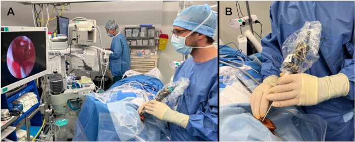
Figure 1: Operating room setup for endoscopic ear surgery. (A) General view showing the monitor and the surgeon using a 0-degree endoscope. (B) Close-up view of the surgeon’s hands manipulating the endoscope. Please click here to view a larger version of this figure.
Disclosures
The authors have no conflicts of interest or competing financial interests to declare.
The manuscript was supported by OpenAI’s ChatGPT-5.5 model for assistance with grammatical editing and language refinement.
The AI tool was employed only for editing support; all ideas and conclusions are the work of the authors.
References
- Anschuetz L, et al. Quantitative Analysis of Surgical Freedom and Area of Exposure in Minimal-Invasive Transcanal Approaches to the Lateral Skull Base. Otol Neurotol. 2018;39(6):785-790.
- Butzer T, et al. Novel Multiportal Approach to the Internal Auditory Canal for Hearing-Preserving Surgery: Feasibility Assessment in Dissections. World Neurosurg. 2022;167:e1376-e1386.
- Yacoub A, et al. Transcanal Transpromontorial Approach to Lateral Skull Base: Maximal Area of Exposure and Surgical Extensions. World Neurosurg. 2020;135:e181-e186.
- Tarabichi M, Arsiwala Z. History of Endoscopic Ear Surgery. Otolaryngol Clin North Am. 2021;54(1):1-9.
- Bennett ML, Zhang D, Labadie RF, Noble JH. Comparison of Middle Ear Visualization With Endoscopy and Microscopy. Otol Neurotol. 2016;37(4):362-6.
- Manna S, Kaul VF, Gray ML, Wanna GB. Endoscopic Versus Microscopic Middle Ear Surgery: A Meta-analysis of Outcomes Following Tympanoplasty and Stapes Surgery. Otol Neurotol. 2019;40(8):983-993.
- Tseng CC, Lai MT, Wu CC, Yuan SP, Ding YF. Comparison of the efficacy of endoscopic tympanoplasty and microscopic tympanoplasty: A systematic review and meta-analysis. Laryngoscope. 2017;127(8):1890-1896.
- Toulouie S, Block-Wheeler NR, Rivero A. Postoperative Pain After Endoscopic vs Microscopic Otologic Surgery: A Systematic Review and Meta-analysis. Otolaryngol Head Neck Surg. 2022;167(1):25-34.
- Gkrinia E, et al. Endoscopic Versus Microscopic Tympanoplasty: A Systematic Review and Metanalysis. Laryngoscope. 2024;134(8):3466-3476.
- Crotty TJ, Cleere EF, Keogh IJ. Endoscopic Versus Microscopic Type-1 Tympanoplasty: A Meta-Analysis of Randomized Trials. Laryngoscope. 2023;133(7):1550-1557.
- Li B, Zhou L, Wang M, Wang Y, Zou J. Endoscopic versus microscopic surgery for treatment of middle ear cholesteatoma: A systematic review and meta-analysis. Am J Otolaryngol. 2021;42(2):102451.
- Anschuetz L, et al. Teaching Middle Ear Anatomy and Basic Ear Surgery Skills: A Qualitative Study Comparing Endoscopic and Microscopic Techniques. Otolaryngol Head Neck Surg. 2021;165(1):174-181.
- Marchioni D, et al. Complications in Endoscopic Ear Surgery. Otol Neurotol. 2018;39(8):1012-1017.
- Takahashi M, Yamamoto Y, Kojima H. Transcanal endoscopic approach for pars flaccida cholesteatoma using a 70-degree angled endoscope. Eur Arch Otorhinolaryngol. 2021;278(4):1283-1288.
- Bonali M, et al. Correlation of Radiologic Versus Endoscopic Visualization of the Middle Ear: Implications for Endoscopic Ear Surgery. Otol Neurotol. 2020;41(9):e1122-e1127.
- Kozin ED, et al. Thermal effects of endoscopy in a human temporal bone model: implications for endoscopic ear surgery. Laryngoscope. 2014;124(8):E332-9.
- Fink R, et al. Endoscopic ossiculoplasty: audiological and surgical outcomes from a multicenter experience with 292 cases. Eur Arch Otorhinolaryngol. 2025;282(11):5571-5580.
- Vachutka J, Trneckova M, Salzman R, Kolarova H, Belakova P. Optimal Light Source Intensity Setting in Endoscopic Ear Surgery. Otol Neurotol. 2022;43(2):e205-e211.
- Fyrmpas G, Tsetsos N, Katotomichelakis M, Rudic M. Lasers in endoscopic middle ear surgery: where do we stand today? Eur Arch Otorhinolaryngol. 2021;278(11):4169-4177.
- Molinari G, et al. "Hot" vs "Cold" endoscopic stapes surgery: a matched case-control study. Eur Arch Otorhinolaryngol. 2023;280(5):2257-2263.
- Anschuetz L, et al. Management of Bleeding in Exclusive Endoscopic Ear Surgery: Pilot Clinical Experience. Otolaryngol Head Neck Surg. 2017;157(4):700-706.
- Alicandri-Ciufelli M, et al. Epinephrine Use in Endoscopic Ear Surgery: Quantitative Safety Assessment. ORL J Otorhinolaryngol Relat Spec. 2020;82(1):1-7.
- Yamauchi D, et al. Closure technique for labyrinthine fistula by "underwater" endoscopic ear surgery. Laryngoscope. 2014;124(11):2616-8.
- Acharya S, Thapa PB, Adhikari D. Underwater endoscopic ear surgery: A systematic review. Eur Arch Otorhinolaryngol. 2026. doi: 10.1007/s00405-025-09997-3. Epub ahead of print.
- Ragonesi T, et al. Digital image enhancement may improve the sensitivity of cholesteatoma detection during endoscopic ear surgery. Clin Otolaryngol. 2023;48(4):595-603.
- Miwa T, Takeda H, Minoda R. Cholesteatoma Imaging Using a Digital Image Enhancement System During Endoscopic Ear Surgery. Otol Neurotol. 2021;42(2):e244.
- Lucidi D, et al. Use of IMAGE1 S technology for detection of cholesteatoma in endoscopic ear surgery: a retrospective case series on 45 patients. Eur Arch Otorhinolaryngol. 2021;278(5):1373-1380.
- Wang TC, et al. Endoscopic Versus Microscopic Type I Tympanoplasty: An Updated Systematic Review and Meta-analysis. Otolaryngol Head Neck Surg. 2024;170(3):675-693.
- Govindan A, et al. Endoscopic Versus Microscopic Tympanoplasty: A Single-Blinded Randomized Comparative Trial. Otol Neurotol. 2025;46(9):1117-1123.
- Kim N, et al. Comparative outcomes between endoscopic and microscopic approaches in pediatric type-1 tympanoplasty. Am J Otolaryngol. 2025;46(5):104677.
- Fink R, Beckmann S, Sheppard SC, Caversaccio M, Anschuetz L. Endoscopic vs. microscopic tympanoplasty in children: a retrospective case-control study. Front Surg. 2025;12:1649552.
- Maran RK, Jain AK, Haripriya GR, Jain S. Microscopic Versus Endoscopic Myringoplasty: A comparative study. Indian J Otolaryngol Head Neck Surg. 2019;71(Suppl 2):1287-1291.
- Lotto C, et al. To detach or not to detach the umbo in type I tympanoplasty: functional results. Eur Arch Otorhinolaryngol. 2024;281(6):2871-2876.
- Beckmann S, Anschuetz L. Minimally invasive tympanoplasty: review of outcomes and technical refinements. Oper Tech Otolaryngol Head Neck Surg. 2021;32(2):143-149.
- Tsetsos N, Vlachtsis K, Stavrakas M, Fyrmpas G. Endoscopic versus microscopic ossiculoplasty in chronic otitis media: a systematic review of the literature. Eur Arch Otorhinolaryngol. 2021;278(4):917-923.
- Lim CY, et al. Outcomes of Endoscopic versus Microscopic Ossicular Chain Reconstruction-A Systematic Review and Meta-analysis. Laryngoscope. 2025;135(6):1899-1907.
- Coleman H, Tikka T, Curran J, Iyer A. Comparison of endoscopic vs microscopic ossiculoplasty: a study of 157 consecutive cases. Eur Arch Otorhinolaryngol. 2023;280(1):89-96.
- Celik O, Ulkumen B. Endoscopic versus microscopic ossiculoplasty: Does the functional outcome vary according to the type of osciculoplasty? Braz J Otorhinolaryngol. 2023;89(2):213-221.
- Fink R, et al. Techniques of Endoscopic Ossiculoplasty. J. Vis. Exp. 2024;203:e66155.
- Soloperto D, et al. Exclusive endoscopic ossiculoplasty with autologous material: step-by-step procedure and functional results. Eur Arch Otorhinolaryngol. 2023;280(11):4869-4878.
- Kwinter A, Purcell PL, Leonard CG, James AL. Comparing Transcanal Endoscopic Ear Surgery to Post-Auricular Microscope-Guided Surgery in Pediatric Ossiculoplasty: Hearing Outcomes and Post-Operative Pain. Otol Neurotol. 2021;42(10):e1648-e1651.
- Molinari G, et al. Endoscopic partial and total ossicular chain reconstruction in children: A multicentric study. Int J Pediatr Otorhinolaryngol. 2025;198:112606.
- Giffoni RB, et al. Endoscopic-Guided Resection of Middle Ear Cholesteatoma: A Systematic Review and Meta-Analysis. Otol Neurotol. 2025;46(4):418-424.
- Basonbul RA, Ronner EA, Kozin ED, Lee DJ, Cohen MS. Systematic Review of Endoscopic Ear Surgery Outcomes for Pediatric Cholesteatoma. Otol Neurotol. 2021;42(1):108-115.
- James AL. Cholesteatoma Severity Determines the Risk of Recurrent Paediatric Cholesteatoma More Than the Surgical Approach. J Clin Med. 2024;13(3):836.
- Jenks CM, et al. Transcanal Endoscopic Ear Surgery for Congenital Cholesteatoma: A Multi-institutional Series. Otolaryngol Head Neck Surg. 2022;167(3):537-544.
- Beckmann S, et al. Endoscopic Cholesteatoma Surgery. J Vis Exp. 2022;179.
- Presutti L, et al. The Impact of the Transcanal Endoscopic Approach and Mastoid Preservation on Recurrence of Primary Acquired Attic Cholesteatoma. Otol Neurotol. 2018;39(4):445-450.
- Ayache S. Transcanal Endoscopic Ear Surgery for Epitympanic Cholesteatoma With Obliteration Using Bioglass. Laryngoscope. 2022;132(2):433-435.
- Canali L, et al. Mastoid Obliteration Using Granules of S53p4 Bioactive Glass: Systematic Review and Meta-Analysis. Otolaryngol Head Neck Surg. 2026;175(1):1-9.
- Simon F, et al. Robot-Assisted Transcanal Endoscopic Ear Surgery for Congenital Cholesteatoma. J Vis Exp. 2023;202.