This prospective multicenter pilot study evaluated the impact of ureteral access sheath size on stone-free outcomes, operative efficiency, and fragment clearance during flexible ureteroscopy for lower-pole stones, using a 6-o’clock working-channel ureteroscope.
Research Article
Impact of Ureteral Access Sheath Size During Flexible Ureteroscopy Using a 6 o’clock Working Channel: A Prospective Multicenter Pilot Study
In This Article
Summary
Abstract
The impact of ureteral access sheath (UAS) size on surgical outcomes in flexible ureteroscopy (f-URS) remains controversial, particularly with modern suction-assisted techniques and evolving definitions of stone-free status. The objective of this study was to evaluate the effect of UAS size on stone-free rate (SFR), operative efficiency, and fragment clearance dynamics during f-URS for lower-pole stones using a 6 o’clock working-channel ureteroscope. This prospective multicenter pilot study included 49 patients undergoing f-URS across three tertiary referral centers. The primary outcome was strict SFR (0 mm), and secondary outcomes included clinical SFR (<2 mm), operative time, laser lithotripsy time, and fragment clearance patterns. Subgroup analysis focused on lower-pole stones. No statistically significant difference in strict SFR was observed between the 11/13 Fr and 12/14 Fr UAS groups. Clinical SFR also did not differ significantly between groups, although it was higher than strict SFR, particularly for lower-pole stones. Mean operative time was comparable between groups. Laser lithotripsy time was numerically longer in the 12/14 Fr group, but the difference was not statistically significant. Given the limited sample size, the findings should be considered exploratory and require confirmation in larger prospective studies. Trends in operative parameters and fragment clearance were observed and should be interpreted cautiously. Operative performance metrics and fragment clearance patterns may complement traditional binary stone-free outcomes when evaluating endourological innovations.
Introduction
Flexible ureteroscopy (f-URS) has become a standard treatment modality for upper urinary tract stones, particularly for lower-pole calculi, owing to its minimally invasive nature and favorable clinical outcomes1. In clinical practice, stone size and location are strongly associated with the likelihood of spontaneous stone passage2. However, despite advances in endoscopic technology, achieving complete stone clearance in lower-pole stones remains challenging because of anatomical constraints, including a narrow infundibulopelvic angle (IPA) and limited fragment evacuation3,4,5.
Traditionally, surgical success has been evaluated using the stone-free rate (SFR), often defined as the complete absence of residual fragments. However, this definition may not fully reflect the clinical effectiveness of f-URS, as small residual fragments (<2 mm) are frequently considered clinically insignificant6,7. Consequently, the concept of “clinical stone-free rate” has gained increasing attention as a more practical measure of postoperative outcomes.
In parallel, technical innovations, including optimization of ureteral access sheath (UAS) size, irrigation systems, and suction-assisted techniques, have aimed to improve intraoperative visibility, reduce intrarenal pressure (IRP), and enhance fragment clearance8,9. Larger UAS diameters may improve irrigation outflow and suction efficiency, thereby facilitating stone fragment evacuation while helping maintain lower intrarenal pressure. However, increasing the sheath diameter should be balanced against potential risks associated with UAS placement10,11.
Recent advances in suction-assisted ureteroscopy have focused on improving stone fragment evacuation while maintaining low IRP and adequate endoscopic visualization. Recently developed suction-assisted UAS have demonstrated promising outcomes, including improved SFR and reduced postoperative infectious complications compared with conventional UAS. However, most published studies have evaluated suction-assisted UAS technology as a whole, whereas the influence of UAS size on newer ureteroscope designs, such as a 6 o’clock working-channel ureteroscope, remains poorly understood. Additionally, a recent study demonstrated that the relative endoscope-sheath diameter (RESD) plays a critical role in modulating intrarenal fluid dynamics and fragment clearance12. A lower RESD, representing a smaller scope relative to a larger sheath, effectively maintains a low IRP and minimizes infectious risks, but may conversely reduce the efficacy of stone fragment evacuation due to altered suction fluid dynamics. Conversely, a higher RESD enhances stone extraction efficiency but may compromise pressure control.
Therefore, further investigation is needed to determine whether UAS size affects surgical outcomes with this ureteroscope design13,14. The hypothesis was that UAS size may influence operative performance and fragment clearance but would not significantly affect SFR when using a 6 o’clock working-channel ureteroscope.
This multicenter pilot study evaluated the impact of UAS size (11/13 Fr vs. 12/14 Fr) on surgical outcomes in patients undergoing retrograde intrarenal surgery using a 6 o’clock working-channel ureteroscope. Operative parameters and fragment clearance were also evaluated, particularly in lower-pole stones, using both strict (0 mm) and clinical (<2 mm) definitions of stone-free status.
Protocol
This study was approved by the Institutional Review Board of Seoul National University Hospital (No. D-2501-093-1608), the Institutional Review Board of Severance Hospital (No. 1-2025-0008), and the Institutional Review Board of Asan Medical Center (No. 2025-0197). The study was conducted in accordance with the principles of the Declaration of Helsinki. Written informed consent was obtained from all participants before enrollment. The tools used in this protocol are listed in the Table of Materials.
1. Study design and population
This study was designed and approved as a prospective multicenter pilot study because insufficient prior evidence was available to support a reliable formal sample-size calculation for comparing surgical outcomes between the two UAS sizes when using a 6 o’clock working-channel ureteroscope. As a pilot study, the findings should be considered preliminary and exploratory and may inform the design of future adequately powered studies.
The study included patients undergoing flexible ureteroscopy for upper urinary tract stones across three tertiary centers. Patients were enrolled consecutively and assigned to undergo surgery using either an 11/13 Fr or 12/14 Fr UAS. A total of 49 patients were included in the final analysis. Stones were located in the lower-pole calyx in 40 patients and in the lower-pole major calyx in the remaining 9 patients.
Participants were allocated within each center using a computer-generated randomization list prepared and maintained by an independent third party. After surgical preparation, an operating-room nurse sequentially revealed the assigned UAS. Investigators and operating surgeons were not involved in sequence generation or allocation. For patient safety, if insertion of the assigned 12/14 Fr UAS was not feasible, the sheath could be replaced with an 11/13 Fr UAS based on intraoperative conditions. Patients whose assigned sheath size was changed intraoperatively were excluded from the comparative analysis.
2. Surgical technique
All procedures were performed under the same protocol using a flexible ureteroscope equipped with a 6 o’clock working-channel configuration designed to enhance lower-pole access. The ureteroscope had a maximum outer diameter of 9.5 Fr. The 11/13 Fr and 12/14 Fr UAS sizes were selected because they are commonly used clinical sizes compatible with this ureteroscope and allow comparison of different scope-to-sheath clearances. A larger clearance is expected to facilitate irrigation outflow, fragment evacuation, and lower intrarenal pressure, although these parameters were not directly measured in the present study.
Laser lithotripsy was performed using standard holmium laser systems at 1 J and 10–20 Hz. Irrigation pressure was maintained between 30 and 80 mmHg. Suction was provided using a hospital wall suction system. Applied negative suction pressure ranged from 60 to 350 mmHg and was adjusted intraoperatively at the surgeon’s discretion according to the clinical situation, including endoscopic visibility, irrigation outflow, stone burden, and collecting-system distension. No standardized suction-pressure protocol was used across the participating centers. UAS size selection followed the assigned group (11/13 Fr vs. 12/14 Fr), although minor intraoperative adjustments were permitted based on ureteral conditions.
3. Outcome measures
The primary outcome was strict SFR, defined as the complete absence of residual fragments (0 mm). Clinical SFR, defined as residual fragments <2 mm in all diameters and consistent with the concept of clinically insignificant residual fragments, was evaluated as a secondary outcome to reflect clinical relevance. Lower-pole stone locations were defined by distinguishing between the lower-pole major calyx and lower-pole minor calyces.
Additional secondary outcomes focused on operative performance and mechanistic efficiency, including operative time, laser lithotripsy time, and fragment clearance patterns, evaluated using residual fragment size distribution. Postoperative stone-free status was assessed using non-contrast computed tomography (CT) performed 3 months after surgery. Postoperative CT images were reviewed separately and formally interpreted by radiologists at each participating institution as part of routine clinical practice, and these radiologic assessments were used directly to determine the status of residual stones. Residual fragments were assessed according to maximal diameter on postoperative imaging. Strict stone-free status was defined as the complete absence of residual fragments, whereas clinical stone-free status was defined as residual fragments measuring <2 mm in all dimensions. Complication rates were also recorded and classified according to the Clavien-Dindo system.
4. Subgroup analysis
A predefined subgroup analysis was performed for patients with lower-pole stones, given the known challenges in fragment clearance in this anatomical location.
5. Statistical analysis
Statistical comparisons of categorical variables were performed using the chi-square test or Fisher’s exact test, as appropriate. Continuous variables were compared using Student’s t-test or the Mann–Whitney U test based on data distribution. Given the exploratory pilot design and limited sample size, no multivariable regression analysis was performed.
Baseline and anatomical variables, including UAS size, stone burden, lower-pole involvement, and anatomical parameters such as the IPA and modified Seoul National University Renal Stone Complexity (S-ReSC) score15, were compared between groups as appropriate. All statistical tests were two-sided, and a p < 0.05 was considered statistically significant. The overall study design and workflow are shown in Figure 1.
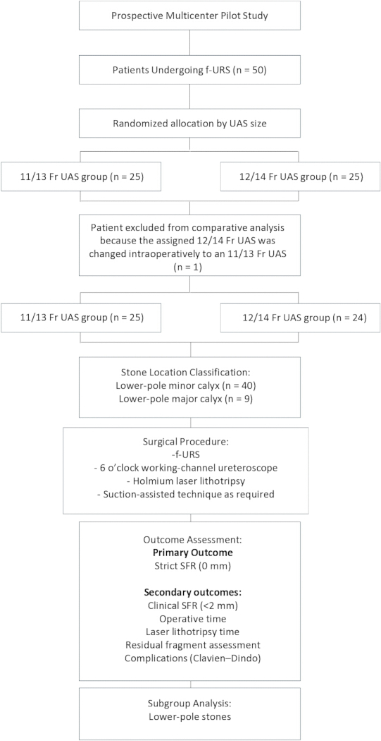
Figure 1: Schematic overview of the prospective multicenter pilot study design and workflow. Patients undergoing flexible ureteroscopy for upper urinary tract stones were prospectively enrolled at three tertiary centers and categorized according to ureteral access sheath size (11/13 Fr or 12/14 Fr). Surgical outcomes, including stone-free status and operative parameters, were evaluated. Please click here to view a larger version of this figure.
Results
Patient characteristics
During the operation, insertion of the assigned sheath was not feasible in one patient initially assigned to the 12/14 Fr group, and the sheath was changed intraoperatively to an 11/13 Fr sheath. This patient was excluded from the analysis. Therefore, 49 patients were included in the final analysis: 25 patients in the 11/13 Fr group and 24 patients in the 12/14 Fr group. Baseline characteristics, including age, stone size, and anatomical parameters, were comparable between the two groups. Lower-pole involvement was present in all cases, reflecting a cohort enriched for anatomically challenging cases. Baseline characteristics are summarized in Table 1.
| Variable | 11/13Fr | 12/14Fr | p-value |
| N | 25 | 24 | |
| Age (years) | 57.72 ± 16.05 | 59.00 ± 10.83 | 0.744 |
| Body mass index (kg/m²) | 22.68 ± 3.16 | 25.50 ± 5.12 | 0.026 |
| Stone size (mm) | 17.65 ± 9.58 | 16.10 ± 4.77 | 0.591 |
| Stone volume (mm³) | 1246.15 ± 1597.59 | 1306.53 ± 1302.08 | 0.914 |
| Number of stones | 2.75 ± 1.94 | 2.30 ± 2.01 | 0.444 |
| Infundibulopelvic angle (°) | 50.08 ± 9.76 | 50.58 ± 14.44 | 0.89 |
| Modified S-ReSC* score | 4.80 ± 2.43 | 4.17 ± 2.37 | 0.361 |
| BMI = Body mass index, IPA = Infundibulopelvic angle, S-ReSC=Seoul National University Renal Stone Complexity | |||
Table 1: Baseline characteristics of the study population. Demographic characteristics, stone-related parameters, and anatomical factors were compared between patients treated with 11/13 Fr and 12/14 Fr ureteral access sheaths. Continuous variables are presented as mean ± standard deviation.
Stone-free rate (Strict vs. Clinical)
In the overall cohort, the strict stone-free rate (defined as no residual fragments, 0 mm) was 52.0% in the 11/13 Fr group and 41.7% in the 12/14 Fr group, with no statistically significant difference between the groups (p = 0.331). When the clinical definition of stone-free status (residual fragments <2 mm) was applied, stone-free rates increased to 84.0% in the 11/13 Fr group and 83.3% in the 12/14 Fr group (p = 0.625). Despite this increase, no difference was observed between the two groups.
Lower-pole subgroup analysis
An additional subgroup analysis was performed in patients with multiple renal stones in whom the stone with the maximal diameter was located in the lower-pole minor calyx (n = 32). The strict stone-free rate (0 mm) was 33.3% in the 11/13 Fr group and 47.1% in the 12/14 Fr group, with no statistically significant difference between the groups (p = 0.430). When the clinical definition of stone-free status (residual fragments <2 mm) was applied, stone-free rates increased to 60.0% in the 11/13 Fr group and 58.8% in the 12/14 Fr group (p = 0.946). Given the small subgroup size, these estimates are statistically imprecise and should be interpreted as exploratory findings rather than evidence of equivalence or of the absence of an effect of UAS size.
Operative efficiency
Stone-free rates did not differ significantly between groups, whereas analysis of operative parameters revealed differences in procedural dynamics. Mean operative time was comparable between the two groups (62.2 min in the 11/13 Fr group vs. 65.8 min in the 12/14 Fr group). Mean laser lithotripsy time was numerically longer in the 12/14 Fr group (17.9 min vs. 13.2 min), but the difference was not statistically significant (p = 0.236). Therefore, no conclusion can be drawn regarding the effect of UAS size on laser lithotripsy time in this pilot study.
Fragment clearance patterns
The number of suction-assisted fragment extraction maneuvers was significantly lower in the 11/13 Fr group than in the 12/14 Fr group. Among patients with a maximal stone diameter ≤20 mm, the mean number of fragment extraction maneuvers was 11.8 ± 8.0 in the 11/13 Fr group and 17.6 ± 9.4 in the 12/14 Fr group (p = 0.039). Similarly, among patients with stones ≤15 mm, the mean number of fragment extraction maneuvers was significantly lower in the 11/13 Fr group than in the 12/14 Fr group (9.1 ± 5.5 vs. 15.3 ± 9.0, p = 0.032). Despite the lower number of fragment extraction maneuvers in the 11/13 Fr group, operative time, laser lithotripsy time, stone-free rates, and residual-fragment outcomes were comparable between the two groups. Operative outcomes are summarized in Table 2.
| Variable | 11/13Fr | 12/14Fr | p-value |
| N | 25 | 24 | |
| Operative Outcomes | |||
| Operative time (min) | 62.16 ± 29.16 | 65.83 ± 35.41 | 0.694 |
| Anesthesia time (min) | 90.72 ± 28.77 | 143.25 ± 23.85 | 0.280 |
| Total lasering time (min) | 13.19 ± 13.41 | 17.93 ± 14.17 | 0.236 |
| Endoscope insertion (count) | 16.60 ± 7.91 | 20.29 ± 9.92 | 0.158 |
| Estimated blood loss (mL) | 3.70 ± 10.47 | 5.21 ± 11.47 | 0.639 |
| Stone-Free Outcomes | |||
| Strict SFR (0 mm), n (%) | 13/25 (52%) | 10/24 (41.7%) | 0.331 |
| Clinical SFR (<2 mm), n (%) | 21/25 (84.0%) | 20/24 (83.3%) | 0.625 |
| Lower Pole Subgroup | |||
| Strict SFR (0 mm), n (%) | 5/15 (33.3%) | 8/17 (47.1%) | 0.430 |
| Clinical SFR (<2 mm), n (%) | 9/15 (60.0%) | 10/17 (58.8%) | 0.946 |
| Fragment Clearance | |||
| Number of suction-assisted fragment extraction | |||
| Stone diameter ≤20 mm | 11.8 ± 8.0 | 17.6 ± 9.4 | 0.039 |
| Stone diameter ≤15 mm | 9.1 ± 5.5 | 15.3 ± 9.0 | 0.032 |
| *SFR = Stone free rate |
Table 2: Operative and clinical outcomes according to ureteral access sheath size. Operative parameters, laser lithotripsy characteristics, and stone-free outcomes using strict (0 mm) and clinical (<2 mm) definitions were compared between the 11/13 Fr and 12/14 Fr ureteral access sheath groups.
Complications
Two patients developed postoperative acute pyelonephritis. Both patients were successfully treated with intravenous antibiotic therapy without requiring additional intervention.
DATA AVAILABILITY:
The de-identified raw dataset underlying the findings of this study is provided as Supplementary File 1.
Supplementary File 1: De-identified raw dataset. Please click here to download this file.
Discussion
The present findings suggest that stone burden and lower-pole anatomy may be important factors influencing fragment clearance during flexible ureteroscopy. However, because this was a small exploratory pilot study, the relative contributions of stone burden, anatomy, and UAS size cannot be determined definitively from the present data. For lower-pole stones exceeding 20 mm, achieving clinical stone-free status using f-URS alone may be limited, regardless of sheath size or irrigation strategy16,17. This multicenter pilot study evaluated the impact of UAS size on surgical outcomes using a 6 o’clock working-channel flexible ureteroscope, with particular focus on operative efficiency and fragment-clearance dynamics. Although conventional analyses often prioritize SFR, this endpoint alone may not adequately reflect the procedural effectiveness of modern f-URS6.
No statistically significant difference was detected in either strict (0 mm) or clinical (<2 mm) stone-free rates between the 11/13 Fr and 12/14 Fr UAS groups. These findings should be interpreted with caution due to the limited sample size. In particular, the lower-pole minor-calyx subgroup was small, and the subgroup analysis was exploratory and statistically imprecise. Therefore, the absence of a statistically significant difference should not be interpreted as evidence of equivalence between the two UAS sizes or as evidence that UAS size has no effect on stone-free outcomes. Final stone clearance may also be influenced by intrinsic factors such as stone burden and anatomical constraints, consistent with previous literature18.
Although larger sheath diameters are associated with improved irrigation and lower intrarenal pressure8, this was not associated with superior operative outcomes in the present cohort. Laser lithotripsy time was numerically longer in the 12/14 Fr group, but the difference was not statistically significant. One possible hypothesis is that increased irrigation flow may promote stone migration during lithotripsy, potentially affecting fragmentation efficiency19. However, this mechanism was not directly evaluated in the present study. Alternative explanations, including differences in surgical technique, laser settings, stone composition, fragment-retrieval strategy, and random variation related to the limited sample size, may also have contributed to the observed numerical difference in laser lithotripsy time. When clinically relevant residual fragments (<2 mm) were considered acceptable, reported stone-free rates increased substantially compared with strict zero-fragment definitions, including in studies of lower-pole stones6. These findings highlight the importance of incorporating clinically meaningful stone-free definitions when interpreting endourological outcomes6.
Although the 12/14 Fr UAS required significantly more suction-assisted fragment-extraction maneuvers, this did not result in shorter operative time, reduced laser lithotripsy time, or improved stone-free rates. The increased frequency of fragment extraction did not translate into greater procedural efficiency or superior clinical outcomes. The reason for the greater number of extraction maneuvers in the 12/14 Fr group cannot be determined from the present study. Importantly, the greater number of maneuvers was not accompanied by statistically significant improvements in operative time or stone-free outcomes. Previous studies have reported improved stone-free rates and perioperative outcomes with suction-assisted ureteral access sheaths compared with conventional ureteroscopy or traditional ureteral access sheaths13,14. These benefits have been attributed to active suction, which enhances fragment evacuation while maintaining low intrarenal pressure. In contrast, the present study specifically evaluated the effect of UAS size (11/13 Fr vs 12/14 Fr) rather than the presence of active suction. This distinction may explain differences from findings reported for suction-assisted systems. The clinical benefits reported for suction-assisted systems may primarily result from the suction mechanism itself rather than from an increase in sheath diameter alone. The present findings are also consistent with previous evidence demonstrating that conventional UAS use does not independently improve stone-free rates, with stone burden remaining the strongest predictor of surgical success20. Emphasis may therefore shift from a binary “stone-free vs not stone-free” framework toward a more nuanced assessment of surgical efficiency and clinically relevant fragment clearance. In this context, UAS characteristics may function more as modulators of intraoperative performance than determinants of ultimate success, consistent with previous reports demonstrating a limited impact of UAS use on final stone-free outcomes20.
The 6 o’clock working-channel configuration further reflects a focus on optimizing intraoperative flow dynamics and fragment-clearance efficiency. This design may improve access to lower poles in minor calyces and help overcome certain anatomical limitations; however, its independent impact could not be evaluated without a comparator scope. Future studies incorporating real-time intrarenal pressure monitoring and quantitative suction metrics may further elucidate the interplay between device design and surgical performance. Several limitations should also be considered. The sample size was relatively small, consistent with the original pilot design, and may have limited the statistical power to detect clinically meaningful differences between groups. Therefore, a type II error, defined as failure to detect a true difference, cannot be excluded. The study was not designed to establish equivalence between the two UAS sizes or to exclude potentially clinically meaningful differences. The lower-pole minor-calyx subgroup was also small, resulting in statistically imprecise estimates. Therefore, the subgroup findings should be considered exploratory and should not be interpreted as evidence of equivalence or absence of an effect of UAS size. Randomization was partially constrained by intraoperative decision-making, introducing potential selection bias. Detailed quantitative measurements of irrigation and suction parameters were not standardized across centers. BMI also differed significantly between the two groups at baseline. Although no direct association between BMI and surgical outcomes was demonstrated in this pilot study, residual confounding related to this baseline imbalance cannot be excluded. Although obesity may substantially increase the technical complexity of percutaneous nephrolithotomy, its impact on flexible ureteroscopy is generally considered less pronounced because the procedure is performed through the natural urinary tract rather than via percutaneous access. The use of wall suction systems also lacked technical standardization across operating rooms and centers, potentially introducing variability in real-time suction efficiency. Furthermore, individual patient factors, such as anatomical variations in the renal collecting system, IPA, and ureteral compliance, can significantly alter surgical difficulty and fluid dynamics, even under identical pressure settings. Despite these limitations, the study provides preliminary evidence that operative-efficiency metrics and fragment-clearance patterns may represent more sensitive indicators of technological performance than traditional SFR alone.
In conclusion, this pilot study did not detect a statistically significant difference in strict or clinical stone-free rates between the 11/13 Fr and 12/14 Fr UAS groups. Given the limited sample size, particularly in subgroup analyses, these findings should be considered exploratory and should not be interpreted as evidence of equivalence between UAS sizes. Differences in fragment-extraction maneuvers warrant further investigation. Larger, adequately powered prospective studies are required to determine whether UAS size meaningfully affects operative performance or stone clearance.
Disclosures
The authors declare no conflicts of interest related to this study.
Acknowledgements
This research was supported by a grant from the Korea Health Technology R&D Project through the Korea Health Industry Development Institute (KHIDI), funded by the Ministry of Health & Welfare, Republic of Korea (grant number: RS-2024-00335457).
Materials
| Name | Company | Catalog Number | Comments |
|---|---|---|---|
| 11/13 Fr ClearPetra ureteral access sheath | Well Lead Medical Co., Ltd. | N/A | Ureteral access sheath for flexible ureteroscopy |
| 12/14 Fr ClearPetra ureteral access sheath | Well Lead Medical Co., Ltd. | N/A | Ureteral access sheath for flexible ureteroscopy |
| Flexible ureteroscope | Dyne Medical Group Inc. | URUS, DMG-SU100R | Single-use flexible ureteroscope |
| MOSES | Boston Scientific | N/A | Laser lithotripsy |
References
- EAU Guidelines. EAU Guidelines on Urolithiasis. Arnhem, the Netherlands: EAU Guidelines Office; 2026. Available from: https://uroweb.org/guidelines/urolithiasis
- Jendeberg J, et al. Size matters: The width and location of a ureteral stone accurately predict the chance of spontaneous passage. Eur Radiol. 2017;27(11):4775–4785.
- Luk A, Geraghty R, Somani B. Endourological options for small (<2 cm) lower pole stones—does the lower pole angle matter? Current Urology Reports. 2023;24(8):365–370.
- Dresner SL, et al. Influence of lower pole infundibulopelvic angle on success of retrograde flexible ureteroscopy and laser lithotripsy for the treatment of renal stones. J Endourol. 2020;34(6):655–660.
- Jessen JP, Honeck P, Knoll T, Wendt-Nordahl G. Flexible ureterorenoscopy for lower pole stones: influence of the collecting system's anatomy. J Endourol. 2014;28(2):146–151.
- Ghani KR, Wolf JS Jr. What is the stone-free rate following flexible ureteroscopy for kidney stones? Nat Rev Urol. 2015;12(5):281–288.
- Chew BH, et al. Natural history, complications and re-intervention rates of asymptomatic residual stone fragments after ureteroscopy: a report from the EDGE Research Consortium. J Urol. 2016;195(4 Pt 1):982–986.
- Rehman J, et al. Characterization of intrapelvic pressure during ureteropyeloscopy with ureteral access sheaths. Urology. 2003;61(4):713–718.
- Tokas T, Skolarikos A, Herrmann TRW, Nagele U. Pressure matters 2: intrarenal pressure ranges during upper-tract endourological procedures. World J Urol. 2019;37(1):133–142.
- Menzies-Wilson R, et al. Flexible and navigable suction access sheaths: balancing sheath and scope size for desired flows. BJU international. 2026 ; 137 Suppl 3(Suppl 3): S60–S70. https://doi.org/10.1111/bju.16865
- Wong VK, et al. Controversies associated with ureteral access sheath placement during ureteroscopy. Investig Clin Urol. 2020;61(5):455–463.
- So SW, et al. Pressure and sheath settings in a flexible suction ureteral access sheath: in-vitro assessment for retrograde intrarenal surgery. World J Urol. 2026;44(1):296. doi:10.1007/s00345-026-06403-3.
- Kayano S, et al. Impact of suction ureteral access sheath in ureteroscopy/retrograde intrarenal surgery: a systematic review and meta-analysis. Transl Androl Urol. 2025;14(5):1315–1326.
- Liu Q, Zeng T, Zhu S. Flexible and navigable suction ureteral access sheath versus traditional ureteral access sheath for flexible ureteroscopy in renal and proximal ureteral stones: a meta-analysis of efficacy and safety. BMC Urol. 2025;25(1):127. doi:10.1186/s12894-025-01817-4.
- Jeong CW, et al. Seoul National University Renal Stone Complexity Score for predicting stone-free rate after percutaneous nephrolithotomy. PLoS One. 2013;8(6):e65888. doi:10.1371/journal.pone.0065888.
- Sampaio FJ, Aragao AH. Inferior pole collecting system anatomy: its probable role in extracorporeal shock wave lithotripsy. J Urol. 1992;147(2):322–324.
- Breda A, Ogunyemi O, Leppert JT, Schulam PG. Flexible ureteroscopy and laser lithotripsy for multiple unilateral intrarenal stones. European Urology. 2009;55(5):1190–1197.
- Resorlu B, et al. The impact of pelvicaliceal anatomy on the success of retrograde intrarenal surgery in patients with lower pole renal stones. Urology. 2012;79(1):61–66.
- Hendlin K, Weiland D, Monga M. Impact of irrigation systems on stone migration. J Endourol. 2008;22(3):453–458.
- Berquet G, et al. The use of a ureteral access sheath does not improve stone-free rate after ureteroscopy for upper urinary tract stones. World J Urol. 2014;32(1):229–232.
Reprints and Permissions
This article has been published
Video Coming Soon