This retrospective historical cohort study examined whether adding a 12-week phased shoulder stability training protocol to standard rehabilitation was associated with functional recovery after posterior cervical laminoplasty. The combined protocol was associated with better cervical and shoulder function and less neck-shoulder pain at 6 and 12 weeks.
Research Article
Shoulder Stability Training and Functional Recovery After Posterior Cervical Laminoplasty: A Retrospective Cohort Study
65 views
⸱
September 11th, 2026
* These authors contributed equally
In This Article
Summary
Abstract
This retrospective cohort study investigated the association between adding a phased shoulder stability training program to conventional rehabilitation and postoperative functional recovery in patients undergoing posterior cervical laminoplasty for cervical radiculopathy. Eighty-one patients were included and divided into an observation group (n = 40), which received phased shoulder stability training in addition to standard 12-week postoperative rehabilitation, and a control group (n = 41), which received standard rehabilitation alone. Cervical spine function, shoulder function, and neck-shoulder pain were evaluated using the Japanese Orthopedic Association (JOA) score, Constant-Murley score, and Visual Analog Scale (VAS) before surgery and at 6 and 12 weeks postoperatively. Repeated-measures analysis of variance was performed to assess between-group differences, time effects, and group-by-time interactions.
Baseline characteristics and preoperative functional scores were comparable between groups (P > 0.05). Significant group-by-time interactions were observed for JOA, Constant-Murley, and VAS scores (all P < 0.001). Compared with conventional rehabilitation alone, phased shoulder stability training was associated with greater improvements in cervical spine and shoulder function at both follow-up time points and significantly greater reductions in neck and shoulder pain (P < 0.05).
These findings suggest that incorporating phased shoulder stability training into conventional postoperative rehabilitation is associated with improved short-term functional recovery and pain relief following posterior cervical laminoplasty. Because of the retrospective study design and historical cohort comparison, prospective randomized controlled trials with larger sample sizes, objective functional assessments, and longer follow-up are needed to confirm these findings.
Introduction
Cervical radiculopathy (CR) is the most common type of cervical spondylosis in clinical practice. Its main pathological basis is nerve root compression caused by cervical intervertebral disc degeneration, osteophyte formation, ossification of the posterior longitudinal ligament, and other factors, which further induce a series of symptoms such as neck and shoulder pain, upper limb radiating pain, numbness, and muscle strength decline, seriously affecting patients' quality of life and work capacity1,2. With the intensification of population aging and changes in lifestyle, the incidence of CR is increasing year by year, and the age of onset is gradually becoming younger3,4.
Posterior cervical surgery is a common surgical method for the treatment of moderate to severe CR. It can quickly relieve patients' clinical symptoms by relieving nerve root compression and reconstructing cervical spine stability1. However, the surgery will cause damage to the soft tissues and muscles around the cervical spine, and postoperative complications such as decreased cervical spine stability, shoulder muscle spasm, and limited shoulder joint activity are common. Some patients even suffer from intractable neck and shoulder pain (axial symptoms), which affects the long-term curative effect5,6. Therefore, scientific and effective postoperative rehabilitation interventions are of great significance for improving patients' cervical and shoulder function, promoting rehabilitation, and reducing complications.
Shoulder stability training is a targeted rehabilitation method that primarily enhances the strength of the surrounding shoulder muscles and improves muscle coordination by exercising the shoulder girdle muscles (such as the deltoid, supraspinatus, infraspinatus, and levator scapulae), thereby improving the stability of the shoulder and cervical spine. At present, shoulder stability training is widely used in the rehabilitation of shoulder diseases such as scapulohumeral periarthritis and rotator cuff injury7,8. However, research on its application in postoperative rehabilitation of cervical spine patients undergoing posterior cervical surgery remains relatively limited. Therefore, this retrospective historical cohort study aimed to investigate the association between the addition of a staged shoulder joint stability training program to conventional rehabilitation protocols and functional recovery in patients following posterior cervical laminoplasty. Specifically, we compared outcomes between patients receiving only conventional rehabilitation versus those receiving conventional rehabilitation combined with staged shoulder joint stability training regarding postoperative cervical function, shoulder joint function, and prognosis of cervicobrachial pain. The results provide preliminary evidence-based insights to optimize postoperative rehabilitation strategies and to support future prospective studies.
Protocol
Study design
This study was designed as a single-center, retrospective historical cohort study. A retrospective analysis was conducted of patients who underwent posterior cervical laminoplasty by the same medical team in the Department of Orthopedics at The Second Affiliated Hospital of Zhejiang University School of Medicine. Since January 2024, the department has implemented a phased shoulder joint stability training program as an optimized postoperative rehabilitation protocol. Patients admitted before the implementation of this program received only conventional rehabilitation therapy and were included in the historical control cohort; those admitted after implementation received conventional rehabilitation combined with the phased shoulder joint stability training program, forming the intervention cohort. Study data were retrospectively collected from electronic medical record systems and standardized rehabilitation follow-up records. Data collection began in June 2024 and continued through September 16, 2024, with the last enrolled patient completing follow-up. Prior to researchers' access to the data, all patient information had been anonymized, and the ethics committee waived the requirement for informed consent (Approval No.: 2024-0311).
Study subjects
Sample
Given that this study employed a retrospective historical cohort design, no prior sample size calculation was performed before data collection. The sample size was determined by the number of consecutive patients who met the inclusion criteria during the predefined study period. A total of 92 patients undergoing posterior cervical surgery were screened during the study period. Eleven patients were excluded due to predefined exclusion criteria or incomplete follow-up data, resulting in 81 patients being included in the analysis (40 in the observation group and 41 in the control group). The primary outcome measure was changes in the Japanese Orthopedic Association (JOA) score postoperatively. To evaluate whether the current sample size would provide sufficient power to detect clinically significant differences, the study also calculated the effect size and 95% confidence interval (CI) for the primary outcome. At 12 weeks postoperatively, the JOA scores were 14.32 ± 1.28 in the observation group and 12.15 ± 1.35 in the control group, indicating an average difference of 2.17 points (95% CI: 1.60–2.74), corresponding to a standardized effect size of Cohen's d = 1.64, suggesting a significant intergroup difference. These results demonstrate that the current sample size adequately estimates intergroup differences in the primary outcome. Although prior sample size calculation was not feasible due to the retrospective observational design, the inclusion of all consecutive eligible patients, combined with the observed effect size and CI results, provides statistical support for the reliability and clinical significance of the findings.
Inclusion criteria
Patients were included if they were (1) 18–70 years old;(2) diagnosed with single or double segment CR by clinical symptoms, physical signs and cervical magnetic resonance imaging (MRI), with ineffective conservative treatment; (3) receiving posterior cervical laminoplasty for the first time; (4) conscious and able to cooperate with postoperative follow-up; (5) had complete medical records and available postoperative follow-up data for at least 12 weeks.
Exclusion criteria
Patients were excluded if they had (1) a history of shoulder joint trauma, rotator cuff injury, scapulohumeral periarthritis, labral injury and other shoulder diseases; (2) complications with systemic diseases such as rheumatoid arthritis and ankylosing spondylitis; (3) Complications with severe cardiac, pulmonary, hepatic and renal insufficiency or malignant tumors; (4) Presence of cognitive impairment or mental diseases, unable to cooperate with training and evaluation.
Exclusion and withdrawal criteria
Patients were excluded and withdrawn from the study of they had (1) severe postoperative complications (such as hematoma compression, or infection) requiring reoperation; (2) severe unrelated diseases or new shoulder trauma during the surgery period; (3) were lost to follow-up or unable to complete the 12-week follow-up for any reason.
Intervention protocol
Patients with incomplete follow-up data were excluded according to predefined criteria. As this study was a retrospective historical cohort study, only patients with complete postoperative evaluation data during the study period were included in the final analysis. All interventions were routinely implemented by clinical healthcare providers based on patients' conditions and rehabilitation needs, rather than being specifically designed for the study. All patients received standardized postoperative rehabilitation care protocols in accordance with departmental clinical practices. The sole difference between cohorts was that the intervention group additionally underwent a phased shoulder stability training program starting in January 2024. This phased program was developed jointly by orthopedic physicians, rehabilitation specialists, and orthopedic nurses. Two orthopedic nurses received standardized training prior to implementation and provided face-to-face, one-on-one guidance to patients before discharge, and distributed illustrated Home Exercise Manuals to guide home-based rehabilitation exercises. Study groups were defined based on actual clinical records.
Control group: routine rehabilitation nursing
Routine nursing care was provided after surgery, including monitoring vital signs, incision care, infection prevention, dietary guidance, and positioning care. On this basis, routine rehabilitation nursing interventions were conducted over a 12 week cycle. Training diaries (including training completion and post-training physical reactions) were recorded, and researchers conducted weekly telephone follow-ups to monitor training implementation and address patients' questions. The specific measures were as follows:
A week after surgery (acute-phase rehabilitation), patients were guided to assume the supine position, and a soft pillow was placed behind the neck to maintain a neutral cervical spine and prevent excessive flexion, extension, and rotation. Patients were encouraged to perform deep breathing and coughing exercises to prevent pulmonary infection. They were instructed to perform finger flexion and extension and given fist-making and fist-loosening training for 10–15 min each time, three times a day, to promote upper-limb blood circulation and prevent muscle atrophy.
Two to four weeks after surgery (recovery phase rehabilitation), neck and shoulder relaxation training was gradually increased, and patients were guided to slowly perform cervical flexion, extension, and lateral flexion training, holding each movement for 15–30 s, 10 times per group, three groups a day, with the movement range based on no obvious pain for patients. At the same time, passive shoulder joint activity training was performed, with the nursing staff assisting patients in performing shoulder joint flexion, extension, abduction, and rotation, holding each movement for 15–30 s, 10 times per group, two groups per day, avoiding excessive force.
Four to twelve weeks after surgery (rehabilitation consolidation phase), patients were instructed to perform active cervical spine activity training, gradually increasing the training intensity and range, and carrying out upper limb muscle strength training (such as light object lifting training, with the weight starting from 0.5 kg and gradually increasing to 2 kg) for 10–15 min each time, 3 times a day, to promote the recovery of cervical spine and upper limb functions.
Observation group: routine rehabilitation nursing + shoulder stability training
The observation group received additional phased shoulder stability training based on the control group's intervention. According to the patients' postoperative recovery, the training was divided into three phases. The shoulder stability training was given one-on-one guidance by two specially trained orthopedic nurses before discharge, and an illustrated Home Training Manual (see Figure 1 and Supplemental File 1) was distributed to the patients. Patients were instructed to exercise 3 times a week, for 20–30 min each time, and to record training diaries (including training completion and physical reactions after training). Researchers conducted weekly telephone follow-ups to monitor training implementation and answer patients' questions. The specific training program was as follows:
A week after surgery (acute-phase rehabilitation: shoulder muscle relaxation and activation training), the focus was on shoulder muscle relaxation and mild activation, with avoidance of excessive shoulder movement.
Shoulder relaxation training: Patients were guided to assume the supine position, relax their shoulders and the surrounding muscles (deltoid, supraspinatus, infraspinatus, etc.), and were slowly massaged with the palm for 5–10 min each time, three times a day, to relieve shoulder muscle spasm.
Scapula activation training: Patients were instructed to take the supine position, the upper limbs placed naturally on both sides of the body, and the scapulae slowly contracted to make them close to the midline, held for 10 s, and then relaxed, 10 times per group, three groups a day to activate the scapula surrounding muscles and lay a foundation for subsequent training.
Shoulder external rotation training: The shoulder joint was rotated externally to 30°, held for 15 s, and then relaxed, 10 times per group, two groups a day, avoiding excessive external rotation angles.
Two to four weeks after surgery (recovery phase/rehabilitation: basic shoulder stability training), the intensity of shoulder training gradually increased, focusing on strengthening the surrounding shoulder muscles and improving shoulder stability.
Wall scapular squeeze training: Patients were asked to stand with their backs against the wall, upper limbs naturally drooping, hands making fists, then slowly squeezing the scapulae backward to make the back fit the wall, hold for 15–30 s, and then relax, 12 times per group, three groups a day.
Resistance band external rotation training: Patients were asked to take a sitting position, upper limbs bending naturally, elbows close to the body, holding one end of the resistance band with the hand and fixing the other end, then slowly rotating the shoulder joint externally to feel the exertion of the posterior shoulder muscles, 12 times per group, three groups a day; the resistance of the resistance band was started from low intensity and gradually increased.
Prone shoulder lifting training: Patients were asked to take a prone position, upper limbs placed naturally on both sides of the body, and then slowly lift their shoulders to make the scapulae leave the bed surface, hold for 10 s, and then relax, 10 times per group, two groups a day, avoiding excessive neck force.
Four to twelve weeks after surgery (rehabilitation consolidation phase: intensive shoulder stability training), the shoulder muscles were strengthened, the coordination between the shoulder and cervical spine improved, and functional recovery was promoted.
Resistance band shoulder abduction training: Patients were asked to stand with upper limbs naturally drooping, holding one end of the resistance band with the hand and fixing the other end in front of the body, then slowly abduct the shoulder joint to 90°, hold for 15 s and then slowly lower it, 12 times per group, three groups a day, gradually increase the resistance of the resistance band.
Scapula stability training: Patients were asked to sit, straighten their upper limbs, place their palms together, slowly lift them to shoulder height, keep the scapulae contracted, and slowly move the upper limbs left and right, 10 times per group, two groups per day.
Wall-standing arm-lifting training: Patients were instructed to stand with their backs against the wall, upper limbs drooping naturally, then slowly lift their upper limbs to shoulder height, hold for 15 s, and lower them, 12 times per group, three groups per day. The neutral position of the cervical spine was emphasized, and neck flexion was to be avoided.
During the training, patients were instructed to stop training immediately if they experienced aggravated neck and shoulder pain, upper-limb numbness, or other discomfort, and to seek medical advice if necessary. The training intensity, frequency, and range were gradually increased according to the patients' rehabilitation status to ensure the safety and effectiveness of the training.
Observation indicators and evaluation methods
Data were collected from the medical record system and rehabilitation follow-up records. All patients were evaluated by the same rehabilitation therapist before surgery and at 6 and 12 weeks after surgery to ensure consistency in evaluation results.
Cervical spine function: Evaluated by the JOA score9, including four dimensions: upper limb motor function (4 points), lower limb motor function (4 points), sensory function (6 points), and bladder function (3 points), with a total score ranging from 0~17 points. A higher score indicated better cervical spine function.
Shoulder joint function: Evaluated by the Constant-Murley shoulder joint score10, including four dimensions: pain (15 points), activities of daily living (20 points), joint range of motion (40 points), and muscle strength (25 points), with a total score of 100 points. A higher score represented better shoulder joint function.
Neck-shoulder pain degree: Evaluated by the VAS, with a total score ranging from 0 to 10 points (0 points for no pain, 10 points for severe pain). A lower score indicated milder pain.
Training compliance: The completion rate of the training regimen in the observation group was calculated based on the training diary using the following formula: Completion Rate = Number of sessions completed at the prescribed intensity (e.g., 50% of total sessions) / Total number of training sessions. A completion rate ≥ 80% was deemed indicative of good adherence.
Data collection and quality control
Prior to initiating this retrospective study, the principal investigator conducted standardized training for all participants, explaining the objectives, content, procedures, and precautions, and administered an assessment to ensure that team members fully mastered the relevant aspects of the study. During implementation, patients undergoing surgery within the same medical team were selected for homogeneous management. Researchers strictly screened subjects according to inclusion and exclusion criteria. Intervention team members did not participate in outcome evaluation or data collection throughout the study. Data were obtained from electronic medical record systems and rehabilitation follow-up records. All patient outcome measures were assessed by the same rehabilitation therapist at preoperative, 6 week postoperative, and 12 week postoperative time points to ensure consistency in evaluations. Key outcome indicators—including general patient characteristics (age, sex, height, weight, education level, disease history, medication history), JOA score, Constant-Murley score, and vascular pain score in the shoulder/neck region—were collected independently by two specialized nursing staff members who were not involved in intervention procedures and were unaware of the study group assignments; these staff members were prohibited from communicating with intervention providers regarding patient care details. Data were entered by the two staff members, who carefully reviewed the information prior to entry and promptly supplemented or excluded any missing data.
Statistical analysis
Statistical significance was defined as a two-sided P value < 0.05. Before selecting the appropriate statistical procedures, the distribution of continuous variables was examined using the Shapiro–Wilk test. Variables satisfying the assumption of normality are reported as mean ± standard deviation (SD), whereas skewed variables are summarized as median and interquartile range (IQR). Frequencies and percentages were used to describe categorical variables.
The choice of statistical test depended on the type and distribution of each variable. Baseline continuous variables were analyzed using either the independent-samples t test or the Mann–Whitney U test, while categorical variables were compared using the chi-square test or Fisher's exact test, as appropriate.
Changes in the Japanese Orthopedic Association (JOA) score, Constant–Murley shoulder function score, and Visual Analog Scale (VAS) score over the study period were evaluated using repeated-measures analysis of variance. The model assessed the effects of study group, follow-up time, and their interaction. The assumption of sphericity was verified using Mauchly's test, and the Greenhouse–Geisser correction was applied whenever this assumption was violated.
Because baseline body mass index (BMI) differed significantly between the two groups, BMI was considered a potential confounding factor. In subsequent adjusted analyses, BMI was included in the statistical model as a covariate to assess whether the between-group differences persisted after controlling for baseline BMI imbalance.
To complement hypothesis testing, the magnitude and precision of treatment effects were also evaluated. For both the primary and secondary outcomes, Cohen's d and the corresponding 95% confidence intervals (CIs) were calculated. Standardized effect sizes were interpreted using conventional benchmarks, with values of approximately 0.2, 0.5, and 0.8 representing small, medium, and large effects, respectively. Where published minimal clinically important differences (MCIDs) were available, the observed changes were additionally interpreted against these thresholds to determine whether the measured improvements were clinically meaningful.
Results
Comparison of general information and training compliance
A total of 92 consecutive patients who met the initial inclusion criteria were screened between June 2023 and June 2024, including 46 patients admitted from June 2023 to December 2023 (control group cohort) and 46 patients admitted from January 2024 to June 2024 (observation group cohort). Among them, 11 patients (6 in the observation group and 5 in the control group) were excluded due to failure to attend follow-up outpatient visits on time and inability to provide complete data. Finally, 81 patients were included in the statistical analysis, with 40 in the observation group and 41 in the control group.
Baseline demographic and clinical characteristics are summarized in Table 1. No statistically significant between-group differences were observed for age, sex, smoking history, disease duration, hypertension, or diabetes mellitus (all P > 0.05). Although BMI differed significantly between the two groups (P = 0.03), subsequent covariance analysis demonstrated that this imbalance did not result in significant differences in the baseline values of the primary outcome measures (JOA, Constant–Murley, and VAS scores) (all P > 0.05). Compliance with the rehabilitation program was high in both groups, reaching 85.0% (34/40) in the observation group and 92.7% (38/41) in the control group, with no statistically significant between-group difference (χ2 = 1.210, P = 0.271).
Comparison of JOA scores between the two groups before and after intervention
The repeated-measures ANOVA results demonstrated that, after controlling for BMI, the JOA score exhibited a group × time interaction effect (F = 15.327, P < 0.001), indicating differences in postoperative recovery trends between the two groups. The JOA scores of both groups at 6 and 12 weeks after intervention were significantly higher than those before intervention (P < 0.05). In addition, the JOA scores of the observation group at 6 and 12 weeks after intervention were significantly higher than those of the control group (P < 0.001). No training-related adverse reactions occurred in the observation group during the intervention period. Details are shown in Table 2.
Comparison of Constant-Murley scores between the two groups before and after intervention
Repeated-measures analysis of covariance revealed a significant group-by-time interaction effect on the Constant–Murley score after adjustment for BMI (F = 21.456, P < 0.001). The Constant-Murley scores of both groups at 6 and 12 weeks after the intervention were significantly higher than their pre-intervention scores (P < 0.05). The Constant-Murley scores of the observation group at 6 and 12 weeks after intervention were significantly higher than those of the control group (P < 0.001). Details are shown in Table 3.
Comparison of neck-shoulder VAS scores between the two groups before and after intervention
Repeated measures analysis of variance showed a significant group-by-time interaction effect on VAS scores after adjustment for BMI (F = 18.742, P < 0.001). The VAS scores of both groups at 6 and 12 weeks after the intervention were significantly lower than at baseline (P < 0.05). The VAS scores of the observation group at 6 and 12 weeks after intervention were significantly lower than those of the control group (P < 0.001). Details are shown in Table 4.
Data Availability Statement: The datasets generated during the current study are available in the Zenodo repository, https://doi.org/10.5281/zenodo.22257885.
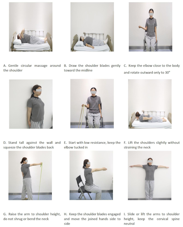
Figure 1. Phased shoulder stability training after posterior cervical laminoplasty. Please click here to view a larger version of this figure.
| Characteristics | Observation group (n=40) | Control group (n=41) | Test statistic | P value |
| Age (years) | 62.32±6.64 | 64.37±5.01 | t=-1.587 | 0.117 |
| Sex (male/female, n) | 32/8 | 29/12 | χ2=0.931 | 0.335 |
| BMI (kg/m²) | 26.71±4.52 | 24.34±5.21 | t=2.212 | 0.030* |
| Course of disease (months) | 4.01±2.06 | 3.72±1.96 | t=0.658 | 0.512 |
| Smoking history (yes/no, n) | 9/31 | 11/30 | χ2=0.200 | 0.655 |
| Hypertension (yes/no, n) | 14/26 | 12/29 | χ2=0.283 | 0.595 |
| Diabetes mellitus (yes/no, n) | 8/32 | 12/29 | χ2=0.914 | 0.339 |
| Training compliance (good/poor, n) | 34/6 | 38/3 | χ2=1.210 | 0.271 |
Table 1: Baseline characteristics and training compliance of the two groups (n, mean ± SD). Abbreviation: BMI = Body Mass Index.
| Groups | n | Before intervention | 6 weeks after intervention | 12 weeks after intervention |
| Observation group | 40 | 8.25±1.32 | 11.86±1.45*# | 14.32±1.28*# |
| Control group | 41 | 8.31±1.29 | 10.23±1.38* | 12.15±1.35* |
| t value | - | -0.198 | 5.238 | 7.542 |
| P value | - | 0.843 | <0.001 | <0.001 |
Table 2: JOA scores between the two groups preoperatively and postoperatively (mean ± SD, points). *P < 0.05, compared with the same group before intervention; #P < 0.05, compared with the control group at the same time point. Abbreviation: JOA = Japanese Orthopedic Association.
| Groups | n | Before intervention | 6 weeks after intervention | 12 weeks after intervention |
| Observation group | 40 | 52.36±5.78 | 68.45±6.23*# | 82.15±5.89*# |
| Control group | 41 | 51.89±5.92 | 60.12±6.15* | 70.32±6.01* |
| t value | - | 0.362 | 6.012 | 8.943 |
| P value | - | 0.718 | <0.001 | <0.001 |
Table 3: Constant-Murley scores between the two groups preoperatively and postoperatively (mean ± SD, points). *P < 0.05, compared with the same group before intervention; #P < 0.05, compared with the control group at the same time point.
| Groups | n | Before intervention | 6 weeks after intervention | 12 weeks after intervention |
| Observation group | 40 | 6.89±1.02 | 3.25±0.86*# | 1.78±0.75*# |
| Control group | 41 | 6.92±1.05 | 4.56±0.92* | 2.89±0.81* |
| t value | - | -0.128 | -6.654 | -6.423 |
| P value | - | 0.898 | <0.001 | <0.001 |
Table 4: Neck-shoulder VAS scores between the two groups preoperatively and postoperatively (mean ± SD, points). *P < 0.05, compared with the same group before intervention; #P < 0.05, compared with the control group at the same time point.
Supplemental File 1: Home training manual for phased shoulder stability training after posterior cervical laminoplasty. Please click here to download this file.
Discussion
The neck and shoulder muscles are important anatomical supports for maintaining cervical spine stability. After posterior cervical surgery, the damage to the neck and shoulder muscle function will lead to the imbalance of cervical spine mechanical balance, which further aggravates symptoms such as neck and shoulder pain and upper limb numbness, and hinders the rehabilitation process of patients11. At present, the importance of early rehabilitation intervention after cervical spondylosis surgery has reached a clinical consensus12,13, but most relevant studies focus on the effects of cervical spine stability training and core muscle training on patients after cervical spondylosis surgery, holding that enhancing the strength of the cervical spine and surrounding muscles is the key to improving postoperative cervical spine function and reducing complications. However, research on the effect of shoulder stability training on postoperative cervical spine function recovery is relatively limited.
The results of this study showed that the JOA scores of the observation group were significantly higher than those of the control group at 6 and 12 weeks after the intervention (P < 0.05), particularly in the recovery of upper-limb motor and sensory functions. At 12 weeks after surgery, the JOA score increased by 6.07 points from baseline in patients who received stage-specific shoulder stability training, compared with 3.84 points in the conventional rehabilitation group. The between-group difference in the magnitude of JOA score improvement was approximately 2.23 points, approaching the minimum clinically important difference reported for the original JOA score after surgery for degenerative cervical myelopathy14,15,16. This finding suggests that the functional improvement observed in the intervention group may represent not only a statistically significant difference but also a clinically meaningful benefit.
Overall, compared with patients who received conventional rehabilitation alone, those who additionally underwent shoulder stability training achieved greater improvements in cervical neurological function, shoulder function, and neck and shoulder pain outcomes. The potential mechanism may be multifactorial: first, phased shoulder stability training can enhance the strength of the surrounding shoulder muscles through targeted exercises, and the shoulder muscles are anatomically and mechanically connected to the cervical spine muscles. Enhancing shoulder strength can indirectly provide stable support for the cervical spine, improve the mechanical balance of the cervical spine, reduce mechanical pressure on the cervical spine's intervertebral discs and nerve roots, and thus promote the recovery of cervical spine function17. In addition, training such as scapula activation and scapular squeeze in shoulder stability training can promote the synergistic contraction of the cervical spine-surrounding muscles, further enhance cervical spine stability, relieve residual nerve root compression, and thus improve patients' upper-limb motor and sensory functions and increase the JOA score.
In contrast, the control group received only routine rehabilitation nursing without targeted shoulder stability training, resulting in a slow recovery of the cervical spine surrounding muscle strength and an insignificant improvement in cervical spine stability, so the effect on cervical spine function recovery was not as good as that of the observation group. Although muscle strength, scapular kinematics, electromyographic activity, and local inflammatory biomarkers were not directly measured in this study, previous research has shown that targeted exercise rehabilitation can substantially improve neuromuscular coordination and functional capacity. These potential mechanisms should therefore be regarded as hypotheses for future investigation and require confirmation through objective biomechanical and physiological measurements. Given the limitations inherent in the retrospective observational design, the present findings should be interpreted as associations rather than definitive evidence of a direct causal relationship.
Axial pain is one of the most common complications after posterior cervical surgery, which is mainly manifested as diffuse dull pain or soreness in the posterior neck, occiput, shoulder, and interscapular area, and the pain is aggravated by neck and shoulder activity, seriously affecting the surgical treatment effect and patients' rehabilitation process18. The occurrence of axial pain will not only exacerbate patients' pain experience but may also lead patients to reduce neck and shoulder activities due to fear of pain, further worsening muscle atrophy and joint stiffness, forming a vicious circle. The occurrence of axial pain is closely related to factors such as posterior cervical muscle injury during surgery, cervical spine mechanical balance, and postoperative long-term immobilization. The results of this study showed that the Constant-Murley scores of the observation group were significantly higher and the VAS scores were significantly lower than those of the control group at 6 and 12 weeks after intervention (P < 0.05), indicating that stage-specific shoulder stability training was associated with improved shoulder function and reduced neck and shoulder pain.
After posterior cervical surgery, patients' shoulder muscles are prone to spasm and atrophy due to surgical injury and long-term immobilization, leading to limited shoulder joint activity and aggravated pain19. The phased shoulder stability training in this study is designed in accordance with the postoperative recovery process of patients: from muscle relaxation and mild activation in the acute phase, to basic strength training in the recovery phase, and then to intensive strength and coordination training in the consolidation phase. This step-by-step training mode can effectively relieve shoulder muscle spasms, enhance shoulder muscle strength and coordination, and gradually improve the range of motion of the shoulder joint. Relevant studies have confirmed that targeted muscle contraction training can promote local muscle blood circulation, accelerate the clearance of inflammatory mediators in the injured area, and stimulate the release of endogenous analgesic substances (such as endorphins), thereby effectively relieving pain symptoms 19. In addition, improved shoulder joint function makes patients' neck and shoulder movements more flexible, which can reduce the aggravation of pain caused by limited joint mobility, form a positive cycle of "pain relief—increased activity—further functional recovery", and further improve the overall rehabilitation effect.
Eleven patients were excluded from this study, some because they met the prespecified exclusion criteria and others because of incomplete follow-up data, which may have introduced potential attrition bias. Although all eligible patients with complete follow-up data during the study period were included to minimize selection bias, differences between the included and excluded patients cannot be entirely ruled out. Future prospective studies with prespecified follow-up protocols and intention-to-treat analyses are needed to further validate the present findings.
The intervention was delivered by professionally trained orthopedic nurses, with continued follow-up support provided after hospital discharge. This finding indicates that nurse-guided rehabilitation programs may offer a feasible approach to extending postoperative functional management from the hospital to the home setting. Nevertheless, whether the observed benefits were attributable to specific exercise components, greater attention and support from healthcare professionals, or higher levels of patient engagement remain to be investigated in future studies.
This study has several strengths. First, the intervention program was systematically structured according to the different stages of postoperative rehabilitation, thereby enhancing its clinical applicability and reproducibility. Second, multiple outcome measures, including cervical spine function, shoulder function, and pain intensity, were evaluated, providing a relatively comprehensive assessment of postoperative rehabilitation outcomes.
Nevertheless, several limitations should be acknowledged. First, this was a single-center retrospective historical cohort study, in which group allocation was determined by the time of hospital admission rather than by randomization. Although baseline characteristics were compared and statistical adjustment was performed for the imbalance in body mass index (BMI), residual selection bias and unmeasured confounding could not be eliminated.
Second, because the intervention group received treatment during a later study period, temporal trends related to advances in surgical techniques, the accumulation of rehabilitation experience, or changes in perioperative nursing protocols may have influenced the between-group differences. In addition, patients receiving the shoulder stability training program received additional rehabilitation guidance and greater therapist contact time, which may have partially contributed to the observed differences between the groups. Future randomized controlled trials with equivalent therapist contact time across groups are warranted to clarify the training program's independent effects.
Third, the outcome assessors were not blinded to group allocation, which may have introduced measurement bias. Future studies should incorporate blinded outcome assessment whenever feasible. Fourth, this study relied primarily on clinical rating scales and lacked objective outcome measures, including shoulder range of motion, muscle strength testing, scapular motion analysis, and electromyography. Future studies should integrate objective functional assessments to provide a more thorough understanding of the potential mechanisms underlying the intervention effects.
Finally, the follow-up period was limited to 12 weeks after surgery; therefore, the long-term sustainability of the observed functional improvements, such as at 6 months postoperatively or beyond, remains uncertain. Future multicenter randomized controlled trials with larger sample sizes and longer follow-up periods are needed to further evaluate the long-term effects of stage-specific shoulder stability training. Moreover, individualized training programs could be tailored to patient characteristics, such as age, preoperative functional status, and BMI, to optimize rehabilitation outcomes and provide more personalized, evidence-based strategies for patients with cervical radiculopathy undergoing posterior cervical surgery.
In conclusion, in this retrospective historical cohort study, conventional rehabilitation combined with a stage-specific shoulder stability training program was associated with improved recovery of cervical spine and shoulder function and greater relief of neck and shoulder pain in patients undergoing unilateral open-door posterior cervical laminoplasty. These findings suggest that shoulder-focused rehabilitation training may represent a promising adjunctive rehabilitation strategy; however, prospective randomized controlled trials are required to further confirm its effectiveness.
Disclosures
During the preparation of this manuscript, artificial intelligence (AI) tools were used solely for language editing, grammatical refinement, and assistance with improving the manuscript structure. AI tools were not used for data collection, statistical analysis, interpretation of the results, generation of scientific content, or preparation of figures and tables. The authors take full responsibility for the accuracy, completeness, and originality of the manuscript. The authors have no conflicts of interest to declare.
Acknowledgements
This work was supported by the Medical and Health Science Program of Zhejiang Province (2025HY0416), and the General Research Project of the Zhejiang Provincial Department of Education (Y202352208).
Materials
| Name | Company | Catalog Number | Comments |
|---|---|---|---|
| Constant-Murley shoulder assessment form | C. R. Constant and A. H. G. Murley; noncommercial assessment instrument | Standard 100-point clinician-administered version | Pain (15 points), activities of daily living (20 points), range of motion (40 points), and strength (25 points). Used preoperatively and at weeks 6 and 12. Product or source URL: pubmed.ncbi.nlm.nih.gov/9250771/ |
| Exercise mat or firm rehabilitation surface | AIREX / Performance Health, Warrenville, IL, USA | Yoga/Pilates 190 | Approximately 190 x 58 x 0.8 cm, dense foam, slip-resistant surface. Used for supine and prone scapular activation and shoulder-lifting exercises; a firm rehabilitation plinth or bed of equivalent stability may be substituted. Product or source URL: performancehealth.com/yoga-pilates-190 |
| Hand-held dynamometer | Lafayette Instrument Company, Lafayette, IN, USA | Model 01165A | Measurement range 0-300 lb (0-136.1 kg; 0-1335 N), accuracy +/-1% of full scale or +/-0.2 lb. Used for objective shoulder abduction strength measurement within the Constant-Murley assessment. Product or source URL: lafayetteevaluation.com/products/01165a-hand-held-dynamometer/ |
| Illustrated Home Training Manual | Nursing Department, The Second Affiliated Hospital, Zhejiang University School of Medicine, Hangzhou, China | Version 1.0; January 2024 | Noncommercial illustrated manual covering the 12-week phased program, exercise frequency and duration, movement instructions, resistance progression, safety stop criteria, and contact instructions. Provided to patients before discharge. Product or source URL: Noncommercial institutional document; available from the corresponding author |
| Japanese Orthopedic Association (JOA) assessment form | Japanese Orthopaedic Association | 17-point clinician-administered version described by Yonenobu et al. (2001) | Four domains: upper-extremity motor function (4 points), lower-extremity motor function (4 points), sensory function (6 points), and bladder function (3 points). Used preoperatively and at weeks 6 and 12. Product or source URL: pubmed.ncbi.nlm.nih.gov/11568701/ |
| Light hand weights / dumbbells | Sammons Preston / Performance Health, Warrenville, IL, USA | Individual neoprene dumbbells; 1-4 lb series | Progressive loads of approximately 0.5, 1.0, 1.5, and 2.0 kg (nearest commercial equivalents acceptable). Used during weeks 4-12 for upper-limb strengthening and light-object lifting exercises. Product or source URL: performancehealth.com/individual-neoprene-dumbbell-sets |
| Progressive resistance bands | THERABAND / Performance Health, Warrenville, IL, USA | Professional Non-Latex Resistance Bands; product code varies by resistance level and roll length | Flat, non-latex band, 10 cm wide; yellow, red, and green levels; cut into approximately 1.5 m lengths. Used for seated shoulder external rotation and standing shoulder abduction, with progression from low to moderate resistance according to tolerance. Product or source URL: performancehealth.com/theraband-professional-non-latex-resistance-bands |
| Resistance-band anchor and handles | THERABAND / Performance Health, Warrenville, IL, USA | Item 081510387; Catalog 713306 | Accessories kit containing an adjustable nylon door anchor, assist strap, and soft-grip handles. Used to secure the free end of the resistance band during home-based exercises. Product or source URL: performancehealth.com/thera-band-accessories-kit |
| Soft cervical support pillow | Institutional standard supply; no brand-specific requirement | Not applicable | Medium-firm foam pillow, approximately 50 x 30 x 8-10 cm. Placed behind the neck during the acute postoperative phase to maintain a neutral cervical position. Product or source URL: Not applicable (non-specialized clinical aid) |
| Stable armless chair | Institutional standard supply; no brand-specific requirement | Not applicable | Non-wheeled chair with a stable backrest and an approximate seat height of 42-46 cm. Used for seated resistance-band external rotation and scapular stability exercises. Product or source URL: Not applicable (non-specialized clinical furniture) |
| Statistical analysis software | IBM Corp., Armonk, NY, USA | IBM SPSS Statistics for Windows, Version 26.0 | Used for descriptive statistics, independent-samples t tests, chi-square or Fisher exact tests, repeated-measures analysis of variance, and covariance adjustment. Product or source URL: ibm.com/support/pages/ibm-spss-statistics-26-documentation |
| Training diary and weekly follow-up checklist | Nursing Department, The Second Affiliated Hospital, Zhejiang University School of Medicine, Hangzhou, China | Version 1.0; January 2024 | Paper-based forms recording session completion, exercise dose, physical reactions, pain or numbness, reasons for missed sessions, and issues discussed during weekly telephone follow-up. Used to calculate training completion and compliance. Product or source URL: Noncommercial institutional document; available from the corresponding author |
| Universal goniometer | Baseline / Fabrication Enterprises, Inc.; distributed by Performance Health | Absolute-Axis 12-inch goniometer; catalog varies by configuration | 360-degree transparent goniometer with two 30-cm arms. Used by the rehabilitation therapist to quantify shoulder flexion, abduction, and rotational range of motion for the Constant-Murley assessment. Product or source URL: performancehealth.com/baseline-absolute-axis-a-a-goniometer |
| Visual Analog Scale (VAS) pain form | Study-specific paper form; noncommercial | Version 1.0; 10-cm horizontal scale | Horizontal 10-cm line anchored by “no pain” and “worst imaginable pain”; measured and converted to a 0-10 score. Used for neck-shoulder pain preoperatively and at weeks 6 and 12. Product or source URL: doi.org/10.1111/j.1365-2702.2005.01121.x |
References
- Kang KC, Lee HS, Lee JH, et al. Cervical radiculopathy: focus on characteristics and differential diagnosis. Asian Spine J. 2020;14(6):921-30.
- Xu X, Ling Y. Manual therapy for cervical radiculopathy: effects on neck disability and pain-a systematic review and network meta-analysis. J Pain Res. 2025;18:2035-45.
- Rissardo JP, Caprara ALF, et al. Management of cervical radiculopathy: current systematic reviews. Indian J Health Sci Biomed Res (KLEU). 2023;16(3):441-8.
- Choi BW, Kim SS, Lee DH, et al. Cervical radiculopathy combined with cervical myelopathy: prevalence and characteristics. Eur J Orthop Surg Traumatol. 2017;27(7):889-93.
- Weisbrod LJ, Ceesay OI, Schmidt C, Haynatzki R, Surdell DL. Analgesic safety and efficacy of perioperative posterior cervical muscle plane blocks in elective posterior cervical spine surgery: a systematic review with meta-analyses. Clin J Pain. 2025;41(3):e1269.
- Lee YN, Shin MMS, Lee W, et al. Effects of shoulder stabilization exercise on pain and function in patients with neck pain. J Phys Ther Sci. 2015;27(12):3619-22.
- Popchak A, Patterson-Lynch B, Christain H, et al. Rehabilitation and return to sports after anterior shoulder stabilization. Ann Joint. 2017;2:62.
- Liaghat B, Pedersen JR, Husted RS, et al. Diagnosis, prevention and treatment of common shoulder injuries in sport: grading the evidence-a statement paper commissioned by the Danish Society of Sports Physical Therapy (DSSF). Br J Sports Med. 2023;57(7):408-16.
- Yonenobu K, Abumi K, Nagata K, et al. Interobserver and intraobserver reliability of the Japanese Orthopaedic Association scoring system for evaluation of cervical compression myelopathy. Spine (Phila Pa 1976). 2001;26(17):1890-4.
- Bankes M, Emery RJH, et al. An evaluation of the Constant-Murley shoulder assessment. J Bone Joint Surg Br. 1997;79-B(4):696-9.
- Deshpande N, Stino A, Smith BW, et al. Defining postoperative C5 palsy and recovery: a systematic review. J Neurosurg Spine. 2023;38(4):457-64.
- Epstein NE, et al. A review article on the benefits of early mobilization following spinal surgery and other medical/surgical procedures. Surg Neurol Int. 2014;5(Suppl 3):S66-73.
- Tong Y, Fernandez L, Bendo JA, et al. Enhanced recovery after surgery trends in adult spine surgery: a systematic review. Int J Spine Surg. 2020;14(4):623-40.
- Kato S, Oshima Y, Matsubayashi Y, et al. Minimum clinically important difference and patient acceptable symptom state of Japanese Orthopaedic Association score in degenerative cervical myelopathy patients. Spine (Phila Pa 1976). 2019;44(10):691-7.
- Oshima Y, Takeshita K, Kato S, et al. Comparison between the Japanese Orthopaedic Association (JOA) score and Patient-Reported JOA (PRO-JOA) score to evaluate surgical outcomes of degenerative cervical myelopathy. Global Spine J. 2022;12(5):795-800.
- Zhou F, Zhang Y, Sun Y, et al. Assessment of the minimum clinically important difference in neurological function and quality of life after surgery in cervical spondylotic myelopathy patients: a prospective cohort study. Eur Spine J. 2015;24(12):2918-23.
- Kim MK, Jeong BC, Yoo KT, et al. The effect of shoulder stabilization exercise and core stabilization exercise on shoulder height and respiratory function in young adults with round shoulder posture. J Korean Soc Phys Med. 2023;18(4):1-17.
- Kibler WB, Sciascia A, Uhl TL, et al. Electromyographic analysis of specific exercises for scapular control in early phases of shoulder rehabilitation. Am J Sports Med. 2008;36(9):1789-98.
- Nijs J, et al. Dysfunctional endogenous analgesia during exercise in patients with chronic pain: to exercise or not to exercise? Pain Physician. 2012;15(3 Suppl):ES205-13.