This prospective cohort study assessed digital wound features, thermography, and local inflammatory markers in burns, chronic ulcers, and hand reconstruction wounds. Multimodal models showed modest internally validated discrimination without significant improvement over clinical assessment, supporting exploratory risk stratification only and requiring external validation before clinical use.
Research Article
Early Digital and Inflammatory Markers of Delayed Healing in Burns, Ulcers, and Hand Reconstruction: A Prospective Cohort Study
56 views
⸱
September 11th, 2026
In This Article
Summary
Abstract
Delayed healing is assessed differently in burns, chronic ulcers, and hand soft-tissue reconstruction wounds, yet these patients are often managed in the same wound service. This prospective single-center cohort study evaluated whether standardized digital wound features, thermography, and local inflammatory markers added to early delayed-healing risk assessment across these three categories. Adults enrolled between 1 January 2024 and 31 December 2025 underwent baseline wound photography, thermal imaging, and wound-fluid sampling. Delayed healing was defined using prespecified wound-type-specific criteria. Clinical, digital-feature, inflammatory-marker, baseline combined, and day-7 updated models were fitted with wound type forced into each model and internally validated with 1000 bootstrap samples. Of 168 patients (57 with burns, 70 with chronic ulcers, and 41 with hand soft-tissue reconstruction wounds), 59 (35.1%) met delayed-healing criteria. Delayed-healing wounds were deeper and had more slough and necrotic tissue, higher exudate scores and wound-reference temperature differences, and higher interleukin-6 (IL-6) concentrations; several other differences were imprecise. Apparent AUCs were 0.691 for the clinical model, 0.717 for the baseline combined model, and 0.715 for the early updated model. The corresponding optimism-corrected AUCs were 0.632, 0.660, and 0.649. The baseline combined model did not significantly improve discrimination over the clinical model (ΔAUC 0.026; DeLong p = 0.400), and adding day-7 area reduction did not improve the baseline combined model (ΔAUC -0.002; p = 0.662). These models therefore showed modest internally validated performance rather than clinical readiness. At present, they serve primarily as a hypothesis-generating framework for early risk stratification. Independent external validation and prospective impact assessment are required before implementation.
Introduction
Delayed wound healing remains a clinical problem across burns, chronic ulcers, and postoperative soft-tissue reconstruction. These conditions differ in cause and time-to-healing expectations, but a tertiary wound service must often make the same early operational decision: which wounds require closer monitoring, earlier specialist review, or escalation of assessment. Burns may require timely excision or grafting1,2, chronic ulcers reflect interacting perfusion, pressure, neuropathy, infection, and metabolic factors3,4, and reconstructive wounds may fail through dehiscence, infection, or flap/graft compromise. A unified model is clinically relevant only if wound type is retained explicitly and heterogeneity is evaluated rather than assumed away.
Chronic ulcers present a different but equally difficult problem. These wounds often persist because of impaired perfusion, infection, neuropathy, pressure, edema, metabolic disease, or excessive local inflammation. Large-scale chronic wound modeling has shown that wound-level factors such as area, depth, duration, location, and etiology contribute substantially to healing prediction3. In diabetic foot ulcers, early percentage change in wound area has been shown to predict subsequent complete healing, with 4-week area reduction serving as a practical benchmark for identifying wounds unlikely to heal with standard care alone4. However, waiting several weeks to determine whether a wound is responding may be too slow for wounds at high risk of infection, tissue loss, or reconstructive failure.
Digital wound assessment offers a way to make early evaluation more objective. Standardized wound photography and calibrated planimetry can quantify wound area and surface change more consistently than visual estimation alone5. Image-based measurement also allows additional wound-bed features to be extracted, including slough percentage, necrotic tissue, erythema, exudate-related appearance, and texture heterogeneity. Smartphone-based and ImageJ-based digital measurement methods have been increasingly evaluated as practical tools for wound area assessment in clinical and near-clinical settings6. Yet most routine wound assessment still relies on area reduction alone, which may miss early tissue-quality changes that precede delayed closure.
Thermal imaging provides another non-invasive dimension of wound assessment. Local temperature variation may reflect inflammatory activity, altered perfusion, bacterial burden, or tissue stress, depending on wound type and timing. Thermal texture analysis has been explored for predicting venous leg ulcer healing trajectories, suggesting that thermal heterogeneity may contain prognostic information beyond wound size7. Recent reviews also describe point-of-care thermography as a promising adjunct for wound monitoring, particularly when interpreted alongside clinical and imaging features rather than as a standalone diagnostic test8. For heterogeneous wound populations, thermal assessment may therefore help capture physiological signals that are not visible in conventional photographs.
Despite different etiologies, the three wound categories share measurable processes relevant to repair: tissue burden, inflammatory activation, matrix turnover, local perfusion, and early change in wound area. Wound-fluid cytokines can reflect local tissue response9,10, while matrix metalloproteinase-9 (MMP-9) reflects protease activity and extracellular-matrix degradation and has been associated with poor diabetic foot-ulcer healing11. These shared domains provide a biological rationale for testing a common model, but they do not establish that predictor effects are identical across wound categories.
Most published studies focus on one wound type or one measurement domain, whereas mixed wound clinics use shared imaging equipment and common assessment workflows. A pooled model could therefore reduce workflow fragmentation, but its clinical value depends on transparent model specification, internal validation, calibration, and explicit subgroup assessment12. Wound-type-specific outcomes and interaction analyses are necessary safeguards because healing timelines are not interchangeable.
Despite shared physiological processes in tissue repair, a gap remains in integrating multimodal assessments across diverse wound populations in routine clinical settings. We hypothesized that a novel pooled, wound-type-adjusted model combining early clinical, digital, thermal, and inflammatory features would improve the early prediction of delayed healing compared with standard clinical evaluation alone, while explicitly preserving etiological heterogeneity. Accordingly, the primary objective was to evaluate the associations of these early features with delayed healing and to compare the prespecified clinical and multimodal models. The analysis also assessed performance within each wound category, wound type-by-predictor interactions, and sensitivity to post-baseline treatment exposure. Given the single-center development design, all performance estimates were considered exploratory and internally validated rather than implementation-ready. If externally validated, this framework could ultimately influence clinical decision-making by triaging high-risk wounds for earlier specialist intervention and optimizing resource allocation across mixed wound services.
Protocol
The study was reviewed and approved before initiation by the Ethics Committee of the General Hospital of Northern Theater Command (approval number Y(2023)455; approval date 11 November 2023). The Ethics Committee waived the requirement for written informed consent because the study was strictly observational and noninterventional. Clinical records, wound photographs, thermal images, wound-fluid or swab specimens, and biomarker data were de-identified and coded with unique study identifiers; the re-identification key was stored separately and was accessible only to authorized study personnel. The details of all the reagents, equipments, thermal-camera model, and software names/versions/RRIDs used are listed in the Table of Materials.
Study design and setting
This prospective observational cohort study was conducted in the Department of Burn and Plastic Surgery and the Hand Surgery Unit of a tertiary teaching hospital between 1 January 2024 and 31 December 2025. Consecutive adults with burn wounds, chronic ulcers, or hand soft-tissue reconstruction wounds were screened at first wound assessment or within 48 h after surgery. Reporting followed the Strengthening the Reporting of Observational Studies in Epidemiology (STROBE) recommendations13. The screening, enrollment, baseline assessment, follow-up, and analysis pathway are shown in Figure 1.
A total of 196 patients with burn wounds, chronic ulcers, or hand soft-tissue reconstruction wounds were screened between 1 January 2024 and 31 December 2025. After exclusion of 28 patients (12 did not meet eligibility criteria, 6 had incomplete baseline imaging, 5 lacked usable fluid or swab samples, and 5 declined follow-up), 168 patients were enrolled and classified into burn wounds, chronic ulcers, or hand soft-tissue reconstruction wounds. All enrolled patients underwent baseline standardized wound photography, thermal imaging, and wound fluid biomarker sampling, followed by scheduled assessments at day 7, day 14, day 21, day 28/week 4, and week 12. Final adjudication classified patients into timely healing and delayed healing groups for analysis, with delayed healing broadly defined as the failure to meet wound-type-specific closure milestones or the need for unplanned surgical intervention.
Participants
Patients were eligible if they were aged 18–85 years, had one clinically identifiable target wound with a measurable wound bed, and were expected to complete at least 4 weeks of follow-up. Three wound categories were included: burn wounds, chronic ulcers, and hand soft-tissue reconstruction wounds. These biologically distinct etiologies were pooled to reflect the operational reality of a mixed-wound tertiary service and to capture shared measurable domains of tissue repair, with etiological heterogeneity explicitly addressed through prespecified subgroup and interaction analyses (detailed in the Statistical analysis section). Burn wounds included superficial partial-thickness, deep partial-thickness, and full-thickness burns managed by conservative dressing, staged debridement, or grafting when clinically required. Chronic ulcers included diabetic foot ulcers, venous leg ulcers, pressure ulcers, and traumatic non-healing ulcers lasting more than 4 weeks. Hand soft-tissue reconstruction wounds included wounds after local flap repair, skin grafting, tendon-exposed wound coverage, or soft-tissue defect reconstruction.
Patients were excluded if they had malignant wounds, active systemic autoimmune disease, systemic corticosteroid or immunosuppressive therapy within the previous 30 days, chemotherapy within the previous 3 months, severe peripheral arterial disease requiring urgent revascularization, incomplete baseline wound imaging, unavailable wound fluid or wound swab samples, or loss to follow-up before the first outcome assessment.
For patients with multiple wounds, one target wound was selected before baseline imaging. The target wound was defined as the wound with the largest area, deepest tissue involvement, or highest clinical concern for delayed closure. The same target wound was followed throughout the study.
Study timeline and assessment schedule
Baseline assessment was defined as T0. For burn and chronic ulcer patients, T0 was the first standardized wound assessment after enrollment. For hand soft-tissue reconstruction patients, T0 was defined as postoperative day 1 to day 3, before routine dressing change whenever feasible.
Follow-up assessments were performed at day 7 ± 2 days, day 14 ± 3 days, day 21 ± 2 days for burn epithelialization assessment, day 28 ± 5 days, and week 12 ± 7 days. Burn wounds were evaluated for epithelialization on day 21. Chronic ulcers were evaluated for early area reduction at week 4 and complete closure by week 12. Hand reconstruction wounds were evaluated for stable closure, wound dehiscence, flap or graft compromise, infection, and unplanned revision by postoperative day 28. The fixed assessment schedule is presented in Table 1.
Standardized wound photography and digital wound measurement
Wound photographs were obtained using a commercial smartphone equipped with a 48-megapixel main camera, positioned approximately 30 cm above and perpendicular to the wound plane. A 2 cm sterile disposable ruler and a color calibration card were placed in the wound plane outside the sterile field. Flash was disabled, fixed clinical lighting was used, and image resolution was at least 3024 pixels × 4032 pixels. Images with motion blur, incomplete wound borders, excessive glare, a missing ruler or color card, or an angle greater than approximately 15° were repeated before dressing application.
Image preprocessing was performed using open-source image analysis software. Images were rotated to a common orientation, cropped to the target wound and surrounding reference skin, and scaled with the 2 cm ruler. Normalize color using the neutral patches of the color calibration card before tissue segmentation. The photograph region of interest was aligned across visits using the ruler, wound-edge landmarks, and the saved polygon mask. Wound margins were traced independently by two trained assessors blinded to healing status. If area estimates differed by more than 10%, a third assessor repeated the measurement and the mean of the two closest estimates was used14. Baseline digital features comprised wound area, perimeter, circularity, slough percentage, necrotic-tissue percentage, erythema index, exudate score, and texture entropy. Slough and necrotic tissue were segmented in the image analysis software after color normalization using the Hue–Saturation–Brightness color thresholding tool. Thresholds were interactively optimized for each image by two trained blinded assessors according to standardized operating procedures, with visual consensus between the assessors against the normalized true-color image serving as the final acceptance criterion. This was followed by manual correction of dressing residue, blood staining, specular glare, and non-wound pixels. All edits were saved in the software's region-of-interest set. Texture entropy was calculated from grayscale images using a gray-level co-occurrence matrix plugin within the software with a pixel distance of 1 and directions of 0°, 45°, 90°, and 135°, and the four values were averaged.
The day-7 wound area reduction rate was calculated as follows:
 (1)
(1)
A negative value indicated wound enlargement. Reliability for baseline wound area was evaluated with the intraclass correlation coefficient [ICC(2,1)], a two-way random-effects, absolute-agreement model, and a subject-level bootstrap 95% confidence interval.
Thermal imaging and temperature-derived features
Thermal images were acquired using a commercial smartphone-compatible thermal camera, connected to the same smartphone. The emissivity setting was fixed at 0.98 for skin surface imaging. The device was calibrated before each imaging session according to the manufacturer’s automatic calibration procedure. Images were captured after the wound had been exposed to room air for 5 min. Room temperature was maintained at 22–25 °C, and relative humidity was maintained at 40–60%. Wound cleansing, irrigation, topical antimicrobial application, and debridement were not performed within 15 min before imaging.
Three regions were recorded: the wound bed, periwound skin within 1 cm of the wound edge, and contralateral or adjacent intact reference skin. The primary thermal variable, the wound-reference temperature difference, was calculated by subtracting the mean pixel temperature of the intact reference-skin region of interest from the mean pixel temperature of the wound-bed region of interest. A difference of ≥2.0 °C was prespecified as a high-temperature-risk pattern. Thermography was treated as an adjunct because temperature is sensitive to inflammation, perfusion, environment, and recent manipulation7,8.
Thermal texture entropy was extracted using standard open-source programming languages and computer vision libraries. The thermal frame was cropped to the same field of view as the photograph; the wound mask was registered using wound-edge landmarks and the camera overlay, then visually checked before extraction. Pixel intensities within the wound region were normalized to the intact-skin reference region. Thermal frames failing the focus, field-of-view, stabilization, or registration checks were repeated or excluded. Thermal texture analysis was exploratory7.
Wound fluid and local inflammatory marker collection
Local inflammatory samples were collected at T0 before wound cleansing, irrigation, debridement, or topical medication. When sufficient exudate was present, wound fluid was collected using a sterile rayon swab placed gently on the wound bed for 30 s without causing bleeding. For relatively dry wounds, the wound bed was moistened with 200 µL sterile normal saline, and the swab was collected after 60 s. Each swab was placed into 1.0 mL phosphate-buffered saline containing 0.05% Tween-20, vortexed for 60 s, and centrifuged at 3,000 x g for 10 min at 4 °C. The supernatant was aliquoted and stored at −80 °C. Samples were thawed only once before assay.
The local inflammatory panel included interleukin-6 (IL-6), interleukin-8 (IL-8/CXCL8), tumor necrosis factor-alpha (TNF-α), and matrix metalloproteinase-9 (MMP-9). All biomarkers were measured using commercially available enzyme-linked immunosorbent assay (ELISA) kits. Assays were performed according to the manufacturers' instructions. Specifically, primary incubations were performed at room temperature (20–25 °C) for 2 h. The standard curve ranges and lower limits of detection were, respectively, 3.1–300 pg/mL and 0.7 pg/mL for IL-6; 1.5 pg/mL and 7.5 pg/mL for IL-8/CXCL8; 0.5 pg/mL and 5.5 pg/mL for TNF-α; and 31.2 pg/mL and 2,000 pg/mL for MMP-9. Samples were assayed in duplicate and repeated when the coefficient of variation exceeded 15%. Cytokines were expressed as pg/mL and MMP-9 as ng/mL. Standardized wound-swab methods were used to characterize the local microenvironment15.
Total protein concentration in each wound fluid eluate was measured using a bicinchoninic acid protein assay. The primary analysis used absolute marker concentrations. A sensitivity analysis used biomarker concentrations normalized to total protein concentration to reduce the influence of exudate dilution.
Peripheral blood samples were collected at T0 for white blood cell (WBC) count, C-reactive protein, serum albumin, hemoglobin, fasting glucose, and glycated hemoglobin when clinically available. Wound bacterial culture was performed when infection was clinically suspected or when purulent exudate was present.
Clinical wound assessment and treatment documentation
At each visit, wound depth, exudate level, necrotic tissue, slough, granulation, epithelialization, periwound erythema, odor, and pain were recorded. Wound depth was graded as follows: grade 1, epidermal or superficial dermal involvement; grade 2, full-thickness skin or subcutaneous involvement without exposed tendon, bone, joint capsule, or implant; and grade 3, deep involvement with exposed tendon, bone, joint capsule, implant, or flap/graft compromise.
Exudate was scored from 0 to 4 (0 = absent; 1 = minimal; 2 = moderate; 3 = heavy; 4 = excessive or requiring an unscheduled dressing change). Baseline clinical infection was defined by purulent discharge or at least two of the following signs: increased pain, warmth, erythema, swelling, malodor, delayed granulation, friable tissue, or systemic inflammatory response.
Treatment was not assigned by the protocol. Follow-up records captured whether a patient received systemic antibiotics for >72 h, negative-pressure wound therapy, post-T0 surgical debridement, skin grafting or flap revision, or unplanned reoperation. Exact start and stop dates were not available in the supplied analytic dataset, so timing relative to individual imaging and sampling visits could not be modeled. These post-baseline indicators were used only in sensitivity analyses, not in the primary baseline models.
Outcome definition and adjudication
The primary outcome was delayed healing, defined using prespecified wound-type-specific criteria. For burn wounds, delayed healing was defined as failure to achieve complete epithelialization by day 21 after injury, unplanned grafting after failed conservative management, or repeated surgical debridement for persistent non-viable tissue. For chronic ulcers, delayed healing was defined as less than 50% reduction in wound area by week 4 or failure to achieve complete closure by week 12. For hand soft-tissue reconstruction wounds, delayed healing was defined as failure to achieve stable wound closure by postoperative day 28, wound dehiscence, flap or graft necrosis requiring additional intervention, new or worsening surgical-site infection after T0 requiring systemic antibiotics, or unplanned revision surgery.
Complete closure was defined as full epithelial coverage without drainage or dressing protection at two consecutive visits. Chronic-ulcer delayed healing was the union of <50% area reduction at week 4 or incomplete closure by week 12. MMP-9 was retained as a candidate predictor because elevated wound-fluid MMP-9 has been associated with poor diabetic foot-ulcer healing11.
Outcome adjudication was performed by two clinicians blinded to digital feature values and biomarker concentrations. Disagreements were resolved by a third senior wound specialist. The prespecified delayed healing criteria are summarized in Table 2.
Prediction time points and candidate predictor control
Two prediction time points were prespecified. The baseline model used only variables available at T0, including clinical variables, baseline digital wound features, baseline thermal features, and baseline inflammatory markers. The early updated model additionally included day-7 wound area reduction. This separation avoided treating day-7 information as a baseline predictor.
The final analytic cohort comprised 168 patients and 59 delayed-healing events. The baseline combined model included six prespecified continuous/ordinal predictors plus two wound-type indicators; the early model added day-7 area reduction. This event-to-parameter ratio is limited, so coefficients and subgroup estimates were interpreted cautiously and bootstrap optimism correction was reported.
Data management and missing data
Clinical data were entered into a secure, web-based electronic data capture platform. Each patient was assigned a study identification number. Identifiable information was stored separately from the analytical dataset. Image files were renamed by study identification number, wound type, and visit time point (e.g., W023_T0_photo and W023_D7_thermal).
Range checks were applied before analysis, and extreme values were retained after verification rather than removed automatically. Missingness in model predictors was below 5%: albumin, day-7 area reduction, erythema, and IL-6 each had three missing values, slough had three, and MMP-9 had six. Continuous predictors were imputed with the cohort median and categorical predictors with the mode. No primary outcomes were missing.
Statistical analysis
Analyses were reproduced using standard open-source statistical and data analysis packages. Continuous variables are reported as mean ± standard deviation (SD) and compared with Welch independent-sample t tests; categorical variables are reported as n (%) and compared with Fisher exact or chi-square tests, as appropriate. p values are two-sided.
Predictors were fixed before model fitting rather than selected by univariable p value. Logistic regression was used throughout. Wound type was represented by chronic-ulcer and hand-reconstruction indicators with burn wounds as reference. Continuous predictors were scaled as prespecified.
Five prespecified models were evaluated: clinical, digital wound-feature, inflammatory-marker, baseline combined, and early updated combined. Discrimination was summarized by AUC with DeLong 95% confidence intervals (CIs). Calibration intercept, calibration slope, and Brier score were calculated. Internal validation used 1000 bootstrap samples to estimate optimism-corrected AUC, calibration, and Brier score. The Transparent Reporting of a multivariable prediction model for Individual Prognosis or Diagnosis (TRIPOD) guidance was followed16.
Empirical decision curves were calculated from patient-level predicted probabilities for thresholds from 0.10 to 0.60. Correlated AUCs were compared with DeLong tests. Model performance was estimated separately within the burn (n = 57), chronic-ulcer (n = 70), and hand-reconstruction (n = 41) subgroups using the pooled-model predictions. Predictor-by-wound-type interactions were added one predictor at a time and assessed by a two-degree-of-freedom likelihood-ratio test. Subgroup and interaction analyses were exploratory.
The reproducible workflow is shown in Figure 2. De-identified patient-level data, a data dictionary, patient-level predictions, coefficients, receiver operating characteristic (ROC)/calibration/decision-curve source tables, and the Python scripts used to regenerate the analyses and figures are provided as supplemental files.
Sensitivity analyses
Five sensitivity analyses were performed: refitting after excluding baseline clinical infection; evaluating pooled-model predictions for 12-week non-closure among chronic ulcers; replacing IL-6 and MMP-9 by log(1 + concentration); dividing these biomarkers by total wound-fluid protein; and adding five post-baseline treatment indicators. The treatment-adjusted analysis was interpreted as a confounding assessment, not as a valid baseline prediction model, because treatments were selected according to clinical severity and some overlapped with outcome criteria.
Results
Patient screening and outcome classification
Between 1 January 2024 and 31 December 2025, 196 patients with burn wounds, chronic ulcers, or hand soft-tissue reconstruction wounds were screened. Twenty-eight patients were excluded: 12 did not meet the eligibility criteria, 6 had incomplete baseline wound imaging, 5 could not provide usable wound fluid or wound swab samples, and 5 declined follow-up. A total of 168 patients were enrolled and included in the final analysis (Figure 1).
Among the 168 included patients, 57 had burn wounds, 70 had chronic ulcers, and 41 had hand soft-tissue reconstruction wounds. Based on the fixed assessment schedule (Table 1) and prespecified outcome criteria (Table 2), the reproducible wound-type-specific outcome algorithm classified 109 patients (64.9%) as timely healing and 59 (35.1%) as delayed healing.
Baseline clinical, digital, and thermal wound characteristics
The prespecified analytical workflow is shown in Figure 2, and the predictor structure used for model fitting is detailed in Table 3. Baseline clinical, digital, and thermal characteristics are summarized in Table 4. The mean age was 54.7 years ± 14.7 years, 94 patients (56.0%) were male, and 62 (36.9%) had diabetes. Baseline clinical infection was more frequent among delayed-healing patients (20.3% vs 8.3%, p = 0.029); age, sex, diabetes, smoking, hemoglobin, and albumin differences were imprecise.
Baseline wound area did not differ between outcome groups (16.0 cm2 ± 12.4 cm2 delayed vs 15.5 cm2 ± 12.0 cm2 timely, p = 0.768). Delayed-healing wounds were deeper (2.20 ± 0.74 vs 1.80 ± 0.70, p < 0.001). The wound-type distribution did not differ significantly by outcome (p = 0.167).
Delayed-healing wounds had more slough (31.2% ± 12.8% vs 24.3% ± 14.2%, p = 0.002), more necrotic tissue (10.8% ± 7.1% vs 8.1% ± 7.3%, p = 0.026), and higher exudate scores (1.90 ± 0.88 vs 1.44 ± 0.90, p = 0.002). Differences in erythema index (p = 0.176) and photographic texture entropy (p = 0.060) were uncertain. Baseline area agreement was ICC(2,1) = 0.997 (bootstrap 95% CI 0.996–0.998); five cases required a third measurement.
All 168 included baseline thermal images were marked as passing quality control. Wound-reference temperature difference was higher in delayed-healing wounds (1.41 °C ± 0.84 °C vs 1.06 °C ± 0.74 °C, p = 0.009), and the ≥2.0 °C pattern occurred in 25.4% versus 11.9% (p = 0.031). Thermal texture entropy did not differ (p = 0.372). Reader-level repeat measurements for non-area digital and thermal features were unavailable, preventing post hoc ICC or kappa estimation.
Day-7 area reduction was observed in 165 patients and was lower on average in the delayed-healing group, but the difference was imprecise (17.9% ± 11.3% vs 21.5% ± 12.2%, p = 0.059). Six patients had wound enlargement by day 7. Patient-level digital and thermal distributions are shown in Figure 3.
Local inflammatory markers and treatment exposures
Local markers and treatment exposures are summarized in Table 5. These specific biomarkers were selected to represent distinct, biologically relevant pathways of impaired tissue repair: IL-6, IL-8/CXCL8, and TNF-α are canonical mediators of early inflammatory activation and leukocyte recruitment, while MMP-9 is a primary driver of excessive extracellular matrix degradation. IL-6 was higher in delayed-healing wounds (65.5 pg/mL ± 49.6 pg/mL vs 50.2 pg/mL ± 30.4 pg/mL, p = 0.034). IL-8/CXCL8 was also numerically higher (238.7 pg/mL ± 149.0 pg/mL vs 195.0 pg/mL ± 112.4 pg/mL), but its confidence was limited (p = 0.052); TNF-α did not differ significantly (p = 0.097).
MMP-9 concentrations were 391.4 ng/mL ± 192.3 ng/mL in delayed-healing wounds and 357.4 ng/mL ± 187.3 ng/mL in timely-healing wounds (p = 0.285). Protein-normalized MMP-9, CRP, WBC count, and positive culture frequency also did not differ significantly.
Follow-up systemic antibiotics, debridement, graft/flap revision, and unplanned reoperation were more common among delayed-healing patients (p = 0.009, p = 0.046, p = 0.026, and p = 0.013, respectively), whereas negative-pressure wound therapy was not (p = 0.126). Exposure frequencies did not differ significantly across the three wound categories (all p ≥ 0.106). While these broad intervention categories were tracked, specific clinical management inherently varied by etiology (e.g., early excision and grafting for burns; offloading, compression, or revascularization for chronic ulcers; and flap salvage for reconstructive wounds). Exact intervention timing was unavailable.
Figure 4 displays all available patient-level biomarker observations and a square, label-aligned Spearman correlation matrix generated from the analytic dataset. Delayed-healing wounds showed higher IL-6 concentrations, while IL-8/CXCL8 and MMP-9 differences were imprecise; the inflammatory markers showed modest correlations with thermal and tissue-burden features.
Univariable and multivariable predictors of delayed healing
Unadjusted group comparisons identified greater wound depth, slough, necrotic tissue, exudate, temperature difference, baseline infection, and IL-6 in the delayed-healing group. These comparisons were descriptive. Following the established analytical workflow (Figure 2), the multivariable models used prespecified predictors (detailed in Table 3) and did not select variables according to univariable significance.
Complete coefficients are reported in Table 6. In the baseline combined model, none of wound area, slough, temperature difference, IL-6, or MMP-9 was independently associated with the outcome at p < 0.05. Wound depth had the largest estimate (adjusted odds ratio [aOR] 1.70 per grade, 95% CI 0.98–2.95; p = 0.061).
Baseline-area, slough, temperature, IL-6, and MMP-9 estimates were respectively aOR 0.99 (95% CI 0.74–1.33), 1.22 (0.90–1.65), 1.44 (0.89–2.32), 1.07 (0.88–1.30), and 0.97 (0.80–1.17). Chronic ulcer versus burn had aOR 0.55 (0.24–1.25), and hand reconstruction versus burn had aOR 1.24 (0.50–3.08).
In the early updated model, day-7 wound area reduction had aOR 0.94 per 10% increase (95% CI 0.68–1.29; p = 0.703), and the remaining estimates were similar to the baseline combined model. Table 6 provides the intercept and all regression coefficients. Individual risk is calculated as p = 1/[1 + exp(-η)], where η is the intercept plus the sum of each listed coefficient multiplied by its scaled predictor value.
These wide confidence intervals and the absence of independent evidence for most predictors indicate substantial uncertainty. The coefficient plots in Figure 5 therefore display estimates and 95% confidence intervals without labeling predictors as established determinants.
Exploratory wound-type-specific estimates and interaction p values are shown in Figure 5 and Supplementary File 1. No wound type-by-predictor interaction was statistically significant (baseline model Pinteraction = 0.271–0.922; day-7 reduction Pinteraction = 0.850), but the subgroup confidence intervals were wide.
Prediction model performance
Model performance is summarized in Table 7. Apparent AUCs were 0.691 (95% CI 0.608–0.773) for the clinical model, 0.719 (0.637–0.801) for the digital-feature model, 0.645 (0.553–0.737) for the inflammatory-marker model, 0.717 (0.632–0.801) for the baseline combined model, and 0.715 (0.630–0.799) for the early updated model.
Optimism-corrected AUCs were 0.632, 0.660, 0.566, 0.660, and 0.649, respectively. The baseline combined model improved apparent AUC over the clinical model by only 0.026 (DeLong p = 0.400); the early updated model differed from the clinical model by 0.024 (p = 0.434) and from the baseline combined model by -0.002 (p = 0.662).
For the baseline combined model, the optimism-corrected Brier score was 0.219, calibration slope 0.713, and calibration intercept 0.005. Corresponding early updated values were 0.222, 0.671, and 0.002. These estimates indicate calibration shrinkage and do not support routine clinical deployment.
Empirical decision curves did not show a clear, stable net-benefit advantage of the multimodal models over the clinical model across all thresholds. Figure 6 therefore presents decision curves as exploratory rather than evidence of clinical utility.
Figure 6 was generated from all 168 patient-level predicted probabilities; receiver-operating-characteristic curves are empirical step functions, calibration uses decile groups with Wilson intervals, and decision curves use the observed predictions without smoothing.
Wound-type-specific performance and sensitivity analyses
It should be explicitly noted that the wound-type-specific analyses are exploratory and statistically underpowered. The baseline-combined-model AUCs were 0.639 (95% CI 0.473–0.805) in burns (n = 57; 20 events), 0.741 (0.596–0.886) in chronic ulcers (n = 70; 20 events), and 0.711 (0.551–0.870) in hand reconstruction (n = 41; 19 events). Early updated AUCs were 0.630, 0.744, and 0.706, respectively.
The interaction analyses did not demonstrate statistically detectable effect differences, but this should not be interpreted as equivalence. For example, the wound-depth aOR was 2.88 (95% CI 1.17–7.09) in chronic ulcers versus 1.17 (0.48–2.83) in burns, with Pinteraction = 0.271. Small subgroup sizes and wide intervals make all wound-type-specific results exploratory.
Sensitivity AUCs were 0.698 after excluding baseline clinical infection, 0.750 for week-12 non-closure among chronic ulcers, 0.715 with log-transformed biomarkers, and 0.716 with protein-normalized biomarkers. Adding five follow-up treatment indicators increased apparent AUC from 0.717 to 0.810 (ΔAUC 0.093; p = 0.005), but the optimism-corrected AUC was 0.743. Because treatment was selected according to severity, occurred after baseline, and partly overlapped outcome criteria, this increase is vulnerable to confounding by indication and temporal/circular bias rather than evidence of improved baseline prediction.
Data from the exploratory wound-type-specific performance analyses, five sensitivity analyses, wound-type-specific adjusted effects with 95% confidence intervals and interaction p values, treatment exposure by wound category, and the treatment-adjusted sensitivity model are provided in Supplementary File 1.
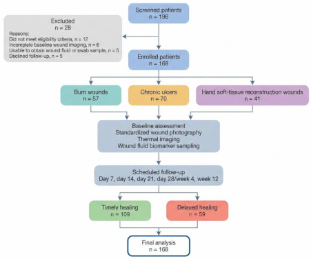
Figure 1: Patient screening, enrollment, follow-up, and analysis pathway. A total of 196 patients with burn wounds, chronic ulcers, or hand soft-tissue reconstruction wounds were screened between 1 January 2024 and 31 December 2025. After exclusion of 28 patients (12 did not meet eligibility criteria, 6 had incomplete baseline imaging, 5 lacked usable fluid or swab samples, and 5 declined follow-up), 168 patients were enrolled and classified into burn wounds, chronic ulcers, or hand soft-tissue reconstruction wounds. All enrolled patients underwent baseline standardized wound photography, thermal imaging, and wound fluid biomarker sampling, followed by scheduled assessments at day 7, day 14, day 21, day 28/week 4, and week 12. Final adjudication classified patients into timely healing and delayed healing groups for analysis, with delayed healing broadly defined as the failure to meet wound-type-specific closure milestones or the need for unplanned surgical intervention. Please click here to view a larger version of this figure.
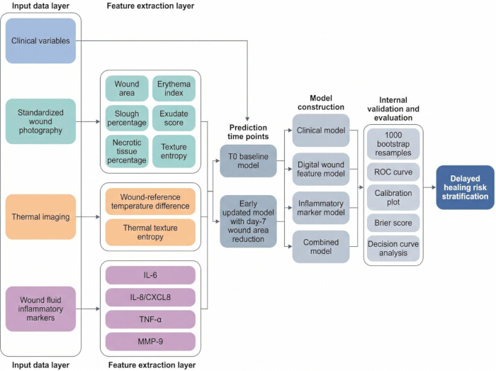
Figure 2: Multimodal analytical workflow for delayed healing prediction. Clinical variables, standardized wound photographs, thermal images, and wound fluid inflammatory markers were collected at baseline. Digital wound features, thermal features, and inflammatory markers were extracted and entered into prespecified prediction models. The baseline model used T0 variables only, whereas the early updated model additionally incorporated day-7 wound area reduction. Clinical, digital wound feature, inflammatory marker, and combined models were evaluated using discrimination, calibration, Brier score, bootstrap internal validation, and decision curve analysis. Please click here to view a larger version of this figure.
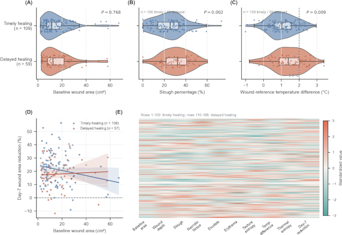
Figure 3: Patient-level digital and thermal wound features. (A) Baseline wound area (n = 109 timely; n = 59 delayed). (B) Slough percentage (n = 106 timely; n = 59 delayed). (C) Wound-reference temperature difference (n = 109 timely; n = 59 delayed). (D) Baseline area versus day-7 area reduction; all available observations are shown (n = 108 timely; n = 57 delayed). (E) Heatmap of all 168 patients ordered by healing status. For panels (A–C), violin plots display data probability density, internal box plots indicate the median (center line) and interquartile range (IQR, hinges), and whiskers extend to a maximum of 1.5 × IQR. In panel (D), shaded error bands represent 95% confidence intervals around the linear regression fits. p values were calculated using two-sided Welch independent-sample t tests. Violin, box, and scatter layers show the full available data; missing day-7 values were imputed only for the model/heatmap as described. Please click here to view a larger version of this figure.
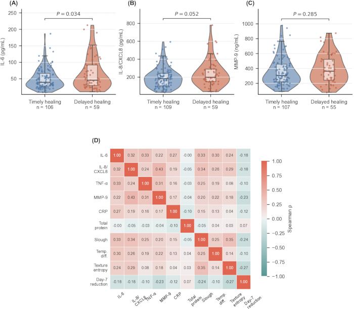
Figure 4: Patient-level inflammatory-marker distributions and correlation matrix. (A) IL-6. (B) IL-8/CXCL8. (C) MMP-9. (D) Square Spearman correlation matrix with identical row and column variables. All available observations are displayed; two-group p values are Welch t tests. Please click here to view a larger version of this figure.
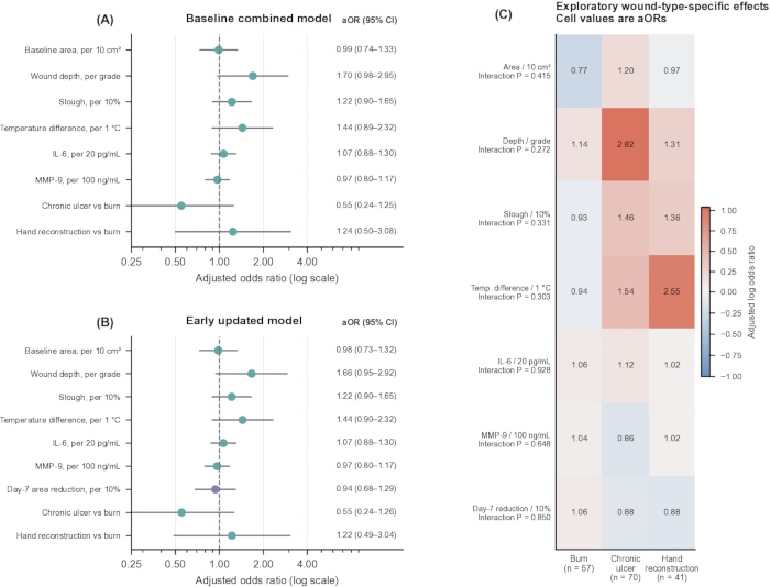
Figure 5: Adjusted model estimates and exploratory wound-type-specific effects. (A) Baseline combined-model adjusted odds ratios with 95% confidence intervals. (B) Early updated-model estimates. (C) Wound-type-specific adjusted odds ratios from one-predictor-at-a-time interaction models; Pinteraction is a two-degree-of-freedom likelihood-ratio test. Burn n = 57, chronic ulcer n = 70, and hand reconstruction n = 41. Please click here to view a larger version of this figure.
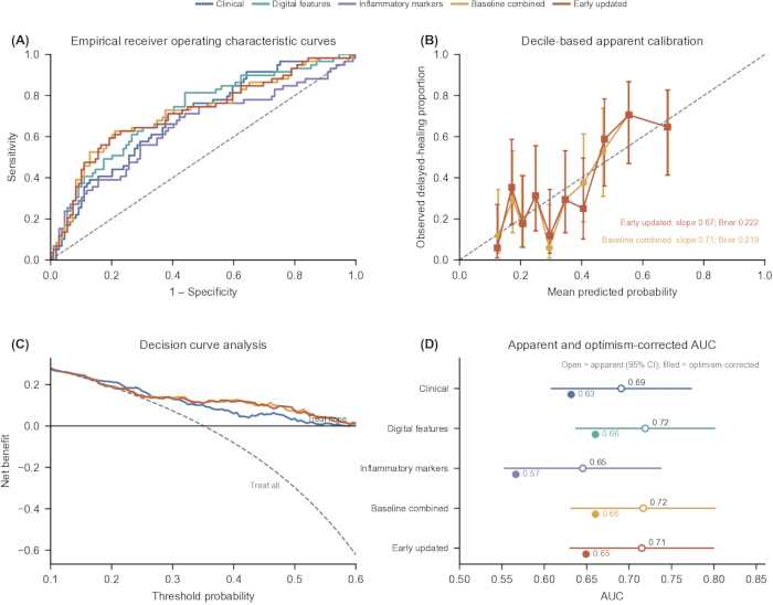
Figure 6: Reproducible model performance from patient-level predictions. (A) Empirical ROC curves with apparent AUC and DeLong 95% CI. (B) Decile-based apparent calibration with Wilson intervals. (C) Decision curves. (D) Apparent AUC with 95% CI and bootstrap optimism-corrected AUC. The baseline combined model did not significantly improve AUC over the clinical model (p = 0.400). Please click here to view a larger version of this figure.
| Assessment item | T0 baseline | Day 7 ± 2 days | Day 14 ± 3 days | Day 21 ± 2 days | Day 28 ± 5 days / Week 4 | Week 12 ± 7 days | |
| Eligibility confirmation | Yes | No | No | No | No | No | |
| Written informed consent | Yes | No | No | No | No | No | |
| Demographic and clinical history | Yes | No | No | No | No | No | |
| Wound type classification | Yes | No | No | No | No | No | |
| Target wound selection | Yes | No | No | No | No | No | |
| Clinical wound assessment | Yes | Yes | Yes | Yes | Yes | Yes | |
| Standardized wound photography | Yes | Yes | Yes | Optional | Yes | Yes | |
| Digital wound area measurement | Yes | Yes | Yes | Optional | Yes | Yes | |
| Slough, necrotic tissue, and erythema assessment | Yes | Yes | Yes | Optional | Yes | Yes | |
| Thermal imaging | Yes | Yes | Optional | Optional | Optional | Optional | |
| Wound fluid or wound swab sampling | Yes | Optional | No | No | No | No | |
| Local inflammatory marker assay | Yes | Optional | No | No | No | No | |
| Peripheral blood inflammatory markers | Yes | Optional | Optional | Optional | Optional | No | |
| Wound bacterial culture when clinically indicated | Yes | Optional | Optional | Optional | Optional | Optional | |
| Treatment documentation | Yes | Yes | Yes | Yes | Yes | Yes | |
| Day-7 wound area reduction calculation | No | Yes | No | No | No | No | |
| Burn wound epithelialization assessment | No | No | No | Yes | Yes if not closed | No | |
| Hand reconstruction outcome assessment | No | No | Optional | Optional | Yes | No | |
| Chronic ulcer area reduction assessment | No | No | No | No | Yes | No | |
| Chronic ulcer complete closure assessment | No | No | No | No | Optional | Yes | |
| Primary delayed healing outcome adjudication | No | No | Optional for burns | Yes for burns | Yes for hand reconstruction and chronic ulcer early response | Yes for chronic ulcer closure | |
Table 1: Fixed assessment schedule. Abbreviations: T0, baseline assessment time point. Note: “Optional” indicates that the assessment was performed when clinically feasible or clinically indicated, but it was not required for the primary baseline model.
| Wound type | Assessment time point | Timely-healing definition | Delayed-healing definition |
| Burn wound | Day 21 ± 2 days | Complete epithelialization by day 21 without unplanned grafting or repeated debridement | No complete epithelialization by day 21, unplanned grafting after failed conservative management, or repeated surgical debridement for persistent non-viable tissue |
| Chronic ulcer | Week 4 and week 12 | ≥50% wound-area reduction by week 4 AND complete closure by week 12 | <50% wound-area reduction by week 4 OR incomplete closure by week 12 |
| Hand soft-tissue reconstruction wound | Postoperative day 28 ± 5 days | Stable wound closure without dehiscence, flap/graft necrosis, new or worsening infection, or unplanned revision | No stable closure by postoperative day 28, wound dehiscence, flap/graft necrosis requiring intervention, new or worsening infection after T0 requiring systemic antibiotics, or unplanned revision surgery |
Table 2: Prespecified wound-type-specific outcome criteria. Chronic-ulcer timely healing required both ≥50% area reduction by week 4 and complete closure by week 12; delayed healing was <50% reduction or incomplete closure. Complete closure required full epithelial coverage without drainage or dressing protection at two consecutive visits.
| Model | Predictors | Wound-type adjustment | Role / evaluation |
| Clinical model | Age (per 10 years), diabetes, serum albumin (per 5 g/L), baseline wound area (per 10 cm²), wound depth grade | Chronic ulcer and hand reconstruction indicators; burn as reference | Comparator; apparent and optimism-corrected AUC, calibration, Brier score |
| Digital wound-feature model | Slough (per 10%), necrotic tissue (per 10%), exudate score, erythema index, photograph texture entropy, wound-reference temperature difference | Chronic ulcer and hand reconstruction indicators; burn as reference | Domain model; apparent and optimism-corrected AUC, calibration, Brier score |
| Inflammatory-marker model | IL-6 (per 20 pg/mL), IL-8/CXCL8 (per 100 pg/mL), TNF-α (per 10 pg/mL), MMP-9 (per 100 ng/mL), CRP (per 10 mg/L), wound-culture result | Chronic ulcer and hand reconstruction indicators; burn as reference | Domain model; apparent and optimism-corrected AUC, calibration, Brier score |
| Baseline combined model | Baseline wound area, wound depth grade, slough, wound-reference temperature difference, IL-6, and MMP-9 | Chronic ulcer and hand reconstruction indicators; burn as reference | Primary baseline model; DeLong comparison with clinical model and decision-curve analysis |
| Early updated combined model | All baseline combined-model predictors plus day-7 wound-area reduction (per 10%) | Chronic ulcer and hand reconstruction indicators; burn as reference | Early trajectory update; DeLong comparisons with clinical and baseline combined models |
Table 3: Exact prediction-model structure. Predictors were prespecified and wound type was forced into every model. Baseline and day-7 updated analyses were kept separate. Abbreviations: AUC, area under the receiver operating characteristic curve; CRP, C-reactive protein; IL, interleukin; MMP-9, matrix metalloproteinase-9.
| Variable | Overall cohort (n = 168) | Timely healing (n = 109) | Delayed healing (n = 59) | P value | |
| Age, years | 54.7 ± 14.7 | 55.6 ± 15.1 | 53.2 ± 14.0 | 0.315 | |
| Male sex, n (%) | 94 (56.0) | 59 (54.1) | 35 (59.3) | 0.626 | |
| Body mass index, kg/m² | 24.8 ± 4.0 | 24.5 ± 4.1 | 25.3 ± 3.7 | 0.184 | |
| Diabetes, n (%) | 62 (36.9) | 43 (39.4) | 19 (32.2) | 0.404 | |
| Current smoking, n (%) | 48 (28.6) | 33 (30.3) | 15 (25.4) | 0.593 | |
| Hemoglobin, g/L | 129.2 ± 16.8 | 129.6 ± 17.2 | 128.3 ± 16.2 | 0.636 | |
| Serum albumin, g/L | 37.6 ± 4.5 | 38.1 ± 4.4 | 36.8 ± 4.7 | 0.085 | |
| Baseline clinical infection, n (%) | 21 (12.5) | 9 (8.3) | 12 (20.3) | 0.029 | |
| Wound type, n (%) | 0.167 | ||||
| Burn wounds | 57 (33.9) | 37 (33.9) | 20 (33.9) | ||
| Chronic ulcers | 70 (41.7) | 50 (45.9) | 20 (33.9) | ||
| Hand soft-tissue reconstruction wounds | 41 (24.4) | 22 (20.2) | 19 (32.2) | ||
| Baseline wound area, cm² | 15.7 ± 12.1 | 15.5 ± 12.0 | 16.0 ± 12.4 | 0.768 | |
| Wound depth grade | 1.94 ± 0.74 | 1.80 ± 0.70 | 2.20 ± 0.74 | <0.001 | |
| Slough area, % | 26.8 ± 14.1 | 24.3 ± 14.2 | 31.2 ± 12.8 | 0.002 | |
| Necrotic tissue area, % | 9.0 ± 7.4 | 8.1 ± 7.3 | 10.8 ± 7.1 | 0.026 | |
| Exudate score | 1.60 ± 0.92 | 1.44 ± 0.90 | 1.90 ± 0.88 | 0.002 | |
| Erythema index | 0.28 ± 0.09 | 0.27 ± 0.08 | 0.30 ± 0.11 | 0.176 | |
| Texture entropy | 4.59 ± 0.44 | 4.54 ± 0.45 | 4.67 ± 0.42 | 0.060 | |
| Wound-reference temperature difference, °C | 1.18 ± 0.79 | 1.06 ± 0.74 | 1.41 ± 0.84 | 0.009 | |
| Temperature difference ≥2.0 °C, n (%) | 28 (16.7) | 13 (11.9) | 15 (25.4) | 0.031 | |
| Thermal texture entropy | 4.76 ± 0.50 | 4.78 ± 0.50 | 4.71 ± 0.52 | 0.372 | |
| Day-7 wound area reduction, % | 20.3 ± 12.0 | 21.5 ± 12.2 | 17.9 ± 11.3 | 0.059 | |
| Wound enlargement by day 7, n (%) | 6 (3.6) | 2 (1.9) | 4 (7.0) | 0.183 | |
Table 4: Baseline clinical, digital, and thermal characteristics. Values are mean ± standard deviation or n (%). p values are Welch t tests, chi-square tests, or Fisher exact tests, as appropriate; available-case n applies to descriptive comparisons.
| Variable | Overall cohort (n = 168) | Timely healing (n = 109) | Delayed healing (n = 59) | P value |
| IL-6, pg/mL | 55.7 ± 38.9 | 50.2 ± 30.4 | 65.5 ± 49.6 | 0.034 |
| IL-8/CXCL8, pg/mL | 210.3 ± 127.7 | 195.0 ± 112.4 | 238.7 ± 149.0 | 0.052 |
| TNF-α, pg/mL | 23.5 ± 11.6 | 22.3 ± 10.8 | 25.6 ± 12.8 | 0.097 |
| MMP-9, ng/mL | 369.0 ± 189.1 | 357.4 ± 187.3 | 391.4 ± 192.3 | 0.285 |
| Total wound-fluid protein, mg/mL | 1.89 ± 0.73 | 1.84 ± 0.68 | 1.99 ± 0.83 | 0.233 |
| MMP-9 normalized to total protein, ng/mg | 230.2 ± 148.2 | 219.8 ± 136.3 | 249.4 ± 167.5 | 0.251 |
| C-reactive protein, mg/L | 13.8 ± 9.9 | 13.3 ± 9.8 | 14.7 ± 9.9 | 0.385 |
| White blood cell count, ×10⁹/L | 7.5 ± 1.9 | 7.3 ± 1.9 | 7.7 ± 2.0 | 0.211 |
| Positive wound culture, n (%) | 72 (42.9) | 46 (42.2) | 26 (44.1) | 0.871 |
| Systemic antibiotics >72 h, n (%) | 44 (26.2) | 21 (19.3) | 23 (39.0) | 0.009 |
| Negative-pressure wound therapy, n (%) | 39 (23.2) | 21 (19.3) | 18 (30.5) | 0.126 |
| Post-baseline surgical debridement, n (%) | 44 (26.2) | 23 (21.1) | 21 (35.6) | 0.046 |
| Skin grafting or flap revision, n (%) | 34 (20.2) | 16 (14.7) | 18 (30.5) | 0.026 |
| Unplanned reoperation, n (%) | 17 (10.1) | 6 (5.5) | 11 (18.6) | 0.013 |
Table 5: Local inflammatory markers and follow-up treatment exposures. Treatment variables are binary ever-exposed indicators; exact start/stop dates were unavailable and the variables were excluded from primary baseline models.
| Predictor | Baseline β | Baseline aOR | Baseline 95% CI | Baseline P | Early β | Early aOR | Early 95% CI | Early P | |
| Intercept | -2.552 | 0.078 | 0.021–0.296 | <0.001 | -2.342 | 0.096 | 0.017–0.531 | 0.007 | |
| Baseline wound area, per 10 cm² increase | -0.010 | 0.990 | 0.738–1.328 | 0.947 | -0.017 | 0.983 | 0.732–1.321 | 0.912 | |
| Wound depth grade, per 1-grade increase | 0.528 | 1.696 | 0.976–2.949 | 0.061 | 0.508 | 1.662 | 0.948–2.916 | 0.076 | |
| Slough area, per 10% increase | 0.199 | 1.220 | 0.900–1.653 | 0.201 | 0.195 | 1.216 | 0.897–1.648 | 0.208 | |
| Wound-reference temperature difference, per 1 °C increase | 0.364 | 1.439 | 0.894–2.316 | 0.134 | 0.366 | 1.442 | 0.895–2.323 | 0.132 | |
| IL-6, per 20 pg/mL increase | 0.069 | 1.071 | 0.884–1.298 | 0.482 | 0.065 | 1.067 | 0.880–1.295 | 0.509 | |
| MMP-9, per 100 ng/mL increase | -0.030 | 0.971 | 0.803–1.174 | 0.759 | -0.035 | 0.966 | 0.797–1.170 | 0.721 | |
| Day-7 wound area reduction, per 10% increase | Not included | Not included | — | — | -0.062 | 0.940 | 0.685–1.291 | 0.703 | |
| Wound type: chronic ulcer vs burn | -0.599 | 0.549 | 0.241–1.252 | 0.154 | -0.594 | 0.552 | 0.242–1.259 | 0.158 | |
| Wound type: hand reconstruction vs burn | 0.216 | 1.241 | 0.500–3.078 | 0.641 | 0.200 | 1.221 | 0.490–3.042 | 0.668 | |
Table 6: Complete logistic-regression specification for the baseline combined and early updated models, including intercepts, coefficients, adjusted odds ratios, 95% confidence intervals, p values, and wound-type indicators. Individual risk is p = 1/[1 + exp(-η)].
| Model | Apparent AUC | 95% CI | Optimism-corrected AUC | Corrected calibration slope | Corrected calibration intercept | Corrected Brier score | ΔAUC vs clinical | DeLong P vs clinical | |
| Clinical model | 0.691 | 0.608–0.773 | 0.632 | 0.695 | 0.004 | 0.227 | — | — | |
| Digital wound feature model | 0.719 | 0.637–0.801 | 0.660 | 0.708 | 0.005 | 0.221 | — | — | |
| Inflammatory marker model | 0.645 | 0.553–0.737 | 0.566 | 0.579 | 0.007 | 0.235 | — | — | |
| Baseline combined model | 0.717 | 0.632–0.801 | 0.660 | 0.713 | 0.005 | 0.219 | 0.026 | 0.400 | |
| Early updated combined model | 0.715 | 0.630–0.799 | 0.649 | 0.671 | 0.002 | 0.222 | 0.024 | 0.434 | |
| Early updated vs baseline combined | — | — | — | — | — | — | -0.002 | 0.662 | |
Table 7: Apparent and 1000-bootstrap optimism-corrected model performance, including ΔAUC and DeLong p values. Corrected calibration and Brier values are reported. Model comparisons use correlated-AUC DeLong tests.
Supplementary File 1: Data supporting the findings of this study.Please click here to download this file.
Discussion
The patient-level analysis yielded a cautious conclusion. Delayed-healing wounds were deeper and showed greater tissue burden, exudate, temperature difference, baseline infection, and IL-6 in descriptive comparisons, but most prespecified multivariable coefficients were imprecise. The baseline and early multimodal models had optimism-corrected AUCs of 0.660 and 0.649 and did not significantly improve on the clinical model. These findings fit the broader view that inflammation, matrix turnover, and tissue remodeling interact during repair17,18,19,20, but they do not establish a clinically deployable predictor.
The imaging findings support standardized measurement while also exposing an important reproducibility gap. Calibrated photography can improve area measurement5,6,14, and thermography may capture physiology not visible in conventional images7,8. However, wound-bed preparation and tissue classification remain sensitive to preprocessing and manual correction21,22. Baseline area agreement was excellent, but reader-level data for slough, necrosis, erythema, texture, and thermal features were not retained. Consequently, the present work cannot claim reproducibility for those non-area features. Wound-fluid findings were also weaker than previously reported; IL-6 differed descriptively, while MMP-9 did not, despite biological plausibility from prior wound-fluid and protease studies9,10,11,15,23,24,25,26.
Practical implementation would require more than statistical discrimination. Smartphone-compatible thermography adds device purchase, calibration, operator training, environmental stabilization, image registration, and quality-control requirements. ELISA adds sample collection and cold-chain handling, duplicate assays, laboratory staff time, batch processing, and delayed turnaround. Exact local costs and turnaround times were not prospectively recorded, and no formal cost-effectiveness analysis was performed. Because the combined model's ΔAUC over the clinical model was small and non-significant, the added technical and laboratory burden is not currently justified for routine use.
Treatment differences complicate causal interpretation. Antibiotics, debridement, grafting/flap revision, and reoperation were more frequent after baseline among delayed-healing patients. Furthermore, the profound heterogeneity in standard-of-care management across these distinct wound categories introduces significant unmeasured confounding. These diverse, etiology-specific clinical pathways likely modify the natural healing trajectory and obscure baseline predictive signals, thereby contributing to the modest discriminative performance of the pooled multimodal model. The treatment-adjusted model had a higher apparent AUC, but these interventions were chosen in response to severity and some formed part of wound-type-specific outcome criteria. Confounding by indication, unavailable treatment timing, and residual confounding therefore prevent interpretation of post-baseline treatment coefficients as intervention effects or valid baseline predictors.
Pooling was motivated by a shared mixed-wound clinical workflow and common measurable repair domains, not by an assumption of biological equivalence. Wound type was forced into every model, and subgroup AUCs, predictor effects, and interactions were reported. Nevertheless, the burn, chronic-ulcer, and hand subgroups contained only 57, 70, and 41 patients, so absence of significant interaction cannot demonstrate consistency. Other limitations include the single-center design, only internal validation, limited event-to-parameter ratio, median/mode imputation, incomplete non-area reliability data, unavailable exact intervention timing, and potentially center-specific imaging/assay workflows. Independent validation, recalibration, and prospective impact testing are required before implementation12,27,28,29.
In conclusion, while specific digital, thermal, and inflammatory features were individually associated with delayed healing, the multimodal prediction models demonstrated only modest internally validated performance, offering no statistically significant discriminative advantage over standard clinical assessment. Consequently, these findings do not currently support the clinical implementation of this pooled model; instead, they provide exploratory groundwork for future, adequately powered multicenter investigations.
Disclosures
The authors have nothing to disclose.
Acknowledgements
The authors thank the clinical staff and nurses of the Department of Burn and Plastic Surgery, General Hospital of Northern Theater Command, and the Second Surgery Department, Liaoning Provincial Corps Hospital of Chinese People's Armed Police Force, for assistance with patient screening and wound imaging. The authors also thank the participating patients. This research received no specific grant from any funding agency in the public, commercial, or not-for-profit sectors.
Materials
| Name | Company | Catalog Number | Comments |
|---|---|---|---|
| BCA Protein Assay Kit | Thermo Fisher Scientific | 23225 | - |
| FLIR ONE Pro Thermal Camera | FLIR Systems | 435-0006-03 | - |
| GLCM Texture plugin for FIJI | FIJI/ImageJ community | v0.4 | - |
| Human IL-6 Quantikine ELISA Kit | R&D Systems | D6050 | - |
| Human IL-8/CXCL8 Quantikine ELISA Kit | R&D Systems | D8000C | - |
| Human MMP-9 ELISA Kit | Abcam | ab100610 | - |
| Human TNF-α Quantikine ELISA Kit | R&D Systems | DTA00D | - |
| iPhone 14 Pro | Apple | — | - |
| Phosphate-buffered saline (PBS) | Thermo Fisher Scientific | 10010023 | - |
| Sterile Rayon Tipped Applicator Swabs | Puritan Medical Products | 25-806 1WR | - |
| Tween-20 | Sigma-Aldrich | P1379 | - |
| X-Rite ColorChecker Classic Mini | Calibrite / X-Rite | MSCCVPR-MINI | - |
| FIJI / ImageJ | National Institutes of Health | 1.54j | SCR_002285 |
| scikit-learn | scikit-learn developers | 1.9.0 | SCR_002577 |
| REDCap | Vanderbilt University | 14.0.9 | SCR_003282 |
| SciPy | SciPy Developers | 1.18.0 | SCR_008058 |
| Python | Python Software Foundation | 3.12.13 | SCR_008394 |
| Matplotlib | Matplotlib Development Team | 3.11.1 | SCR_008624 |
| NumPy | NumPy Developers | 2.5.1 | SCR_008628 |
| OpenCV | OpenCV team | 4.9.0 | SCR_015526 |
| Statsmodels | Statsmodels developers | 0.14.6 | SCR_016074 |
| Seaborn | Seaborn developers | 0.13.2 | SCR_018132 |
| pandas | pandas Development Team | 3.0.5 | SCR_018214 |
| scikit-image | scikit-image developers | 0.22.0 | SCR_021340 |
References
- Rowan MP, et al. Burn wound healing and treatment: review and advancements. Crit Care. 2015;19(1):243.
- Ji S, Xiao S, Xia Z. Consensus on the treatment of second-degree burn wounds (2024 edition). Burns Trauma. 2024;12:tkad061.
- Cho SK, Mattke S, Gordon H, Sheridan M, Ennis W. Development of a model to predict healing of chronic wounds within 12 weeks. Adv Wound Care. 2020;9(9):516–24.
- Sheehan P, Jones P, Caselli A, Giurini JM, Veves A. Percent change in wound area of diabetic foot ulcers over a 4-week period is a robust predictor of complete healing in a 12-week prospective trial. Diabetes Care. 2003;26(6):1879–82.
- Kientzy M, et al. Accuracy of digital measurement for quantitative and qualitative indicators of wound healing and repair: a systematic review protocol. BMJ Open. 2024;14(5):e085969.
- Tanner J, et al. Digital wound monitoring with artificial intelligence to prioritise surgical wounds in cardiac surgery patients for priority or standard review: protocol for a randomised feasibility trial (WISDOM). BMJ Open. 2024;14(9):e086486.
- Monshipouri M, et al. Thermal imaging potential and limitations to predict healing of venous leg ulcers. Sci Rep. 2021;11(1):13239.
- Fridberg M, Bafor A, Iobst CA, Laugesen B. The role of thermography in assessment of wounds: a scoping review. Injury. 2024;55(11):111833.
- Saleh K, Sonesson A, Persson K, Riesbeck K, Schmidtchen A. Inflammation biomarkers and correlation to wound status after full-thickness skin grafting. Front Med. 2019;6:159.
- Rembe JD, et al. Immunomarker profiling in human chronic wound swabs reveals IL-1β/IL-1RA and CXCL8/CXCL10 ratios as potential biomarkers for wound healing, infection status and regenerative stage. J Transl Med. 2025;23(1):407.
- Liu Y, et al. Increased matrix metalloproteinase-9 predicts poor wound healing in diabetic foot ulcers. Diabetes Care. 2009;32(1):117–9.
- Efthimiou O, et al. Developing clinical prediction models: a step-by-step guide. BMJ. 2024;386:e078276.
- von Elm E, et al. The Strengthening the Reporting of Observational Studies in Epidemiology (STROBE) statement: guidelines for reporting observational studies. Ann Intern Med. 2007;147(8):573–7.
- Sharun K, Dhama K, Tiwari R. Comparative assessment of smartphone-based digital wound area measurement and ImageJ-based digital image analysis. Narra J. 2025.
- Rembe JD, et al. Assessment and monitoring of the wound micro-environment in chronic wounds using standardized wound swabbing for individualized diagnostics and targeted interventions. Biomedicines. 2024;12(10):2187.
- Collins GS, Reitsma JB, Altman DG, Moons KGM. Transparent reporting of a multivariable prediction model for individual prognosis or diagnosis: the TRIPOD statement. Ann Intern Med. 2015;162(1):55–63.
- Wallace HA, Basehore BM, Zito PM. Wound healing phases. StatPearls. Treasure Island (FL): StatPearls Publishing; 2023.
- Landén NX, Li D, Ståhle M. Transition from inflammation to proliferation: a critical step during wound healing. Cell Mol Life Sci. 2016;73(20):3861–85.
- Schultz GS, Ladwig G, Wysocki A. Extracellular matrix: review of its roles in acute and chronic wounds. Principles of Wound Healing. Adelaide: University of Adelaide Press; 2011.
- Almadani YH, Vorstenbosch J, Davison PG, Murphy AM. Wound healing: a comprehensive review. Semin Plast Surg. 2021;35(3):141–4.
- Sibbald RG, Elliott JA, Persaud-Jaimangal R. Wound bed preparation 2021. Adv Skin Wound Care. 2021;34(4):183–95.
- Harries RL, Bosanquet DC, Harding KG. Wound bed preparation: TIME for an update. Int Wound J. 2016;13(Suppl 3):8–14.
- Gao M, et al. Regulation of inflammation during wound healing: the function of mesenchymal stem cells and strategies for therapeutic enhancement. Front Pharmacol. 2024;15:1345779.
- Mikosiński J, Kruk-Jeromin J, Drobnik J. Longitudinal evaluation of biomarkers in wound fluids from chronic venous leg ulcers. Acta Derm Venereol. 2022;102:adv00667.
- Trengove NJ, et al. Analysis of the acute and chronic wound environments: the role of proteases and their inhibitors. Wound Repair Regen. 1999;7(6):442–52.
- Fu K, Zheng Q, Wei H. Role of matrix metalloproteinases in diabetic foot ulcers. Front Pharmacol. 2022;13:1050630.
- Collins GS, et al. Evaluation of clinical prediction models: from development to external validation. BMJ. 2024;384:e074819.
- Binuya MAE, Engelhardt EG, Schats W, Schmidt MK, Steyerberg EW. Methodological guidance for the evaluation and updating of clinical prediction models: a systematic review. BMC Med Res Methodol. 2022;22(1):316.
- Saelmans A, et al. Implementation and updating of clinical prediction models: a systematic review. Mayo Clin Proc Digit Health. 2025;3(3):100228.