Here, we describe an approach to produce genetically modified embryos using CRISPR-Cas9 gene editing and somatic cell nuclear transfer for generating sheep models of human disease. This approach is ideal for precise gene editing in donor cells and producing cloned embryos without genetic mosaicism.
Method Article
Generation of Genetically Engineered Embryos Using Gene Editing and Somatic Cell Nuclear Transfer for Production of Sheep Models of Human Disease
In This Article
Summary
Abstract
Large animal models are valuable tools for investigating human disease. Sheep, pigs, and goats often better recapitulate the anatomy and physiology of human organs and the complexity of human disease, thereby enhancing their clinical relevance compared to rodents. CRISPR-Cas9 and somatic cell nuclear transfer (SCNT) enable the generation of large animal models with greater precision, versatility, and genetic uniformity. The primary benefit of this approach, compared with zygote microinjection, is the ability to confirm in vitro whether the desired genetic modification and potential off-target mutations are present in gene-edited cells prior to animal production. Moreover, SCNT eliminates the chance of genetic mosaicism, which frequently results from zygote microinjection. Here, we describe the generation of gene-edited ovine cells through non-homologous end-joining (NHEJ) and homology-directed repair (HDR), followed by the production of cloned embryos carrying the mutations of interest. Genetic modifications are introduced by transfecting cultured somatic cells, typically fetal fibroblasts, with the CRISPR-Cas9 system. Mutation efficiency in pooled cells is assessed by polymerase chain reaction (PCR) and Sanger sequencing of edited genes and analyzed using Tracking of Indels by DEcomposition (TIDE)/Tracking of Insertions, Deletions, and Recombination events (TIDER) software. Limiting dilution of the pooled cells is performed to obtain single-cell-derived colonies, which are screened by PCR and DNA sequencing of edited genes. Donor cells with the edit(s) of interest are subsequently expanded and used for the generation of embryos by SCNT. After limiting dilution and cell screening, 22/114 (19.3%) of colonies modified through NHEJ contained knockout (KO) mutations and 4/56 (7.1%) of colonies modified with HDR contained the F508del mutation. A total of 370 genetically modified embryos were created from four colonies. These methods are successfully used for precise gene editing in fetal fibroblasts and generation of genetically engineered embryos to produce ovine models of human disease.
Introduction
Animal models are essential tools for understanding the biological mechanisms of human diseases and development of new therapeutics. In vitro models, such as cell or organoid culture, do not reproduce the complexity of cellular subtypes and cell-cell interactions that occur in vivo in organs1,2. Thus, animals provide whole-organism models with greater detail of cellular and system-level interactions, disease progression, and the safety of therapeutics. Rodents are practical model organisms given their low cost, quick generation time, and comprehensive genetic characterization. However, rodent models for human diseases are limited by physiological and anatomical differences with humans, as well as their faster metabolism and shorter lifespans3,4. This is evidenced by the high proportion of therapies tested in animal models that fail in clinical trials, the majority of which use rodents5,6,7,8. Sheep, pigs, goats, dogs, and non-human primates have been used to produce models for a wide variety of human diseases, which may help increase the clinical translation rate for new therapies and medicines3,9,10,11,12. These species also offer many advantages over rodent models due to their increased similarity to humans. Large animal models for human disease thus provide the most accurate models available and play a critical role in translating animal studies into clinical applications.
Clustered regularly interspaced short palindromic repeats (CRISPR)-Cas9 is a versatile and programmable method for genetic modification of animal cells and introduction of precise disease-causing mutations. CRISPR-Cas9-mediated gene editing employs a guide RNA (gRNA) complementary to the target site, allowing CRISPR-associated protein 9 (Cas9) to form a double-stranded break (DSB) in a targeted manner13. Cellular repair of DSBs occurs primarily through two mechanisms: non-homologous end joining (NHEJ) and homology-directed repair (HDR). The NHEJ pathway repairs a DSB by joining DNA ends, an error-prone mechanism that may result in the formation of random insertions or deletions (indels) at the break site14. These mutations may knock out (KO) the gene by creating a frameshift mutation or a premature stop codon, which disrupt the gene’s function. HDR, occurring mainly during the S and G2 phases of the cell cycle, is when the cell repairs a DSB using a homologous allele as a repair template15,16. Incorporation of specific insertions, deletions, or substitutions, known as knock-ins (KI), is possible by introducing a synthetic single-stranded oligodeoxynucleotide (ssODN) or other template DNA with the edit of interest, which the cell can use to repair the break. CRISPR-Cas9 may also be used for multiplexed editing to introduce multiple mutations at once for modeling multigenic diseases3. Compared to previous gene-editing techniques such as zinc-finger nucleases (ZFNs) or transcription activator-like effector nucleases (TALENs), CRISPR-Cas9 is much simpler and more resource efficient to use in large animal cells17. Different loci can be targeted easily by designing a new gRNA, which has helped accelerate the introduction of new large animal models using CRISPR-Cas93,13. However, several limitations remain, including the editing efficiency of CRISPR-Cas9, which can vary considerably depending on the species, cellular environment, and nature of the edit18,19. HDR rates, in particular, are lower than NHEJ rates and depend greatly on the cell type, genomic loci, and cell cycle stage20,21. Moreover, the introduction of unintended edits at off-target loci, or even on-target loci, remains an issue22. Even so, CRISPR-Cas9 has become the primary gene editing platform for producing genetically engineered large animal models.
Somatic cell nuclear transfer (SCNT) facilitates the creation of genetically modified embryos with high precision and homogeneity. This process, first successfully performed in mammals in 1997, involves the transfer of a cell nucleus into an enucleated oocyte23. SCNT possesses many advantages over other techniques, such as zygotic microinjection, in which genetic material or DNA-editing systems are directly injected into the zygote or early embryo24,25,26. Zygotic microinjection carries the inherent risk of genetic heterogeneity, or mosaicism, between cells in the embryo, which can occur if DNA replication or cell division occurs prior to genetic modification27,28,29. The value of such mosaic embryos for animal models is reduced due to the difficulty of genotyping and phenotypic characterization29. In SCNT, gene editing is decoupled from embryo production; each cloned embryo is derived from a single cell, eliminating the risk of mosaicism and ensuring the embryo is genetically homogeneous30,31,32. Additionally, donor cells are edited and carefully screened in vitro to confirm clonality and the presence of the desired modification prior to the SCNT procedure, saving time and resources25,26. This is especially important when introducing specific KIs. Cell screening also allows the identification of potential mutations in off-target sites, thus reducing the risk of genetic disruption at other loci. These advantages address many of the concerns associated with CRISPR-Cas9. Therefore, SCNT provides greater control over the genetics of the resulting embryos compared to zygote microinjection, especially for more complex edits such as multiplexed edits, large insertions, and humanized alleles33. Although, SCNT is limited by low cloning efficiency, technical requirements, and abnormal epigenetic reprogramming, the latter of which can lead to molecular and phenotypic disruptions in clones, potentially obscuring the effects of genetic modification in founder animals. However, these epigenetic alterations are not passed on to the offspring32,33. Despite these limitations, the combination of SCNT with CRISPR-Cas9 enables precise genome engineering with strict control over embryo genetics, supporting the reliable production of large animal disease models without mosaicism. Here, we describe a detailed approach for genetic modification of sheep fetal fibroblasts (SFF) using either NHEJ or HDR, followed by SCNT for cloned embryo production, with the ultimate goal of generating sheep models of human disease. This approach is largely applicable to other livestock species, though gene editing may require optimization in different cell types, with this protocol acting as a useful starting point.
Protocol
All animal procedures were approved by the Institutional Animal Care and Use Committee (IACUC: #15508 and #10089) at Utah State University and performed in accordance with National Institutes of Health (NIH) guidelines. In this study, domestic Romney sheep (Ovis aries) were used to isolate sheep fetal fibroblasts (Supplementary File 134). See Figure 1 for an overview of the protocol.
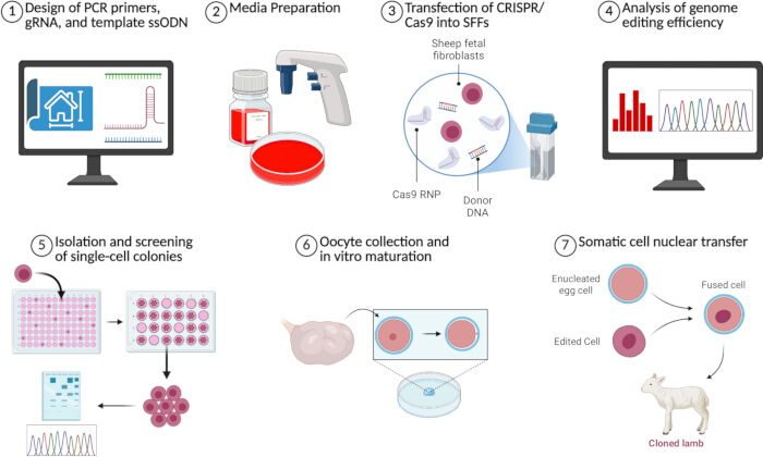
Figure 1: Overview of CRISPR-Cas9 and SCNT methods used to generate genetically modified embryos. A summary of each of the seven major steps of the protocol is depicted. Created in BioRender. Perisse, I. (2026). Please click here to view a larger version of this figure.
1. Design of PCR primers, gRNA, and template ssODN
- Open the genomic region to be edited in Benchling (https://www.benchling.com/academic).
- Design PCR primers to amplify and sequence the genomic region of interest.
- Highlight the target region to be amplified, plus 200 base pairs upstream and downstream of the target region to ensure ample distance for quality trace calls and accurate analysis of mutation efficiency with TIDE software.
- On the right-hand navigation bar click Primers > Create Primers > Wizard.
- Under Region, click Use Selection. Under Amplicon, adjust Size accordingly and adjust any other parameters as desired.
- Click Generate Primers. Select one or more primer pairs with low penalty scores to order.
NOTE: Depending on the edit being introduced, it is recommended to design primers that will bind only to correctly edited DNA to allow for quicker screening of positive colonies.
- Design gRNA to edit DNA with CRISPR-Cas9.
- In Benchling, highlight the target region to be edited, then on the right-hand navigation bar, click CRISPR > Design and analyze guides.
- Click the Genome dropdown menu and select or import the model organism genome. Adjust any other parameters as desired.
- Click Finish > +.
- Under Genome Region, click No region set and select or input the genome region containing the target site. Click Set genome region.
- Select one or more guides with both high on-target scores and high off-target scores to order.
- If performing KI edits, design an ssODN template.
- On the right-hand navigation bar, click CRISPR > Design HR template (ssODN).
- Click the Genome dropdown menu and select or import the model organism genome. Click Create.
- Click on the SEQUENCE MAP tab, highlight the bases to be edited, and create the modified version of the DNA sequence by typing the desired edits into the keyboard. Go back to the DESIGN HR TEMPLATE tab and click Next.
- Adjust the Template region to include at least 50 base pairs of homology arms on each side of the edit, then click Next.
- Input the sequence of the gRNA selected in step 1.3.5 in the Guide box to introduce Protospacer Adjacent Motif (PAM)-disrupting silent mutations. Click Next.
- Click Copy the template and order the ssODN template generated.
NOTE: Sequences of PCR primers, CRISPR-Cas9 gRNAs, and template ssODNs designed using Benchling for the generation of a sheep cystic fibrosis model carrying either a knockout (KO) mutation or the F508del mutation are presented in Table 1.
2. Media preparation
- Prepare cell culture medium by supplementing Dulbecco’s modified Eagle’s medium (DMEM) high Glucose with 15% fetal bovine serum (FBS) and 1% Penicillin-Streptomycin (Pen/Strep) (v/v). Pre-warm in a water bath at 37 °C before use.
- Prepare freezing medium by supplementing DMEM with 45% FBS and 10% dimethyl sulfoxide (DMSO).
- Prepare oocyte collection medium by supplementing M199 medium with 1% Pen/Strep, 0.3% bovine serum albumin (BSA) fraction V, 1% FBS, and 0.06% heparin sodium salt.
- Order In Vitro Maturation (IVM) and In Vitro Culture (IVC) media, which are commercially available serum-free media formulated for bovine oocyte maturation and embryo culture, respectively.
NOTE: Post-fusion and IVC dishes are prepared with seven 40 µL drops of IVC medium and covered with embryo-tested mineral oil. - Prepare cell starvation medium by supplementing DMEM high Glucose with 0.5% FBS and 1% Pen/Strep (v/v). Pre-warm in a water bath at 37 °C before use.
- Prepare HEPES-supplemented synthetic oviductal fluid (HSOF) medium as previously described35.
- Prepare enucleation medium by supplementing 500 µL of HSOF medium with 4.5 mM sucrose and 10 µg/mL Cytochalasin B (CB).
- Prepare injection media by supplementing 500 µL of HSOF medium with 10 µg/mL Cytochalasin B (CB).
NOTE: Enucleation and injection media are prepared fresh on the day of SCNT. One drop (200 µL) of each medium is placed in 100 mm dishes and covered with embryo-tested mineral oil. - Prepare fusion medium as previously described35. Remove 5–10 mL of fusion medium from 4 °C and keep at room temperature until use.
- Prepare CB Medium by supplementing IVC medium with 10 µg/mL CB.
NOTE: A CB dish is prepared with seven 40 µL drops of CB medium and covered with embryo-tested mineral oil. - Prepare the ionomycin medium by supplementing the HSOF medium with 5 µM ionomycin calcium salt.
- Prepare activation medium by supplementing IVC medium with 2 mM 6-Dimethylaminopurine (DMAP) and 10 µg/mL Cycloheximide (CHX).
NOTE: An activation dish is prepared with seven 40 µL drops of activation medium and covered with embryo-tested mineral oil.
3. Transfection of CRISPR-Cas9 into SFFs
- (Conditions) Perform all cell culture procedures in a biosafety cabinet or laminar flow hood. Culture SFFs in a T-25 flask with cell culture medium at 37 °C in 5% CO2, 5% O2, and 90% N2 until approximately 70%–80% confluent prior to transfection.
- Remove all media from the flask and wash the cells once with 5 mL of Dulbecco’s phosphate-buffered saline (DPBS). Incubate with 1 mL of trypsin-like dissociation enzyme for 5–10 min until the cells dissociate.
- Dilute the trypsin-like dissociation enzyme using an equal volume of cell culture medium and centrifuge the cells at 200 × g for 5 min.
- (Timing) Prepare the gRNA: Cas9 ribonucleoprotein (RNP) complex by incubating 2.5 µL of 100 µM gRNA with 2.5 µL of 5 µg/µL Cas9 protein for 10 min at room temperature.
NOTE: Cas9 RNP: gRNA ratios may require optimization depending on the cell type, target gene, and number of targets. - Prepare the nucleofection solution by combining 82 µL of nucleofection solution with 18 µL of supplement solution.
- Combine the nucleofection solution with the RNP complex and if using an ssODN, add 2 µL of 200 µM ssODN, then gently mix by pipetting.
- After centrifugation, carefully discard the supernatant and place the tube upside down on a lint-free tissue to remove residual medium.
NOTE: Residual culture medium may affect transfection efficiency and cell viability. Ensure that excess liquid is completely removed before resuspending the cell pellet in the nucleofection solution. - (Critical) Resuspend fibroblasts in the nucleofection solution, then transfer the mixture into a 100 µL nucleocuvette, avoiding bubble formation. Gently tap the nucleocuvette on a hard surface to get the mixture to the bottom of the cuvette.
NOTE: Bubbles inside the nucleocuvette may interfere with the electroporation. - (Relevant setting) Electroporate the cells using program EH-100, or according to the manufacturer’s recommendations. Allow the cells to recover for 10 min at room temperature.
- Transfer the electroporated cells using a transfer pipette into 5 mL pre-equilibrated cell culture medium supplemented with HDR enhancer at a final concentration of 1–2 µM, which may require optimization in other cell types.
- (Conditions) Culture the cells in cell culture medium at 37 °C in 5% CO2, 5% O2, and 90% N2 for 48 h prior to genomic DNA isolation.
- Remove the medium from the cells, wash once with DPBS, and add 1 mL of trypsin-like dissociation enzyme.
- After the cells detach, add 1 mL of cell culture medium to dilute the trypsin-like dissociation enzyme.
- Allocate approximately two-thirds of the cell suspension for cryopreservation and one-third for genomic DNA extraction into labeled 1.5 mL tubes. Centrifuge all tubes at 200 × g for 5 min.
- Resuspend the cell pellet designated for cryopreservation in 500 µL of freezing medium. Transfer the resuspended cells into cryovials and place them in a controlled-rate freezing container at -80 °C, minimizing the time cells remain in the freezing medium before transfer to -80 °C.
- Resuspend the cell pellet designated for DNA extraction in 200 µL of DPBS. Extract genomic DNA using a genomic DNA purification kit according to the manufacturer’s instructions.
4. Analysis of genome editing efficiency
- Amplify the target loci from the isolated DNA using high-fidelity DNA polymerase and the previously designed PCR primers.
- Confirm PCR amplification by agarose gel electrophoresis in a 1% agarose gel with a 1 kb DNA ladder.
- Purify the PCR products using a PCR purification kit and submit for Sanger sequencing.
- If a KO was performed, use TIDE software (https://apps.datacurators.nl/tide) to assess editing efficiency36. Use high-quality chromatograms with minimal background signal for accurate editing efficiency analysis.
- Open TIDE and name the sample in the Title plot box. Under Guide sequence, input the sequence of the gRNA used.
- Under Control Sample Chromatogram, click Browse and upload the chromatogram of the unedited control sample.
- Under Test Sample Chromatogram, click Browse and upload the chromatogram of the edited test sample.
- Navigate to the Decomposition tab to view the indel spectrum and mutation efficiency. Check the R2 value in the upper-right-hand corner of the indel spectrum.
- If the R2 value is less than 0.9, check the Advanced settings box under Parameters and adjust the sliders for the alignment window, decomposition window, and indel size until the R2 value is greater than 0.9 and proper alignment is achieved.
- If a KI was performed, use TIDER software (https://apps.datacurators.nl/tider) to assess editing efficiency37.
- Open TIDER. Name the sample in the Title plot box. Under Guide sequence, input the sequence of the gRNA used.
- Under Control Sample Chromatogram, click Browse and upload the chromatogram of the unedited control sample.
- Under Reference Chromatogram, click Browse and upload a chromatogram of a reference sample carrying the desired edit.
NOTE: If no such sequence exists, one may be generated by PCR amplification using extra primers designed to introduce the mutation into a sequence of DNA38. - Under Test Sample Chromatogram, click Browse and upload the chromatogram of the edited test sample.
- Navigate to the Decomposition tab to view the indel spectrum, mutation efficiency, and HDR efficiency. Check the R2 value in the upper-right-hand corner of the indel spectrum.
- If the R2 value is less than 0.9, check the Advanced settings box under Parameters and adjust the sliders for the alignment window, decomposition window, distance upstream from break site, and indel size until the R2 value is greater than 0.9 and proper alignment is achieved.
NOTE: (Critical) A new sequencing may be required if the file sequencing quality is poor.
5. Isolation and screening of single-cell colonies
- (Conditions) Thaw and culture transfected SFFs in a T-25 flask overnight at 37 °C with 5% CO2, 5% O2, and 90% N2, or until the cells reach approximately 80% confluence.
- Remove the media, wash the cells once with 5 mL of DPBS, and dissociate the cells with 1 mL of trypsin-like dissociation enzyme.
- Add cell culture medium to dilute the trypsin-like dissociation enzyme and transfer the cell suspension to a 15 mL conical tube. Centrifuge the cells at 200 × g for 5 min.
- Discard the supernatant and resuspend the cell pellet in 1 mL of cell culture medium. Mix gently but thoroughly to obtain a homogeneous single-cell suspension.
- (Critical) Prepare a 1:10 dilution of the cell suspension, then count the cells using a hemocytometer with Trypan blue.
- Calculate the volume of diluted cell suspension required to seed approximately one cell per well.
NOTE: For five 96-well plates, prepare approximately 500 cells. - Prepare 10 mL of cell culture medium per 96-well plate in a 50 mL conical tube.
- Add the calculated volume of the 1:10 diluted cell suspension to the culture medium and mix well by gentle pipetting and inversion.
- Pour the diluted cell suspension into a sterile reagent reservoir.
- Dispense 100 µL of the cell suspension into each well of a 96-well plate.
- (Conditions) Incubate the plates at 37 °C in 5% CO2, 5% O2, and 90% N2 for 5 days.
- Examine each well under a microscope and mark wells containing one colony. Exclude wells containing more than one colony.
NOTE: Single-cell-derived colonies are usually round, compact, and morphologically homogeneous. - Return the plates to the incubator and culture for an additional 2 days under the same conditions.
- After 7 total days of culture in 96-well plates, remove the medium from wells containing a single growing colony. Wash each selected well once with DPBS.
- Add 100 µL of trypsin-like dissociation enzyme to each selected well and incubate until the cells detach. Add 100 µL of cell culture medium to each well to dilute the trypsin-like dissociation enzyme.
- Transfer the entire volume from each well into one well of a 24-well plate.
- Wash the original 96-well plate with additional cell culture medium and transfer the wash to the corresponding 24-well plate well to maximize cell recovery. Bring the final volume in each 24-well plate well to 500 µL with cell culture medium.
- (Conditions) Incubate the 24-well plates at 37 °C in 5% CO2, 5% O2, and 90% N2 for 3 days.
- Examine the wells under a microscope and select colonies near confluence.
- Remove the medium from wells selected for collection, wash once with DPBS, and add 500 µL of trypsin-like dissociation enzyme.
- After the cells detach, add 1 mL of cell culture medium to dilute trypsin-like dissociation enzyme.
- Freeze cells and isolate genomic DNA according to steps 3.14–3.16.
- Amplify the target loci by PCR and confirm the genotype by Sanger sequencing.
- Identify wild-type, KO, and KI colonies based on sequencing results. Use TIDE/TIDER analysis as described in steps 4.4 and 4.5 to estimate HDR rates and indel spectrums for validation of correct editing and identification of non-single-cell-derived colonies.
NOTE: Off-target analysis may also be performed at this step (Supplementary File 1). - Expand only colonies with the desired genotype and normal cellular morphology for downstream SCNT experiments.
6. Ovine oocyte collection and in vitro maturation
- Collect sheep ovaries from a local abattoir and transport to the laboratory in 0.9% saline at 25 °C within 4 h of collection.
- Mature oocytes in vitro.
- Release cumulus-oocyte complexes (COCs) from ovaries using a scalpel blade by slicing the ovarian surface while it’s submerged in oocyte collection medium.
- (Timing) Select COCs with at least 1–3 layers of compact cumulus cells and uniform cytoplasm for IVM. Wash COCs in fresh oocyte collection medium and culture in pre-equilibrated IVM medium, with up to 40 COCs per well of 500 µL of IVM medium in a 4-well dish and incubate at 38.5 °C with 5% CO2 for 21–22 h.
- Check the maturation of the oocytes.
- Move the desired number of COCs using a 200 µL pipette into a 1.5 mL microcentrifuge tube, adding the same volume (1:1) of 0.6 mg/mL hyaluronidase into the tube.
- Pipette the mixed solution up and down until all the cumulus cells have been removed. Move the oocytes into a 1 mL HSOF drop and wash the oocytes through three additional HSOF drops.
- Using a mouth pipette or a 10 µL pipette and tip, roll and select the oocytes presenting a polar body under a stereomicroscope. The acceptable polar body extrusion rates are 50%–80%.
7. Somatic cell nuclear transfer
- Prepare donor cells before SCNT.
- Thaw cryopreserved cells 3–5 days prior to SCNT in a water bath at 37 °C for 1–2 min until no ice remains in the tube.
- Remove the cell suspension and add an equal volume of cell culture medium into a 15 mL centrifuge tube and centrifuge at 406 x g for 5 min.
- Remove the supernatant and resuspend the cell pellet in 500 µL of cell culture medium.
- (Conditions) Seed the cells in various concentrations in a 4-well dish in a final volume of 500 µL per well and culture at 37 °C with 5% CO2, 5% O2, and 90% N2.
- Replace the cell culture medium every 48 h. After the cells reach full confluency, replace with cell starvation medium 24 h–48h before SCNT.
NOTE: Cell starvation over 48 h may increase the proportion of apoptosis39,40.
- Prepare donor cells (day of SCNT).
- Remove the starvation medium from the well and wash one time with 500 µL of DPBS (-).
- Add 250 µL of trypsin-like dissociation enzyme and incubate at 37 °C for 5 min, or until the cells detach from the dish.
- Add 500 µL of cell culture medium to dilute the trypsin-like dissociation enzyme.
- Move the cells to a 15 mL centrifuge tube and centrifuge at 406 x g for 5 min.
- Remove the supernatant and resuspend the cells in 60–80 µL of HSOF medium and keep at 4 °C until use.
- Enucleate the oocytes.
- Prepare holding pipettes by pulling thin-wall glass capillaries with a micropipette puller and shaping with a microforge to achieve an inner diameter of 30 µm and an outer diameter of 100 µm.
NOTE: Both pipettes can also be purchased from commercial suppliers. - Prepare injection pipettes by pulling thin-wall glass capillaries with a micropipette, beveling at a 45° angle with a microgrinder, and shaping with a microforge to achieve an inner diameter of 20–25 µm. The parameters for the micropipette puller, microforge, and microgrinder can be found in a previous report41.
- Move the metaphase II (MII)-staged oocytes that presented a polar body, into the drop of enucleation medium. Immobilize the oocyte using a holding pipette while the polar body is in the 4–5 o’clock position.
NOTE: Standard operating procedures for micromanipulation can be referred to the previous report41. - (Critical) Use a beveled injection pipette with a sharp tip to pierce the zona pellucida and aspirate the polar body and 10%–20% of the cytoplasm under the polar body.
- Push the removed cytoplasm into the drop and release the oocyte from the holding pipette.
- Repeat steps 7.3.2–7.3.4 for all the oocytes until all have been enucleated. Return enucleated oocytes to IVM medium and incubate at 38.5 °C and 5% CO2 for 30 min.
- Prepare holding pipettes by pulling thin-wall glass capillaries with a micropipette puller and shaping with a microforge to achieve an inner diameter of 30 µm and an outer diameter of 100 µm.
- Inject donor cells into enucleated oocytes.
- Add the prepared cell suspension into the bottom of the injection medium drop and move enucleated oocytes to the same drop.
- Select 1–10 round, healthy cells and aspirate them into the injection pipette.
- Use the holding pipette to immobilize an oocyte and use the injection pipette to pierce into the zona pellucida and put one cell between the zona and the ooplasm.
- Repeat steps 7.4.2–7.4.3 for all enucleated oocytes and move them to 2–3 mL of HSOF medium in a 35 mm dish before cell fusion.
- Fuse enucleated oocytes and donor cells.
- Connect the electrofusion pulse generator cables to the wires of the microslide chamber (0.5 mm) and place the chamber onto the microscope stage.
- Add 600 µL of fusion medium to the chamber.
- (Critical/Relevant setting) Align 4–6 cytoplasm-cell pairs between the two wires of the chamber with the donor cell located at the 6 or 12 o’clock position. Apply two DC electric fusion pulses of 2.0 kV/cm for 40 µs.
- Remove the cytoplasm-cell pairs from the fusion chamber and keep them in a new 35 mm dish containing 2–3 mL of HSOF medium.
- (Conditions) Repeat steps 7.5.3–7.5.4 until all injected oocytes have undergone the fusion process. Wash the couplets in a post-fusion dish and incubate at 38.5 °C with 5% CO2, 5% O2, and 90% N2 for 30 min. Replace the fusion medium in the chamber every 15–20 min.
- Activate and culture embryos.
- (Conditions) 30 min after fusion, all fused embryos are first incubated in CB medium at 38.5 °C with 5% CO2, 5% O2, and 90% N2 for 45 min.
- Afterward, couplets are activated in 5 µM ionomycin diluted in HSOF for 5 min, followed by incubation in activation medium for 4 h.
NOTE: Ionomycin medium is freshly prepared before use. Activation at 24–26 h post-maturation is recommended to improve full-term development. - (Conditions) After activation, the reconstructed embryos are washed and cultured in IVC medium, with a maximum of 40 embryos per 40 µL drop at 38.5 °C with 5% CO2, 5% O2, and 90% N2 overnight or until embryo transfer.
Results
The major steps of the protocol are summarized in Figure 1. We used CRISPR-Cas9 and SCNT to develop a sheep model for cystic fibrosis (CF) containing either a KO indel mutation produced through NHEJ, or the F508del mutation through HDR. Parts of these results have been previously published42,43. We designed gRNAs and PCR primers for both strategies to target Exon 11 of the Cystic Fibrosis Transmembrane Conductance Regulator (CFTR) gene based on the sheep GenBank sequence (NC_019461.2.0). An ssODN was also designed for KI of the F508del mutation, a three base pair deletion that is the most common mutation to cause CF in human patients. Figure 2A depicts Exon 11 of CFTR and both the KO and KI targeting strategies, including relative locations of the gRNAs, PCR primers, and mutations. SFFs were first isolated from a male and a female domestic Romney sheep (O. aries) fetus at day 45 of gestation. Transfection with gRNA 1 for KO of CFTR was then performed via electroporation of pX330-U6-Chimeric_BB-CBh-hSpCas9 plasmid (Addgene plasmid 42230) as previously described43. Transfection of CRISPR-Cas9 RNP: gRNA complexes with gRNA 2 and ssODN 1 was performed via electroporation for KI of the F508del mutation. TIDE analysis revealed 46.9% of cells in the pool contain the 3 nucleotide (nt) deletion, with an R2 of 0.96. The indel spectrum and estimated efficiency are shown in Figure 2B. Limiting dilution was used to isolate a total of 170 single cell-derived colonies which were screened by PCR and Sanger sequencing. There were 114 colonies isolated from transfection without an ssODN, 22 (19.3%) of which contained KO mutations. There were 56 colonies produced by transfection of the ssODN with the F508del mutation, with four of these (7.1%) being homozygous for F508del. Two colonies with KO mutations were selected and used as donor cells to produce CFTR-/- embryos through SCNT. The KO mutations were 2 and 7 nt deletions, respectively. The Sanger sequencing results from these colonies are shown in Figure 2C. Two colonies with the F508del mutation were selected to produce CFTRF508del/F508del embryos, and their Sanger sequencing results are shown in Figure 2D. A total of 370 embryos were produced, with 233 embryos containing a KO mutation and 137 containing the F508del mutation (Table 2). For in vitro development, the blastocyst rate of ovine SCNT embryos was approximately 10%–15%. This protocol was successfully implemented to edit the CFTR gene in SFF and produce embryos for the generation of an ovine CF model.

Figure 2: Generation of CFTR -/- and CFTRF508del/F508del SFF cells. (A) Schematic diagram of Exon 11 of the CFTR gene and CRISPR-Cas9 target sites used to introduce KO mutations using NHEJ (left) and KI F508del mutations using HDR (right). The arrows represent Forward and Reverse Primer 1 (blue) and Forward and Reverse Primer 2 (pink) used to amplify Exon 11. (B) Graph depicting the indel spectrum and overall estimated editing efficiency based on Sanger sequencing data from pooled cells transfected with Cas9: gRNA 2 and ssODN1 for introduction of the F508del mutation. The rate of 3 nt deletions in these cells was 46.9% which corresponds with the F508del mutation, with an R2 of 0.96. (C) Sanger sequencing results for the CF19F and CF49 cell colonies containing random biallelic KO mutations. Both colonies have short deletions (CF19F, -2 nt; CF49, -7 nt). (D) Sanger sequencing results for cell colonies containing the F508del mutation, with ‘CTT’ removed. Created in BioRender. Perisse, I. (2026) https://BioRender.com/v74agcd. Please click here to view a larger version of this figure.
| Name | Mutation | Sequence |
| Forward Primer 1 | KO | GCATAGCAGCATACCCAA |
| Reverse Primer 1 | KO | GTAACCAAACCAGCCCAC |
| gRNA 1 | KO | GGGAGAATTGGAACCTTCAG |
| Forward Primer 2 | F508del | TGAACTCAGCACCCCATCTCTG |
| Reverse Primer 2 | F508del | TGCAGGCTTCTTATAGCAGGGG |
| gRNA 2 | F508del | ATTAAAGATAACATCATCTT |
| ssODN 1 | F508del | T*G*CTCTCAGTATTCCTGGATCATG CCTGGAACCATTAAAGATAACATCA TTGGTGTTTCCTATGATGAATATAGA TATAGGAGTGTCATCAAAGCATG*C*C |
Table 1: Sequence of designed oligos. Sequences of PCR primers, CRISPR-Cas9 gRNAs, and template ssODNs designed in Benchling and used for the generation of a sheep cystic fibrosis model containing either a KO mutation or the F508del mutation. An “*” indicates a phosphorothioate modification.
| Colony | Genotype | Number of Embryos |
| CF19F | -/- | 134 |
| CF49 | -/- | 99 |
| Fd78 | F508del/F508del | 110 |
| Fd92 | F508del/F508del | 27 |
| Total | 370 |
Table 2: Summary of colonies used as donor cells and embryos produced. A total of four colonies were used to produce 370 embryos with mutations in the CFTR gene. Two colonies contained KO mutations, and two contained the F508del KI mutation. All colonies are derived from male cells, except for CF19F, which is from a female cell line.
Supplementary File 1: Protocol for the isolation of sheep fetal fibroblasts and off-target analysis.Please click here to download this file.
Discussion
In this procedure, the design of effective primers, gRNAs, and ssODNs is crucial for successful gene editing. Poor binding of these molecules can hinder gene editing and identification of desirable colonies, inhibiting progression to SCNT. It is recommended to purchase multiple variants of each sequence for testing and optimization prior to proceeding with single-cell colony isolation. Transfection of Cas9 plasmids for KO in fibroblasts typically results in suboptimal editing efficiency, and use of Cas9 RNP is recommended for higher efficiency (70%–90%)44,45,46. Successful limiting dilution should result in a similar proportion of colonies with a KO. The expected editing efficiency for KI depends heavily on the type of edit being introduced; deletions are typically more efficient than insertions or replacements in our experience. The proportion of colonies with the KI mutation after limiting dilution will therefore vary as well. In our results, the rate of colonies containing the F508del mutation (7.1%) was much lower than the estimated editing rate (46.9%), which is not ideal. Even with low rates of correct edits in single cell-derived colonies, many embryos can still be produced. However, the higher the rate of useful colonies, the less time and resources are required, making optimization important. To troubleshoot low gene editing efficiency, ensure no air bubbles are present in the cuvette during electroporation, optimize the Cas9: gRNA ratio by titration, and test alternative gRNAs or ssODNs. Additionally, the use of Cas9 RNP is important for editing efficiency, as the results demonstrate that delivery of plasmids leads to editing with lower efficiency43. For poor viability and colony growth during limiting dilution, consider increasing the percentage of serum in the media, adding growth factor supplements, or adjusting the seeding density. Ensure accurate counting of cells prior to seeding and mix the cell suspension thoroughly.
As a valuable tool for generating animal models, the SCNT technique also involves several critical steps that can influence developmental outcomes. First, oocyte quality is typically affected by the donor age and seasons, as most sheep breeds are seasonal breeders. However, our previous results showed that season did not affect in vitro embryo development when ovaries/oocytes were collected from slaughterhouse-derived prepubertal sheep47. In addition, when adult oocytes were used, in vivo development was comparable to that observed with prepubertal oocytes35. The activation time of sheep SCNT embryos is crucial for full-term development. Early activation time between 24–26 h post maturation has been shown to reduce early pregnancy loss and increase full-term development35. This may be related to an environment with high maturation-promoting factor (MPF), associated with early activation, that promotes an increased rate of premature chromosome condensation (PCC) of the somatic nucleus, which is believed to be beneficial for reprogramming48.
CRISPR-Cas9 has many advantages over other gene editing techniques due to its versatility, programmability, and simplicity17. Despite its strength, there are also limitations. CRISPR-Cas9 editing efficiency and off-target activity can vary depending on species, cell type, genomic loci, gRNA design, and the intended edit, and efficiency is often optimized for rodent or human cells18,49. Additionally, not all edits may be targetable using the traditional NGG PAM of SpCas9. Many strategies exist to overcome these limitations, including optimizing gRNA design and RNP delivery of Cas950. Numerous Cas nuclease variants have been discovered or engineered with higher editing activity, greater fidelity, and a wider targeting range to address these limitations. Furthermore, several alternative genome editing systems have been developed, including base editors, prime editors, and recombinase-based editors, many of which rely on CRISPR-guided proteins50. Recombinase editing is particularly useful for introducing large inserts. Any of these strategies could potentially be employed to improve editing outcomes with this protocol.
Compared to zygotic microinjection, SCNT enables greater control over embryo genetics due to the ability to screen donor cells in vitro for clonality and both on-target and off-target mutations prior to embryo production. This allows for confirmation of desired genetic modification and eliminates the risk of mosaicism in the resulting embryos24,25,26. Notably, under high-MPF conditions, G0/G1 donor nuclei can undergo PCC with more normal DNA replication, whereas S- and G2-phase donor nuclei are prone to DNA damage and chromosomal abnormalities51,52. Therefore, coordinating donor and recipient cell cycle phases is essential for successful reprogramming. In addition, IVM and IVC media can affect SCNT embryo development. Large offspring syndrome (LOS) often occurs in in vitro fertilized and SCNT embryos in cattle and sheep. One possible contributing factor is the presence of serum in oocyte maturation and embryo culture media. Therefore, using serum-free medium may decrease the incidence of LOS in sheep SCNT animal production and improve overall SCNT efficiency. SCNT remains limited by low efficiency, high costs, specialized equipment, technical expertise in embryo manipulation, and a large number of recipients, although several strategies have been developed to improve cloning efficiency by regulating epigenetic reprogramming53. These strategies include epigenome-modifying drugs or knockdowns of epigenetic regulators controlling DNA methylation, histone modifications, or X-chromosome inactivation. Though there has been limited success in translating these results from mice to livestock species, several of these techniques have been used to improve cloning efficiency in pigs and goats54,55,56,57.
Nevertheless, CRISPR-Cas9 and SCNT are an effective combination of techniques for the production of genetically modified embryos. CRISPR-Cas9 is simpler and more programmable than ZFNs or TALENs, which has led to its dominance in the field of gene editing. SCNT enhances the precision of embryo production by enabling genetic screening prior to nuclear transfer and eliminating the risk of mosaicism. Beyond animal disease models, this method has broad applications in agriculture and biomedicine. In livestock, it can be used to improve disease resistance, climate tolerance, and production traits25,58. Furthermore, there is potential for animals to grow immune-engineered organs for xenotransplantation into humans or to serve as bioreactors by growing valuable antibodies, enzymes, or therapeutic proteins for medical or industrial purposes33,59. In summary, CRISPR-Cas9 and SCNT are powerful approaches that enable precise and versatile genome editing coupled with strict control of embryo genetics to generate large animal models of human disease.
Disclosures
The authors employed Grammarly (Grammarly Inc.) and ChatGPT (OpenAI) to improve the language and readability of the manuscript per accepted uses by scientific publishers. These tools were not used to generate original text, research concepts, data, images, or figures in this proposal. After using these tools, the authors reviewed and edited the content as needed and take full responsibility for this content.
Acknowledgements
This work was supported in part by the Utah Agricultural Experiment Station (UTAO1328) and the Cystic Fibrosis Foundation (award POLEJA23PO).
Materials
| Name | Company | Catalog Number | Comments |
|---|---|---|---|
| 1 kb DNA ladder | Thermo Fisher Scientific | SM0313 | DNA size marker |
| 15 mL centrifuge tube | Thermo Fisher Scientific | 12-565-269 | |
| 1.5 mL microcentrifuge tube | Thermo Fisher Scientific | 05-408-129 | |
| 4D-Nucleofector Core Unit | Lonza | AAF-1003B | |
| 4D-Nucleofector X Unit | Lonza | AAF-1003X | |
| 4-well dish | Nunc | 144444 | |
| 50 mL centrifuge tube | Thermo Fisher Scientific | 12-565-270 | |
| 6-DMAP | Sigma-Aldrich | D2629 | |
| 24-well plate | Corning | 3524 | Cell expansion |
| 96-well plate | Corning | 3596 | Single-cell colony isolation |
| Agarose | Thermo Fisher Scientific | BP160-100 | Agarose gel electrophoresis |
| Beaker (250 mL) | Kimble | 14000-250 | |
| Benchling software | Benchling | https://www.benchling.com/academic | Sequence alignment and analysis |
| IVC medium | IVF Bioscience | 71005 | |
| IVM medium | IVF Bioscience | 71001 | |
| BSA | Gold Bio | A-421-250 | |
| BTX ECM | BTX | ECM-200 | Equipment (Discontinued) |
| BTX microslide chamber | BTX | 450103 | |
| Cas9 protein | Integrated DNA Technologies | Custom | CRISPR-Cas9 genome editing |
| Chlorhexidine Gluconate (2%) | Aspen Vet | 11584915 | |
| CHX | Sigma-Aldrich | C7698 | |
| CRISPOR software | CRISPOR | https://crispor.gi.ucsc.edu/ | Off-target prediction software |
| Cryovial | Thermo Fisher Scientific | 12-565-163N | |
| Cytochalasin B | Sigma-Aldrich | C6762 | |
| Dimethyl sulfoxide | Fisher Scientific | BP231-100 | Cryopreservation reagent |
| DMEM high glucose | Cytiva | SH30022 | Fibroblast culture medium |
| DNA purification Kit | Thermo Fisher Scientific | K0781 | Whole Blood Genomic DNA purification |
| DPBS without Ca/Mg | Cytiva | SH30256 | Used for washing cells and tissues |
| Ethanol | Pharco | 111000200CSGL | |
| Fetal bovine serum | Cytiva | SH30396 | Cell culture supplement |
| Forceps | Skylar | 66-6112 | |
| Genomic DNA purification kit | Qiagen | 69504 | Genomic DNA extraction |
| Gentamicin solution | Gibco | 15750-060 | Used in rinsing medium |
| Guide RNA | Integrated DNA Technologies | Custom | CRISPR guide RNA |
| HDR enhancer | Integrated DNA Technologies | 10007921 | Enhances homology-directed repair |
| Hemocytometer | Hausser Scientific | 02-671-10 | Cell counting |
| Hemostats | Roboz Surgical | RS-7884 | |
| Heparin sodium salt | Sigma-Aldrich | H3149 | |
| Hot-Start DNA Polymerase | Promega | M5123 | PCR amplification |
| Hyaluronidase | Sigma-Aldrich | H3506 | |
| Ionomycin | Sigma-Aldrich | I0634 | |
| M199 Medium | Cytiva | SH30253.01 | |
| Mesh Screen (40 mesh) | Sigma-Aldrich | S-0770 | Opening size 380 μm |
| Microforge | Narishige | MF-9 | |
| Microgrinder | Narishige | EG-4 | |
| Micropipette Puller | Sutter Instrument Co | P-87 | |
| Mineral Oil | Kitazato/Dibimed | 96014 | |
| Mouth Pipette | Thermo Fisher Scientific | 13-678-20D | Disposable pasteur pipettes |
| Mr. Frosty Freezing Container | Thermo Fisher Scientific | 15-350-50 | Controlled-rate freezing container |
| Nucleocuvette | Lonza | V4XP-3032 | Electroporation cuvette |
| P3 Primary Cell 4D-Nucleofector X Kit L | Lonza | V4XP-3024 | Includes nucleofection solution, supplement solution, nucleocuvettes, and transfer pipettes |
| PCR purification kit | Qiagen | 28006 | PCR product purification |
| Penicillin-streptomycin | Gibco | 15070-063 | Antibiotic supplement |
| Petri dish (100 mm) | Nunc | 172958 | |
| Petri dish (35 mm) | Nunc | 150460 | |
| Reagent Reservoir | CELLTREAT | 3054-1003 | |
| Scalpel | MedBlades | MB 2-22 | |
| Scissors | Roboz Surgical | RS-6802 | |
| Single-stranded oligodeoxynucleotide donor template | Integrated DNA Technologies | Custom | HDR donor template |
| Sodium Chloride | Thermo Fisher Scientific | BP358-212 | For saline solution |
| Sucrose | Sigma-Aldrich | S1888 | |
| Surgical Tray | Approved Vendor | 74262 | |
| SYBR Safe DNA Gel Stain | Thermo Fisher Scientific | S33102 | DNA gel stain |
| T-25 Flasks | Thermo Fisher Scientific | 156367 | Cell culture flask |
| T-75 flask | Corning | 430641U | Cell culture flask |
| Thin wall glass capillary | World Precision Instruments | TW100-6 | |
| TIDE/TIDER software | TIDE/TIDER | https://tide.nki.nl/ | Genome editing analysis software |
| Trypan blue | Cytiva | SV30084 | |
| TrypLE Express | Gibco | 12605-010 | Trypsin-like dissociation enzyme |
References
- Chang MCJ, Grieder FB. The continued importance of animals in biomedical research. Lab Anim (NY). 2024;53(11):295-297.
- Bhaduri A, et al. Cell stress in cortical organoids impairs molecular subtype specification. Nature. 2020;578(7793):142-148.
- Lin Y, et al. Application of CRISPR/Cas9 system in establishing large animal models. Front Cell Dev Biol. 2022;10:919155.
- Seok J, et al. Genomic responses in mouse models poorly mimic human inflammatory diseases. Proc Natl Acad Sci U S A. 2013;110(9):3507-3512.
- Ineichen BV, et al. Analysis of animal-to-human translation shows that only 5% of animal-tested therapeutic interventions obtain regulatory approval for human applications. PLoS Biol. 2024;22(6):e3002667.
- Schuh JCL. Trials, tribulations, and trends in tumor modeling in mice. Toxicol Pathol. 2004;32(1):53-66.
- Mak IW, Evaniew N, Ghert M. Lost in translation: animal models and clinical trials in cancer treatment. Am J Transl Res. 2014;6(2):114-118.
- Domínguez-Oliva A, et al. The importance of animal models in biomedical research: current insights and applications. Animals (Basel). 2023;13(7):1223.
- Li XJ, Lai L. A booming field of large animal model research. Zool Res. 2024;45(2):311-313.
- Pinnapureddy AR, et al. Large animal models of rare genetic disorders: sheep as phenotypically relevant models of human genetic disease. Orphanet J Rare Dis. 2015;10(1):107.
- Polejaeva IA, Rutigliano HM, Wells KD. Livestock in biomedical research: history, current status and future prospective. Reprod Fertil Dev. 2015;28(2):112-124.
- Kelly OM, et al. Towards clinical translation of nanomedicines: formulation scale-up and model systems. Adv Drug Deliv Rev. 2026;234:115862.
- Jinek M, et al. A programmable dual-RNA-guided DNA endonuclease in adaptive bacterial immunity. Science. 2012;337(6096):816-821.
- Pfeiffer P, Vielmetter W. Joining of nonhomologous DNA double strand breaks in vitro. Nucleic Acids Res. 1988;16(3):907-924.
- Thomas KR, Capecchi MR. Site-directed mutagenesis by gene targeting in mouse embryo-derived stem cells. Cell. 1987;51:503-512.
- Takata M, et al. Homologous recombination and non-homologous end-joining pathways of DNA double-strand break repair have overlapping roles in the maintenance of chromosomal integrity in vertebrate cells. EMBO J. 1998;17(18):5497-5508.
- Jiang S, Shen QW. Principles of gene editing techniques and applications in animal husbandry. 3 Biotech. 2019;9(1):28.
- Javaid N, Choi S. CRISPR/Cas system and factors affecting its precision and efficiency. Front Cell Dev Biol. 2021;9:761709.
- Wang J, et al. Application of gene editing technology in livestock: progress, challenges, and future perspectives. Agriculture (Basel). 2025;15(20):2155.
- Saleh-Gohari N, Helleday T. Conservative homologous recombination preferentially repairs DNA double-strand breaks in the S phase of the cell cycle in human cells. Nucleic Acids Res. 2004;32(12):3683-3688.
- Miyaoka Y, et al. Systematic quantification of HDR and NHEJ reveals effects of locus, nuclease, and cell type on genome-editing. Sci Rep. 2016;6:23549.
- Wen W, Zhang XB. CRISPR-Cas9 gene editing induced complex on-target outcomes in human cells. Exp Hematol. 2022;110:13-19.
- Schnieke AE, et al. Human factor IX transgenic sheep produced by transfer of nuclei from transfected fetal fibroblasts. Science. 1997;278(5346):2130-2133.
- Tang L, González R, Dobrinski I. Germline modification of domestic animals. Anim Reprod. 2015;12(1):93-104.
- Perisse IV, et al. Improvements in gene editing technology boost its applications in livestock. Front Genet. 2021;11:614688.
- Kwon DH, Gim GM, Yum SY, Jang G. Current status and future of gene engineering in livestock. BMB Rep. 2024;57(1):50-59.
- Mehravar M, Shirazi A, Nazari M, Banan M. Mosaicism in CRISPR/Cas9-mediated genome editing. Dev Biol. 2019;445(2):156-162.
- Niu Y, et al. Generation of gene-modified cynomolgus monkey via Cas9/RNA-mediated gene targeting in one-cell embryos. Cell. 2014;156(4):836-843.
- Yen ST, et al. Somatic mosaicism and allele complexity induced by CRISPR/Cas9 RNA injections in mouse zygotes. Dev Biol. 2014;393(1):3-9.
- Ni W, et al. Efficient gene knockout in goats using CRISPR/Cas9 system. PLoS One. 2014;9(9):e106718.
- Polejaeva IA. 25th anniversary of cloning by somatic cell nuclear transfer: generation of genetically engineered livestock using somatic cell nuclear transfer. Reproduction. 2021;162(1):F11-F22.
- Hodges CA, Stice SL. Generation of bovine transgenics using somatic cell nuclear transfer. Reprod Biol Endocrinol. 2003;1:81.
- Galli C. Current techniques of gene editing in pigs for xenotransplantation. Transpl Int. 2025;38:13807.
- Concordet JP, Haeussler M. CRISPOR: intuitive guide selection for CRISPR/Cas9 genome editing experiments and screens. Nucleic Acids Res. 2018;46(W1):W242-W245.
- Liu Y, et al. A retrospective analysis of sheep generated by somatic cell nuclear transfer. Theriogenology. 2024;227:102-111.
- Brinkman EK, Chen T, Amendola M, van Steensel B. Easy quantitative assessment of genome editing by sequence trace decomposition. Nucleic Acids Res. 2014;42(22):e168.
- Brinkman EK, et al. Easy quantification of template-directed CRISPR/Cas9 editing. Nucleic Acids Res. 2018;46(10):e58.
- Brinkman EK, Van Steensel B. Rapid quantitative evaluation of CRISPR genome editing by TIDE and TIDER. Methods Mol Biol. 2019;1961:29-44.
- Dalman A, et al. Synchronizing cell cycle of goat fibroblasts by serum starvation causes apoptosis. Reprod Domest Anim. 2010;45(5):e46-e53.
- Sadeghian-Nodoushan F, et al. Mimosine as well as serum starvation can be used for cell cycle synchronization of sheep granulosa cells. Chin J Biol. 2014;2014(1):851736.
- Page RL, Malcuit CM. Micromanipulation techniques for cloning. In: Principles of Cloning. Elsevier; 2014. p. 39-51.
- Viotti Perisse I, et al. Sheep models of F508del and G542X cystic fibrosis mutations show cellular responses to human therapeutics. FASEB Bioadv. 2021;3(10):841-854.
- Fan Z, et al. A sheep model of cystic fibrosis generated by CRISPR/Cas9 disruption of the CFTR gene. JCI Insight. 2018;3(19).
- Berthault C, et al. Highly efficient ex vivo correction of COL7A1 through ribonucleoprotein-based CRISPR/Cas9 and homology-directed repair to treat recessive dystrophic epidermolysis bullosa. J Invest Dermatol. 2024;144(6):1322-1333.e13.
- Martufi M, et al. Single-step, high-efficiency CRISPR-Cas9 genome editing in primary human disease-derived fibroblasts. CRISPR J. 2019;2(1):31-40.
- Bloomer H, Khirallah J, Li Y, Xu Q. CRISPR/Cas9 ribonucleoprotein-mediated genome and epigenome editing in mammalian cells. Adv Drug Deliv Rev. 2022;181:114087.
- Blocher R, et al. Effect of laparoscopic ovum pick-up on ovine in vitro oocyte maturation and somatic cell nuclear transfer embryo development. Anim Reprod Sci. 2026;292:108251.
- Campbell KH, Loi P, Otaegui PJ, Wilmut I. Cell cycle co-ordination in embryo cloning by nuclear transfer. Rev Reprod. 1996;1(1):40-46.
- Chen Y, Niu Y, Ji W. Genome editing in nonhuman primates: approach to generating human disease models. J Intern Med. 2016;280(3):246-251.
- Liu D, Cao D, Han R. Recent advances in therapeutic gene-editing technologies. Mol Ther. 2025;33(6):2619-2644.
- Campbell K, Alberio R. Reprogramming the genome: role of the cell cycle. Biosci Proc. 2019.
- Chia G, et al. Genomic instability during reprogramming by nuclear transfer is DNA replication dependent. Nat Cell Biol. 2017;19(4):282-291.
- Wang X, et al. Epigenetic reprogramming during somatic cell nuclear transfer: recent progress and future directions. Front Genet. 2020;11:205.
- Loi P, Iuso D, Czernik M, Ogura A. A new, dynamic era for somatic cell nuclear transfer? Trends Biotechnol. 2016;34(10):791-797.
- Zhao J, et al. Histone deacetylase inhibitors improve in vitro and in vivo developmental competence of somatic cell nuclear transfer porcine embryos. Cell Reprogram. 2010;12(1):75-83.
- Huang J, et al. BIX-01294 increases pig cloning efficiency by improving epigenetic reprogramming of somatic cell nuclei. Reproduction. 2016;151(1):39-49.
- Han C, et al. Overexpression of TET3 in donor cells enhances goat somatic cell nuclear transfer efficiency. FEBS J. 2018;285(14):2708-2723.
- Wang S, et al. Application of gene editing technology in resistance breeding of livestock. Life (Basel). 2022;12(7):1070.
- Houdebine LM. Production of pharmaceutical proteins by transgenic animals. Comp Immunol Microbiol Infect Dis. 2009;32(2):107-121.
Reprints and Permissions
This article has been published
Video Coming Soon